|
Tendinosis
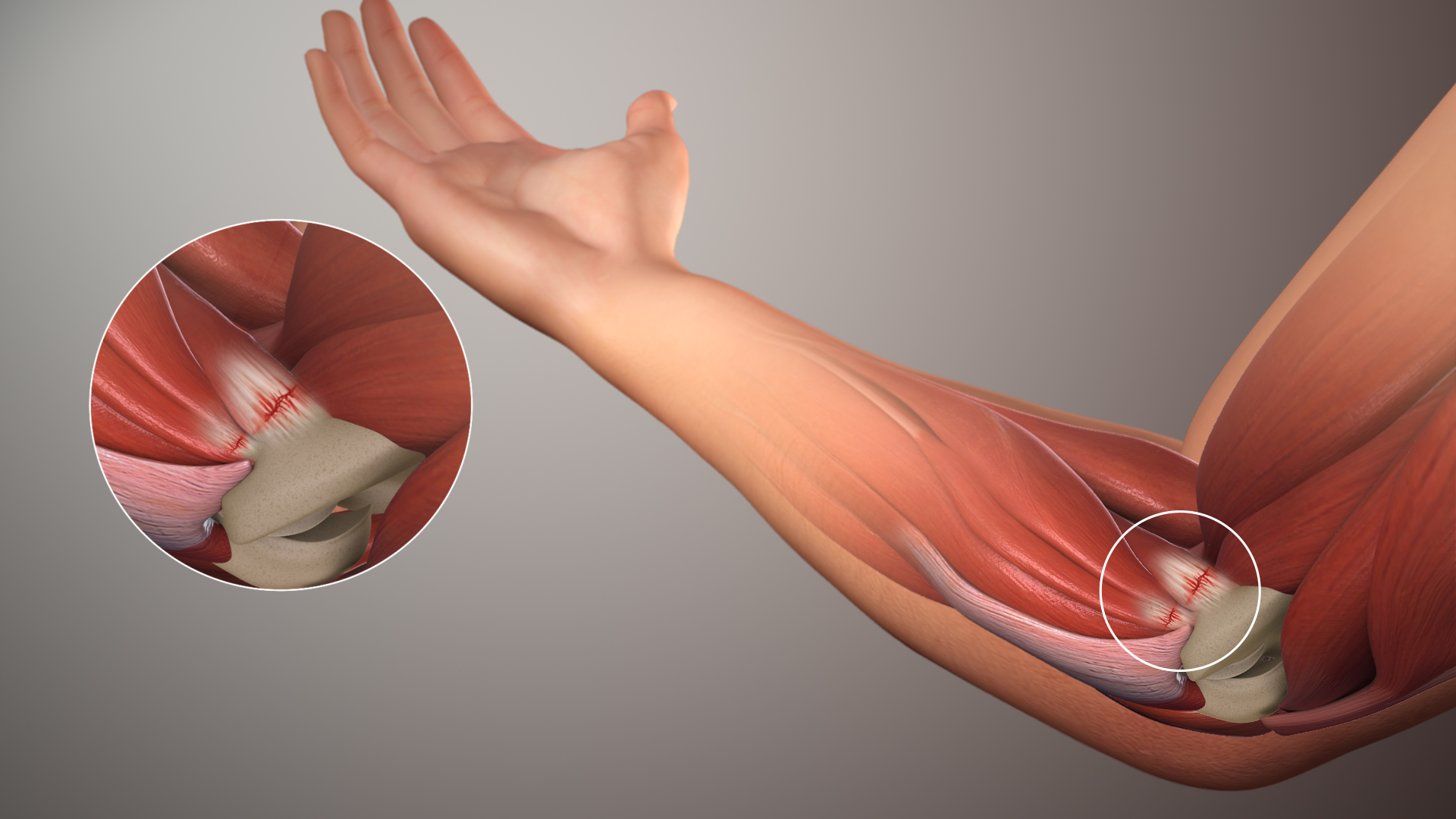
Tendinopathy is a type of tendon disorder that results in pain, swelling, and impaired function. The pain is typically worse with movement. It most commonly occurs around the shoulder ( rotator cuff tendinitis, biceps tendinitis), elbow (tennis elbow, golfer's elbow), wrist, hip, knee (jumper's knee, popliteus tendinopathy), or ankle (Achilles tendinitis). Causes may include an injury or repetitive activities. Less common causes include infection, arthritis, gout, thyroid disease, diabetes and the use of quinolone antibiotic medicines. Groups at risk include people who do manual labor, musicians, and athletes. Diagnosis is typically based on symptoms, examination, and occasionally medical imaging. A few weeks following an injury little inflammation remains, with the underlying problem related to weak or disrupted tendon fibrils. Treatment may include rest, NSAIDs, splinting, and physiotherapy. Less commonly steroid injections or surgery may be done. About 80% of overuse ... [...More Info...] [...Related Items...] OR: [Wikipedia] [Google] [Baidu] |
Tennis Elbow
Tennis elbow, also known as lateral epicondylitis is an enthesopathy (attachment point disease) of the origin of the extensor carpi radialis brevis on the lateral epicondyle. It causes pain and tenderness over the bony part of the lateral epicondyle. Symptoms range from mild tenderness to severe, persistent pain. The pain may also extend into the back of the forearm. It usually has a gradual onset, but it can seem sudden and be misinterpreted as an injury. Tennis elbow is often idiopathic. Its cause and pathogenesis are unknown. It likely involves tendinosis, a degeneration of the local tendon. It is thought this condition is caused by excessive use of the muscles of the back of the forearm, but this is not supported by evidence. It may be associated with work or sports, classically racquet sports (including paddle sports), but most people with the condition are not exposed to these activities. The diagnosis is based on the symptoms and examination. Medical imaging is not ... [...More Info...] [...Related Items...] OR: [Wikipedia] [Google] [Baidu] |
Achilles Tendon
The Achilles tendon or heel cord, also known as the calcaneal tendon, is a tendon at the back of the lower leg, and is the thickest in the human body. It serves to attach the plantaris, gastrocnemius (calf) and soleus muscles to the calcaneus (heel) bone. These muscles, acting via the tendon, cause plantar flexion of the foot at the ankle joint, and (except the soleus) flexion at the knee. Abnormalities of the Achilles tendon include inflammation ( Achilles tendinitis), degeneration, rupture, and becoming embedded with cholesterol deposits ( xanthomas). The Achilles tendon was named in 1693 after the Greek hero Achilles. History The oldest-known written record of the tendon being named after Achilles is in 1693 by the Flemish/Dutch anatomist Philip Verheyen. In his widely used text he described the tendon's location and said that it was commonly called "the cord of Achilles." The tendon has been described as early as the time of Hippocrates, who described it as th ... [...More Info...] [...Related Items...] OR: [Wikipedia] [Google] [Baidu] |
Achilles Tendinitis
Achilles tendinitis, also known as Achilles tendinopathy, is soreness of the Achilles tendon. It is accompanied by alterations in the tendon's structure and mechanical properties. The most common symptoms are pain and swelling around the back of the ankle. The pain is typically worse at the start of exercise and decreases thereafter. Stiffness of the ankle may also be present. Onset is generally gradual. Achilles tendinopathy is idiopathic, meaning the cause is not well understood. Theories of causation include overuse such as running, a lifestyle that includes little exercise, high-heel shoes, rheumatoid arthritis, and medications of the fluoroquinolone or steroid class. Diagnosis is generally based on symptoms and examination. Proposed interventions to treat tendinopathy have limited or no scientific evidence to support them, such as pre-exercise stretching, strengthening calf muscles, avoiding over-training, adjustment of running mechanics, and selection of footwear. Tre ... [...More Info...] [...Related Items...] OR: [Wikipedia] [Google] [Baidu] |
Calcific Tendinitis
Calcific tendinitis is a common condition where deposits of calcium phosphate form in a tendon, sometimes causing pain at the affected site. Deposits can occur in several places in the body, but are by far most common in the rotator cuff of the shoulder. Around 80% of those with deposits experience symptoms, typically chronic pain during certain shoulder movements, or sharp acute pain that worsens at night. Calcific tendinitis is typically diagnosed by physical exam and X-ray imaging. The disease often resolves completely on its own, but is typically treated with non-steroidal anti-inflammatory drugs to relieve pain, rest and physical therapy to promote healing, and in some cases various procedures to breakdown and/or remove the calcium deposits. Adults aged 30–50 are most commonly affected by calcific tendinitis. It is twice as common in women as men, and is not associated with exercise. Calcifications in the rotator cuff were first described by Ernest Codman in 1934. The name, ... [...More Info...] [...Related Items...] OR: [Wikipedia] [Google] [Baidu] |
Jumper's Knee
Patellar tendinitis, also known as jumper's knee, is an overuse injury of the tendon that straightens the knee. Symptoms include pain in the front of the knee. Typically the pain and tenderness is at the lower part of the kneecap, though the upper part may also be affected. Generally there is no pain when the person is at rest. Complications may include patellar tendon rupture. Risk factors include being involved in athletics and being overweight. It is particularly common in athletes who are involved in jumping sports such as basketball and volleyball. Other risk factors include sex, age, occupation, and physical activity level. It is increasingly more likely to be developed with increasing age. The underlying mechanism involves small tears in the tendon connecting the kneecap with the shinbone. Diagnosis is generally based on symptoms and examination. Other conditions that can appear similar include infrapatellar bursitis, chondromalacia patella and patellofemoral syn ... [...More Info...] [...Related Items...] OR: [Wikipedia] [Google] [Baidu] |
Golfer's Elbow
Golfer's elbow, or medial epicondylitis, is tendinosis (or more precisely enthesopathy) of the medial common flexor tendon on the inside of the elbow. It is similar to tennis elbow, which affects the outside of the elbow at the lateral epicondyle. The tendinopathy results from overload or repetitive use of the arm, causing an injury similar to ulnar collateral ligament injury of the elbow in "pitcher's elbow". Description The anterior-medial forearm contains several muscles that flex the wrist and pronate the forearm. These muscles have a common tendinous attachment at the medial epicondyle of the humerus at the elbow joint. The flexor and pronator muscles of the forearm include the pronator teres, flexor carpi radialis, palmaris longus, and flexor digitorum superficialis, all of which originate on the medial epicondyle and are innervated by the median nerve. The flexor carpi ulnaris muscle also inserts on the medial epicondyle and is innervated by the ulnar nerve. Together, ... [...More Info...] [...Related Items...] OR: [Wikipedia] [Google] [Baidu] |
Patellar Tendinitis
Patellar tendinitis, also known as jumper's knee, is an overuse injury of the tendon that straightens the knee. Symptoms include pain in the front of the knee. Typically the pain and tenderness is at the lower part of the kneecap, though the upper part may also be affected. Generally there is no pain when the person is at rest. Complications may include patellar tendon rupture. Risk factors include being involved in athletics and being overweight. It is particularly common in athletes who are involved in jumping sports such as basketball and volleyball. Other risk factors include sex, age, occupation, and physical activity level. It is increasingly more likely to be developed with increasing age. The underlying mechanism involves small tears in the tendon connecting the kneecap with the shinbone. Diagnosis is generally based on symptoms and examination. Other conditions that can appear similar include infrapatellar bursitis, chondromalacia patella and patellofemoral syn ... [...More Info...] [...Related Items...] OR: [Wikipedia] [Google] [Baidu] |
Tendon
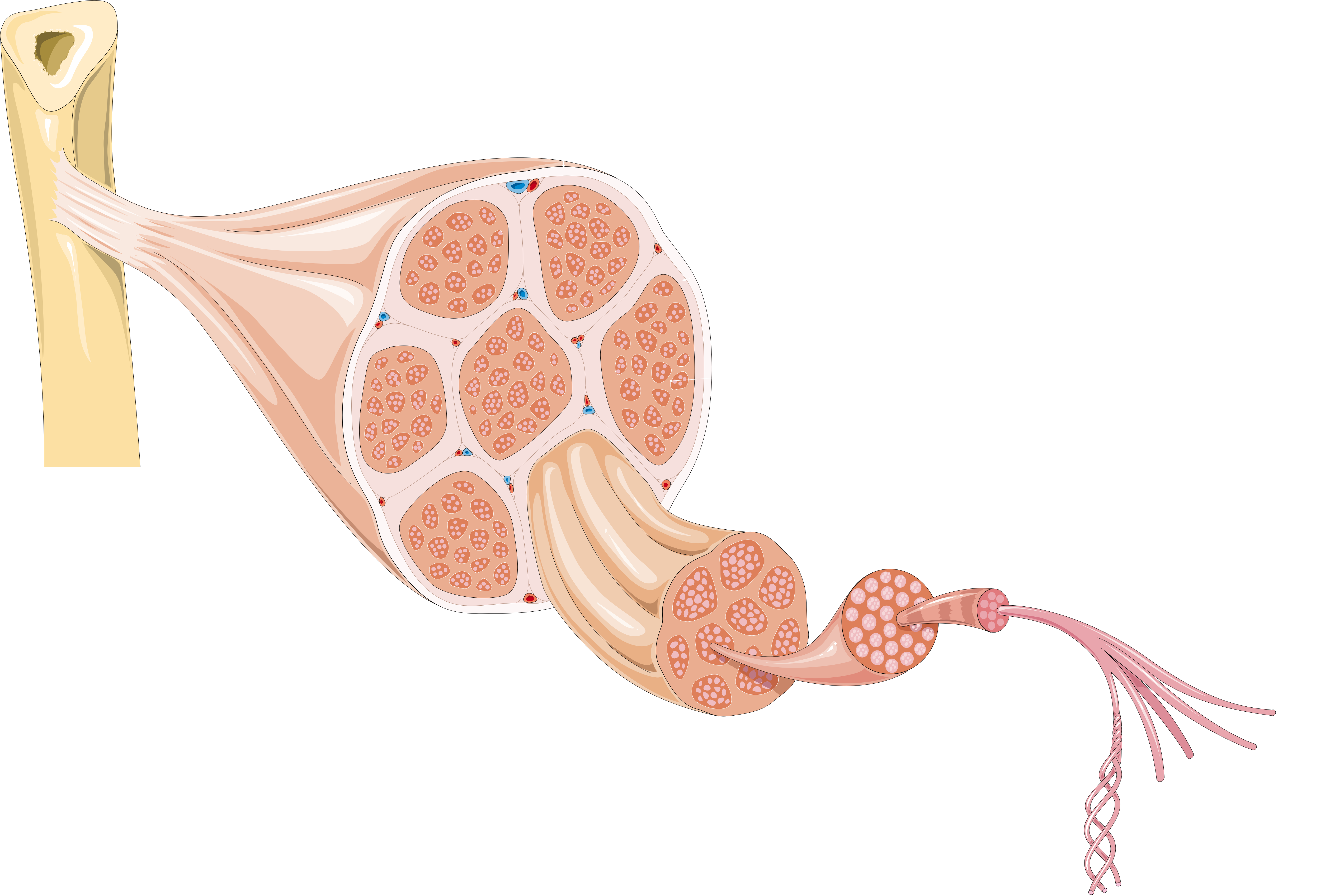
A tendon or sinew is a tough band of fibrous connective tissue, dense fibrous connective tissue that connects skeletal muscle, muscle to bone. It sends the mechanical forces of muscle contraction to the skeletal system, while withstanding tension (physics), tension. Tendons, like ligaments, are made of collagen. The difference is that ligaments connect bone to bone, while tendons connect muscle to bone. There are about 4,000 tendons in the adult human body. Structure A tendon is made of dense regular connective tissue, whose main cellular components are special fibroblasts called tendon cells (tenocytes). Tendon cells synthesize the tendon's extracellular matrix, which abounds with densely-packed collagen fibers. The collagen fibers run parallel to each other and are grouped into fascicles. Each fascicle is bound by an endotendineum, which is a delicate loose connective tissue containing thin collagen fibrils and elastic fibers. A set of fascicles is bound by an epitenon, whi ... [...More Info...] [...Related Items...] OR: [Wikipedia] [Google] [Baidu] |
Tenocyte
In animal and Human biology, a tendon cell is a cell that makes up tendons, the bands of connective tissue that connects muscles to bones. Tendon cells, also known as tenocytes or tendon fibroblasts, are specialized cells that contribute to the structure, function, and repair of tendons in the body. Tendons are fibrous tissues that connect muscles to bones, and tendon cells play a vital role in maintaining tendon homeostasis and facilitating healing following injury. Function Tendon cells are primarily responsible for the production and maintenance of the tendon extracellular matrix (ECM), which consists mainly of collagen fibers. These cells are involved in synthesizing collagen and other ECM components that provide tendons with tensile strength. Tendon cells also participate in remodeling the ECM in response to mechanical stress and injury. Structure Tendon cells are typically elongated, spindle-shaped cells that align along the axis of tendon fibers. They contain large am ... [...More Info...] [...Related Items...] OR: [Wikipedia] [Google] [Baidu] |
Pathophysiology
Pathophysiology (or physiopathology) is a branch of study, at the intersection of pathology and physiology, concerning disordered physiological processes that cause, result from, or are otherwise associated with a disease or injury. Pathology is the medical discipline that describes conditions typically ''observed'' during a disease state, whereas physiology is the biological discipline that describes processes or mechanisms ''operating'' within an organism. Pathology describes the abnormal or undesired condition (symptoms of a disease), whereas pathophysiology seeks to explain the functional changes that are occurring within an individual due to a disease or pathologic state. Etymology The term ''pathophysiology'' comes from the Ancient Greek πάθος (''pathos'') and φυσιολογία (''phisiologia''). History Early Developments The origins of pathophysiology as a distinct field date back to the late 18th century. The first known lectures on the subject were delivered ... [...More Info...] [...Related Items...] OR: [Wikipedia] [Google] [Baidu] |
Fibrils
Fibrils () are structural biological materials found in nearly all living organisms. Not to be confused with fibers or filaments, fibrils tend to have diameters ranging from 10 to 100 nanometers (whereas fibers are micro to milli-scale structures and filaments have diameters approximately 10–50 nanometers in size). Fibrils are not usually found alone but rather are parts of greater hierarchical structures commonly found in biological systems. Due to the prevalence of fibrils in biological systems, their study is of great importance in the fields of microbiology, biomechanics, and materials science. Structure and mechanics Fibrils are composed of linear biopolymers, and are characterized by rod-like structures with high length-to-diameter ratios. They often spontaneously arrange into helical structures. In biomechanics problems, fibrils can be characterized as classical beams with a roughly circular cross-sectional area on the nanometer scale. As such, simple beam bend ... [...More Info...] [...Related Items...] OR: [Wikipedia] [Google] [Baidu] |


