|
Teeth (human)
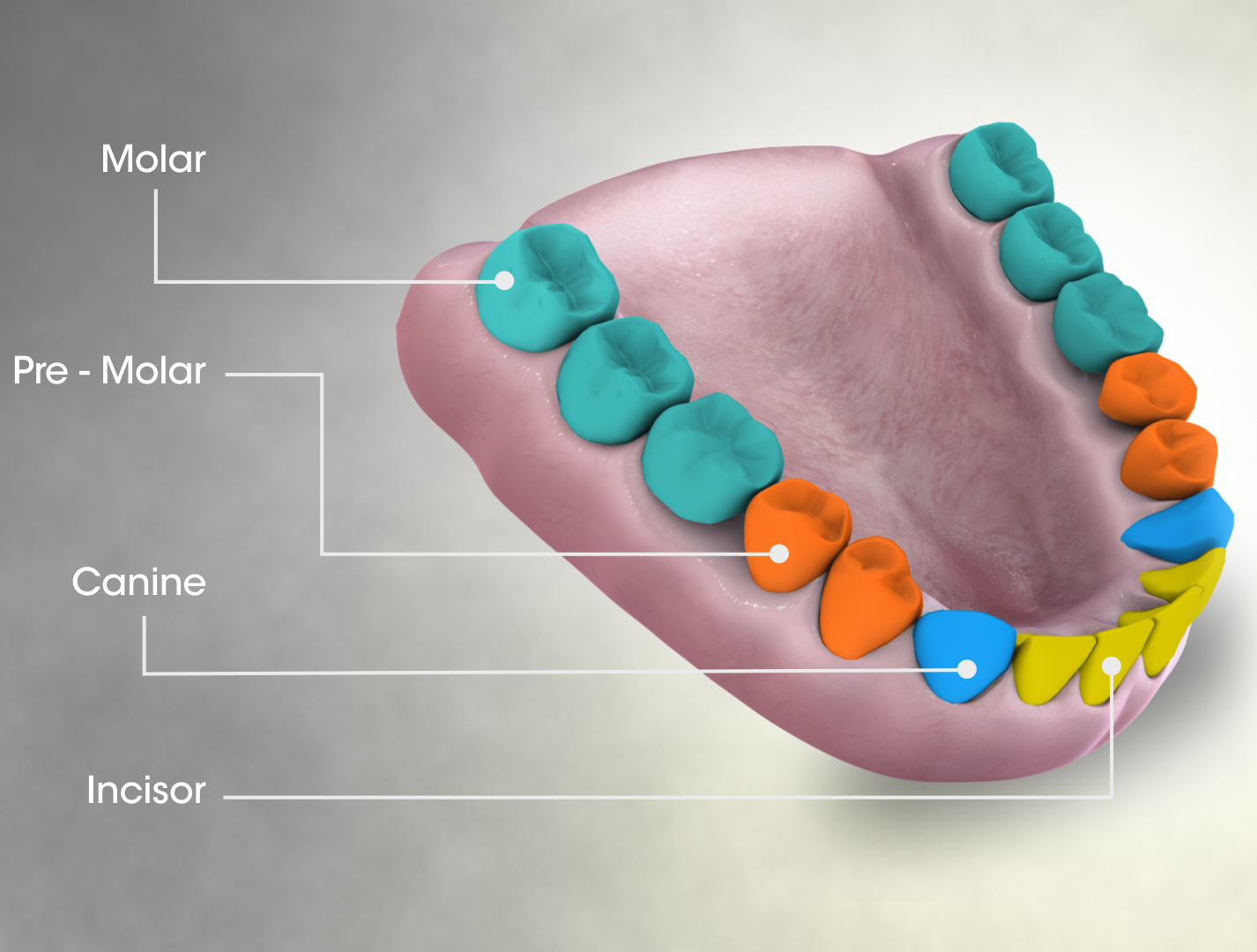
The human teeth function to Mastication, mechanically break down items of food by cutting and crushing them in preparation for swallowing and digesting. As such, they are considered part of the human digestive system. Humans have four types of tooth, teeth: incisors, Canine tooth, canines, premolars, and Molar (tooth), molars, which each have a specific function. The incisors cut the food, the canines tear the food and the molars and premolars crush the food. The roots of teeth are embedded in the maxilla (upper jaw) or the Human mandible, mandible (lower jaw) and are covered by gums. Teeth are made of multiple tissues of varying density and hardness. Humans, like most other mammals, are diphyodont, meaning that they develop two sets of teeth. The first set, deciduous teeth, also called "primary teeth", "baby teeth", or "milk teeth", normally eventually contains 20 teeth. Primary teeth typically start to appear ("tooth eruption, erupt") around six months of age and this may be ... [...More Info...] [...Related Items...] OR: [Wikipedia] [Google] [Baidu] |
Incisor
Incisors (from Latin ''incidere'', "to cut") are the front teeth present in most mammals. They are located in the premaxilla above and on the mandible below. Humans have a total of eight (two on each side, top and bottom). Opossums have 18, whereas armadillos have none. Structure Adult humans normally have eight incisors, two of each type. The types of incisor are: * maxillary central incisor (upper jaw, closest to the center of the lips) * maxillary lateral incisor (upper jaw, beside the maxillary central incisor) * mandibular central incisor (lower jaw, closest to the center of the lips) * mandibular lateral incisor (lower jaw, beside the mandibular central incisor) Children with a full set of deciduous teeth (primary teeth) also have eight incisors, named the same way as in permanent teeth. Young children may have from zero to eight incisors depending on the stage of their tooth eruption and tooth development. Typically, the mandibular central incisors erupt first, followed ... [...More Info...] [...Related Items...] OR: [Wikipedia] [Google] [Baidu] |
Dental Anatomy
Dental anatomy is a field of anatomy dedicated to the study of human tooth structures. The development, appearance, and classification of teeth fall within its purview. (The function of teeth as they contact one another falls elsewhere, under dental occlusion.) Tooth formation begins before birth, and the teeth's eventual morphology is dictated during this time. Dental anatomy is also a taxonomical science: it is concerned with the naming of teeth and the structures of which they are made, this information serving a practical purpose in dental treatment. Usually, there are 20 primary ("baby") teeth and 32 permanent teeth, the last four being third molars or "wisdom teeth", each of which may or may not grow in. Among primary teeth, 10 usually are found in the maxilla (upper jaw) and the other 10 in the mandible (lower jaw). Among permanent teeth, 16 are found in the maxilla and the other 16 in the mandible. Each tooth has specific distinguishing features. Growing of tooth ... [...More Info...] [...Related Items...] OR: [Wikipedia] [Google] [Baidu] |
Dental Notation
Dental professionals, in writing or speech, use several different dental notation systems for associating information with a specific tooth. The three most common systems are the FDI World Dental Federation notation (ISO 3950), the Universal Numbering System, and the Palmer notation. The FDI notation is used worldwide, and the Universal is used widely in the United States. The FDI notation can be easily adapted to computerized charting. Another system is used by paleoanthropologists. History A committee of the American Dental Association (ADA) recommended the use of the Palmer notation method in 1947. Since Palmer notation method required the use of symbols, its use was difficult on keyboards. As a result, the association officially supported the Universal system in 1968. The World Health Organization and the Fédération Dentaire Internationale officially uses the two-digit numbering system of the FDI system. However, in 1996, the ADA adopted the ISO System as an alternati ... [...More Info...] [...Related Items...] OR: [Wikipedia] [Google] [Baidu] |
Supernumerary Roots
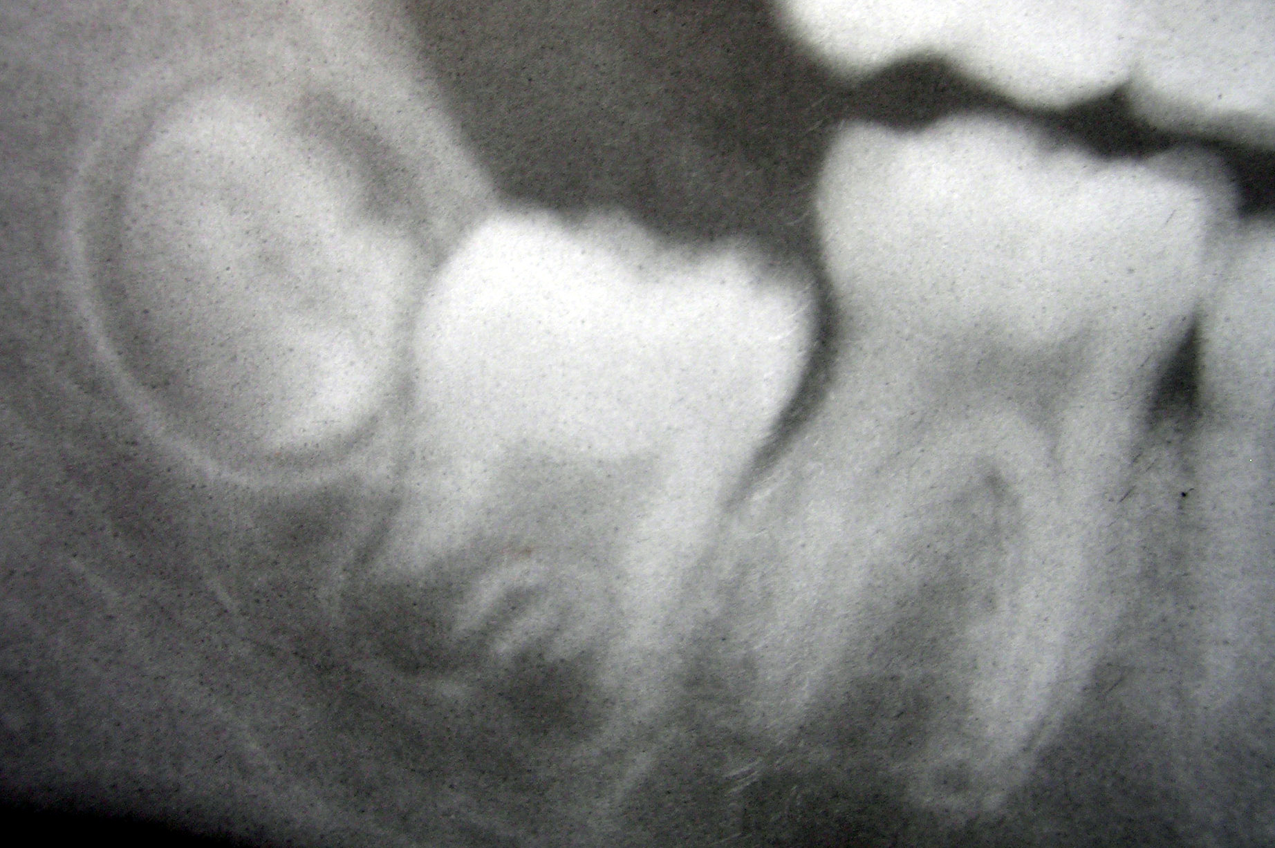
Supernumerary roots is a condition found in teeth when there may be a larger number of roots than expected. The most common teeth affected are mandibular (lower) canines, premolars, and molars, especially third molars. Canines and most premolars, except for maxillary (upper) first premolars, usually have one root. Maxillary first premolars and mandibular molars usually have two roots. Maxillary molars usually have three roots. When an extra root is found on any of these teeth, the root is described as a supernumerary root. The clinical significance of this condition is associated with dentistry when accurate information regarding root canal anatomy is required when root canal treatment Root canal treatment (also known as endodontic therapy, endodontic treatment, or root canal therapy) is a treatment sequence for the infected pulp of a tooth which is intended to result in the elimination of infection and the protection of ... is required. References External link ... [...More Info...] [...Related Items...] OR: [Wikipedia] [Google] [Baidu] |
Mandible
In anatomy, the mandible, lower jaw or jawbone is the largest, strongest and lowest bone in the human facial skeleton. It forms the lower jaw and holds the lower tooth, teeth in place. The mandible sits beneath the maxilla. It is the only movable bone of the skull (discounting the ossicles of the middle ear). It is connected to the temporal bones by the temporomandibular joints. The bone is formed prenatal development, in the fetus from a fusion of the left and right mandibular prominences, and the point where these sides join, the mandibular symphysis, is still visible as a faint ridge in the midline. Like other symphyses in the body, this is a midline articulation where the bones are joined by fibrocartilage, but this articulation fuses together in early childhood.Illustrated Anatomy of the Head and Neck, Fehrenbach and Herring, Elsevier, 2012, p. 59 The word "mandible" derives from the Latin word ''mandibula'', "jawbone" (literally "one used for chewing"), from ''wikt:mandere ... [...More Info...] [...Related Items...] OR: [Wikipedia] [Google] [Baidu] |
Maxillary (other)
{{disambig ...
Maxillary means "related to the maxilla (upper jaw bone)". Terms containing "maxillary" include: *Maxillary artery *Maxillary nerve *Maxillary prominence *Maxillary sinus The pyramid-shaped maxillary sinus (or antrum of Highmore) is the largest of the paranasal sinuses, and drains into the middle meatus of the nose through the osteomeatal complex.Human Anatomy, Jacobs, Elsevier, 2008, page 209-210 Structure It is ... [...More Info...] [...Related Items...] OR: [Wikipedia] [Google] [Baidu] |
Root Canal
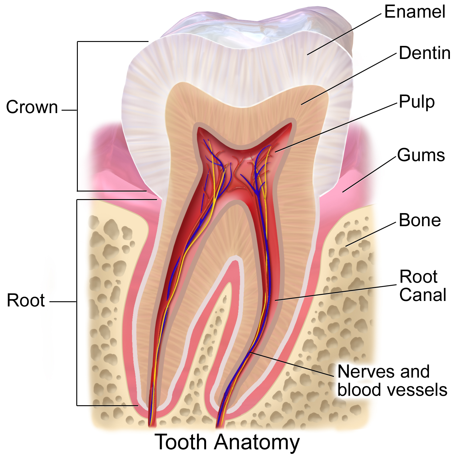
A root canal is the naturally occurring anatomic space within the root of a tooth. It consists of the pulp chamber (within the coronal part of the tooth), the main canal(s), and more intricate anatomical branches that may connect the root canals to each other or to the surface of the root. Structure At the center of every tooth is a hollow area that houses soft tissues, such as the nerve, blood vessels, and connective tissue. This hollow area contains a relatively wide space in the coronal portion of the tooth called the pulp chamber. These canals run through the center of the roots, similar to the way graphite runs through a pencil. The pulp receives nutrition through the blood vessels, and sensory nerves carry signals back to the brain. A tooth can be relieved from pain if there is irreversible damage to the pulp, via root canal treatment. Root canal anatomy consists of the pulp chamber and root canals. Both contain the dental pulp. The smaller branches, referred to as '' ... [...More Info...] [...Related Items...] OR: [Wikipedia] [Google] [Baidu] |
Cementum
Cementum is a specialized calcified substance covering the root of a tooth. The cementum is the part of the periodontium that attaches the teeth to the alveolar bone by anchoring the periodontal ligament.Illustrated Dental Embryology, Histology, and Anatomy, Bath-Balogh and Fehrenbach, Elsevier, 2011, page 170. Structure The cells of cementum are the entrapped cementoblasts, the cementocytes. Each cementocyte lies in its lacuna, similar to the pattern noted in bone. These lacunae also have canaliculi or canals. Unlike those in bone, however, these canals in cementum do not contain nerves, nor do they radiate outward. Instead, the canals are oriented toward the periodontal ligament and contain cementocytic processes that exist to diffuse nutrients from the ligament because it is vascularized. After the apposition of cementum in layers, the cementoblasts that do not become entrapped in cementum line up along the cemental surface along the length of the outer covering of the perio ... [...More Info...] [...Related Items...] OR: [Wikipedia] [Google] [Baidu] |
Dentin
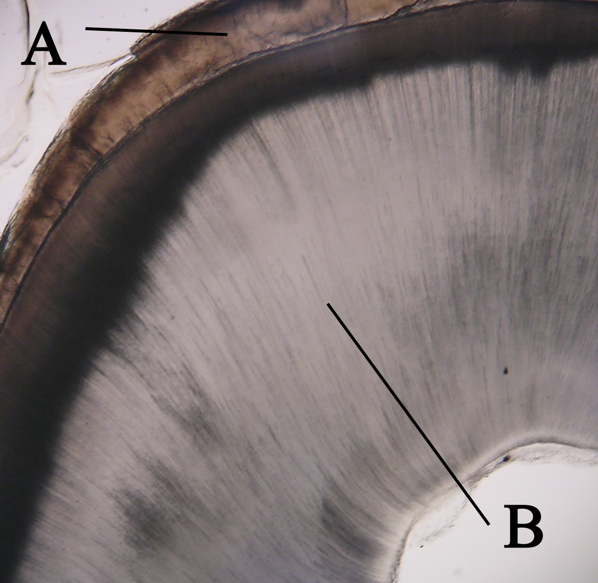
Dentin () (American English) or dentine ( or ) (British English) ( la, substantia eburnea) is a calcified tissue of the body and, along with enamel, cementum, and pulp, is one of the four major components of teeth. It is usually covered by enamel on the crown and cementum on the root and surrounds the entire pulp. By volume, 45% of dentin consists of the mineral hydroxyapatite, 33% is organic material, and 22% is water. Yellow in appearance, it greatly affects the color of a tooth due to the translucency of enamel. Dentin, which is less mineralized and less brittle than enamel, is necessary for the support of enamel. Dentin rates approximately 3 on the Mohs scale of mineral hardness. There are two main characteristics which distinguish dentin from enamel: firstly, dentin forms throughout life; secondly, dentin is sensitive and can become hypersensitive to changes in temperature due to the sensory function of odontoblasts, especially when enamel recedes and dentin channels becom ... [...More Info...] [...Related Items...] OR: [Wikipedia] [Google] [Baidu] |
Cementoenamel Junction
The cementoenamel junction, frequently abbreviated as the CEJ, is a slightly visible anatomical border identified on a tooth. It is the location where the enamel, which covers the anatomical crown of a tooth, and the cementum, which covers the anatomical root of a tooth, meet. Informally it is known as the neck of the tooth. The border created by these two dental tissues has much significance as it is usually the location where the gingiva attaches to a healthy tooth by fibers called the gingival fibers The gingival fibers are the connective tissue fibers that inhabit the gingival tissue adjacent to teeth and help hold the tissue firmly against the teeth. They are primarily composed of type I collagen, although type III fibers are also involved .... Active recession of the gingiva reveals the cementoenamel junction in the mouth and is usually a sign of an unhealthy condition. There exists a normal variation in the relationship of the cementum and the enamel at the cementoe ... [...More Info...] [...Related Items...] OR: [Wikipedia] [Google] [Baidu] |
Tooth Enamel
Tooth enamel is one of the four major Tissue (biology), tissues that make up the tooth in humans and many other animals, including some species of fish. It makes up the normally visible part of the tooth, covering the Crown (tooth), crown. The other major tissues are dentin, cementum, and Pulp (tooth), dental pulp. It is a very hard, white to off-white, highly mineralised substance that acts as a barrier to protect the tooth but can become susceptible to degradation, especially by acids from food and drink. Calcium hardens the tooth enamel. In rare circumstances enamel fails to form, leaving the underlying dentin exposed on the surface. Features Enamel is the hardest substance in the human body and contains the highest percentage of minerals (at 96%),Ross ''et al.'', p. 485 with water and organic material composing the rest.Ten Cate's Oral Histology, Nancy, Elsevier, pp. 70–94 The primary mineral is hydroxyapatite, which is a crystalline calcium phosphate. Enamel is formed o ... [...More Info...] [...Related Items...] OR: [Wikipedia] [Google] [Baidu] |
Crown (tooth)
In dentistry, crown refers to the anatomical area of teeth, usually covered by enamel. The crown is usually visible in the mouth after developing below the gingiva The gums or gingiva (plural: ''gingivae'') consist of the mucosal tissue that lies over the mandible and maxilla inside the mouth. Gum health and disease can have an effect on general health. Structure The gums are part of the soft tissue lin ... and then erupting into place. If part of the tooth gets chipped or broken, a dentist can apply an artificial crown. Crowns are used most commonly to entirely cover a damaged tooth or cover an implant. Bridges are also used to cover a space if one or more teeth is missing. They are cemented to natural teeth or implants surrounding the space where the tooth once stood. There are various materials that can be used including a type of cement or stainless steel. The cement crowns look like regular teeth while the stainless steel crowns are silver or gold. References ... [...More Info...] [...Related Items...] OR: [Wikipedia] [Google] [Baidu] |


