|
Superior Thyroid Vein
The superior thyroid vein begins in the substance and on the surface of the thyroid gland, by tributaries corresponding with the branches of the superior thyroid artery, and ends in the upper part of the internal jugular vein. It receives the superior laryngeal vein, superior laryngeal and cricothyroid veins. Additional images File:Gray577.png, The venae cavae and azygos veins with their tributaries. File:Gray1178.png, The thymus of a full-time fetus. References Veins of the head and neck Thyroid {{circulatory-stub ... [...More Info...] [...Related Items...] OR: [Wikipedia] [Google] [Baidu] |
Thyroid Gland
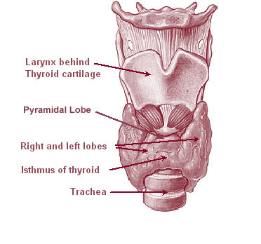
The thyroid, or thyroid gland, is an endocrine gland in vertebrates. In humans it is in the neck and consists of two connected lobe (anatomy), lobes. The lower two thirds of the lobes are connected by a thin band of Connective tissue, tissue called the thyroid isthmus. The thyroid is located at the front of the neck, below the Adam's apple. Microscopically, the functional unit of the thyroid gland is the spherical Thyroid follicular cell#Location, thyroid follicle, lined with thyroid follicular cell, follicular cells (thyrocytes), and occasional parafollicular cells that surround a follicular lumen, lumen containing colloid. The thyroid gland secretes three hormones: the two thyroid hormonestriiodothyronine, triiodothyronine (T3) and thyroid hormone, thyroxine (T4)and a peptide hormone, calcitonin. The thyroid hormones influence the basal metabolic rate, metabolic rate and protein biosynthesis, protein synthesis, and in children, growth and development. Calcitonin plays a role in ... [...More Info...] [...Related Items...] OR: [Wikipedia] [Google] [Baidu] |
Internal Jugular Vein
The internal jugular vein is a paired jugular vein that collects blood from the brain and the superficial parts of the face and neck. This vein runs in the carotid sheath with the common carotid artery and vagus nerve. It begins in the posterior compartment of the jugular foramen, at the base of the skull. It is somewhat dilated at its origin, which is called the ''superior bulb''. This vein also has a common trunk into which drains the anterior branch of the retromandibular vein, the facial vein, and the lingual vein. It runs down the side of the neck in a vertical direction, being at one end lateral to the internal carotid artery, and then lateral to the common carotid artery, and at the root of the neck, it unites with the subclavian vein to form the brachiocephalic vein (innominate vein); a little above its termination is a second dilation, the ''inferior bulb''. Above, it lies upon the rectus capitis lateralis, behind the internal carotid artery and the nerves passing ... [...More Info...] [...Related Items...] OR: [Wikipedia] [Google] [Baidu] |
Superior Thyroid Artery
The superior thyroid artery arises from the external carotid artery just below the level of the greater cornu of the hyoid bone and ends in the thyroid gland. Structure From its origin under the anterior border of the sternocleidomastoid the superior thyroid artery runs upward and forward for a short distance in the carotid triangle, where it is covered by the skin, platysma, and fascia; it then arches downward beneath the omohyoid, sternohyoid, and sternothyroid muscles. To its medial side are the inferior pharyngeal constrictor muscle and the external branch of the superior laryngeal nerve. Branches It distributes twigs to the adjacent muscles, and numerous branches to the thyroid gland, connecting with its fellow of the opposite side, and with the inferior thyroid arteries. The branches to the gland are generally two in number. One, the larger, supplies principally the anterior surface; on the isthmus of the gland it connects with the corresponding artery of the opposite ... [...More Info...] [...Related Items...] OR: [Wikipedia] [Google] [Baidu] |
Internal Jugular Vein
The internal jugular vein is a paired jugular vein that collects blood from the brain and the superficial parts of the face and neck. This vein runs in the carotid sheath with the common carotid artery and vagus nerve. It begins in the posterior compartment of the jugular foramen, at the base of the skull. It is somewhat dilated at its origin, which is called the ''superior bulb''. This vein also has a common trunk into which drains the anterior branch of the retromandibular vein, the facial vein, and the lingual vein. It runs down the side of the neck in a vertical direction, being at one end lateral to the internal carotid artery, and then lateral to the common carotid artery, and at the root of the neck, it unites with the subclavian vein to form the brachiocephalic vein (innominate vein); a little above its termination is a second dilation, the ''inferior bulb''. Above, it lies upon the rectus capitis lateralis, behind the internal carotid artery and the nerves passing ... [...More Info...] [...Related Items...] OR: [Wikipedia] [Google] [Baidu] |
Superior Laryngeal Vein
The superior laryngeal vein is a vein which drains the larynx into the superior thyroid vein The superior thyroid vein begins in the substance and on the surface of the thyroid gland, by tributaries corresponding with the branches of the superior thyroid artery, and ends in the upper part of the internal jugular vein. It receives the supe .... Veins of the head and neck {{circulatory-stub ... [...More Info...] [...Related Items...] OR: [Wikipedia] [Google] [Baidu] |
Cricothyroid Veins
Cricothyroid may refer to: * Cricothyroid muscle The cricothyroid muscle is the only tensor muscle of the larynx aiding with phonation. It is innervated by the superior laryngeal nerve. Its action tilts the thyroid forward to help tense the vocal cords. Structure The cricothyroid muscle o ... * Cricothyroid ligament {{disambig ... [...More Info...] [...Related Items...] OR: [Wikipedia] [Google] [Baidu] |
Venae Cavae
In anatomy, the venae cavae (; singular: vena cava ; ) are two large veins (great vessels) that return deoxygenated blood from the body into the heart. In humans they are the superior vena cava and the inferior vena cava, and both empty into the right atrium. They are located slightly off-center, toward the right side of the body. The right atrium receives deoxygenated blood through coronary sinus and two large veins called venae cavae. The inferior vena cava (or caudal vena cava in some animals) travels up alongside the abdominal aorta with blood from the lower part of the body. It is the largest vein in the human body. MadSci Network: Anatomy. Retrieved 19 September 2013. The superior vena cava (or cranial vena cava in animals) is above the heart, and ... [...More Info...] [...Related Items...] OR: [Wikipedia] [Google] [Baidu] |
Azygos Vein
The azygos vein is a vein running up the right side of the thoracic vertebral column draining itself towards the superior vena cava. It connects the systems of superior vena cava and inferior vena cava and can provide an alternative path for blood to the right atrium when either of the venae cavae is blocked. Structure The azygos vein transports deoxygenated blood from the posterior walls of the thorax and abdomen into the superior vena cava. It is formed by the union of the ascending lumbar veins with the right subcostal veins at the level of the 12th thoracic vertebra, ascending to the right of the descending aorta and thoracic duct, passing behind the right crus of diaphragm, anterior to the vertebral bodies of T12 to T5 and right posterior intercostal arteries. At the level of T4 vertebrae, it arches over the root of the right lung from behind to the front to join the superior vena cava. The trachea and oesophagus is located medially to the arch of the azygous vein. The ... [...More Info...] [...Related Items...] OR: [Wikipedia] [Google] [Baidu] |
Thymus
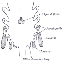
The thymus is a specialized primary lymphoid organ of the immune system. Within the thymus, thymus cell lymphocytes or ''T cells'' mature. T cells are critical to the adaptive immune system, where the body adapts to specific foreign invaders. The thymus is located in the upper front part of the chest, in the anterior superior mediastinum, behind the sternum, and in front of the heart. It is made up of two lobes, each consisting of a central medulla and an outer cortex, surrounded by a capsule. The thymus is made up of immature T cells called thymocytes, as well as lining cells called epithelial cells which help the thymocytes develop. T cells that successfully develop react appropriately with MHC immune receptors of the body (called ''positive selection'') and not against proteins of the body (called ''negative selection''). The thymus is largest and most active during the neonatal and pre-adolescent periods. By the early teens, the thymus begins to decrease in size and a ... [...More Info...] [...Related Items...] OR: [Wikipedia] [Google] [Baidu] |
Fetus
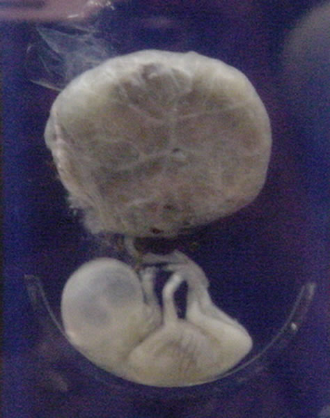
A fetus or foetus (; plural fetuses, feti, foetuses, or foeti) is the unborn offspring that develops from an animal embryo. Following embryonic development the fetal stage of development takes place. In human prenatal development, fetal development begins from the ninth week after fertilization (or eleventh week gestational age) and continues until birth. Prenatal development is a continuum, with no clear defining feature distinguishing an embryo from a fetus. However, a fetus is characterized by the presence of all the major body organs, though they will not yet be fully developed and functional and some not yet situated in their final anatomical location. Etymology The word ''fetus'' (plural ''fetuses'' or '' feti'') is related to the Latin '' fētus'' ("offspring", "bringing forth", "hatching of young") and the Greek "φυτώ" to plant. The word "fetus" was used by Ovid in Metamorphoses, book 1, line 104. The predominant British, Irish, and Commonwealth spelling is '' ... [...More Info...] [...Related Items...] OR: [Wikipedia] [Google] [Baidu] |
Veins Of The Head And Neck
Veins are blood vessels in humans and most other animals that carry blood towards the heart. Most veins carry deoxygenated blood from the tissues back to the heart; exceptions are the pulmonary and umbilical veins, both of which carry oxygenated blood to the heart. In contrast to veins, arteries carry blood away from the heart. Veins are less muscular than arteries and are often closer to the skin. There are valves (called ''pocket valves'') in most veins to prevent backflow. Structure Veins are present throughout the body as tubes that carry blood back to the heart. Veins are classified in a number of ways, including superficial vs. deep, pulmonary vs. systemic, and large vs. small. *Superficial veins are those closer to the surface of the body, and have no corresponding arteries. *Deep veins are deeper in the body and have corresponding arteries. * Perforator veins drain from the superficial to the deep veins. These are usually referred to in the lower limbs and feet. *Communic ... [...More Info...] [...Related Items...] OR: [Wikipedia] [Google] [Baidu] |


