|
Superior Cerebral Veins
The superior cerebral veins, numbering eight to twelve, drain the superior, lateral, and medial surfaces of the hemispheres. They are predominantly found in the sulci between the gyri, but can also be found running across the gyri. Individually they drain into the superior sagittal sinus The superior sagittal sinus (also known as the superior longitudinal sinus), within the human head, is an unpaired area along the attached margin of the falx cerebri. It allows blood to drain from the lateral aspects of anterior cerebral hemispher .... The anterior veins run at near right angles to the sinus while the posterior and larger veins are directed at oblique angles, opening into the sinus in a direction opposed to the current (anterior to posterior) of the blood contained within it. Additional images File:Slide6Neo.JPG, Meninges and superficial cerebral veins. Deep dissection. Superior view. References External links Diagram Veins of the head and neck {{circulatory-stub ... [...More Info...] [...Related Items...] OR: [Wikipedia] [Google] [Baidu] |
Superior Sagittal Sinus
The superior sagittal sinus (also known as the superior longitudinal sinus), within the human head, is an unpaired area along the attached margin of the falx cerebri. It allows blood to drain from the lateral aspects of anterior cerebral hemispheres to the confluence of sinuses. Cerebrospinal fluid drains through arachnoid granulations into the superior sagittal sinus and is returned to venous circulation. Structure Commencing at the foramen cecum, through which it receives emissary veins from the nasal cavity, it runs from anterior to posterior, grooving the inner surface of the frontal, the adjacent margins of the two parietal lobes, and the superior division of the cruciate eminence of the occipital lobe. Near the internal occipital protuberance, it drains into the confluence of sinuses and deviates to either side (usually the right). At this point it is continued as the corresponding transverse sinus. The superior sagittal sinus is usually divided into three parts: anterior ... [...More Info...] [...Related Items...] OR: [Wikipedia] [Google] [Baidu] |
Cerebral Arteries
The cerebral arteries describe three main pairs of arteries and their branches, which perfuse the cerebrum of the brain. The three main arteries are the: * ''Anterior cerebral artery'' (ACA) * ''Middle cerebral artery'' (MCA) * ''Posterior cerebral artery'' (PCA) Both the ACA and MCA originate from the cerebral portion of internal carotid artery, while PCA branches from the intersection of the posterior communicating artery and the anterior portion of the basilar artery. The three pairs of arteries are linked via the anterior communicating artery and the posterior communicating arteries In human anatomy, the left and right posterior communicating arteries are arteries at the base of the brain that form part of the circle of Willis The circle of Willis (also called Willis' circle, loop of Willis, cerebral arterial circle, and W .... All three arteries send out arteries that perforate brain in the medial central portions prior to branching and bifurcating further. The arteries ... [...More Info...] [...Related Items...] OR: [Wikipedia] [Google] [Baidu] |
Sulcus (neuroanatomy)
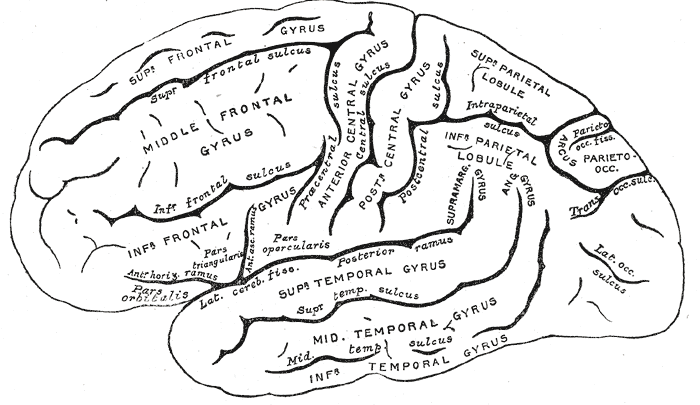
In neuroanatomy, a sulcus (Latin: "furrow", pl. ''sulci'') is a depression or groove in the cerebral cortex. It surrounds a gyrus (pl. gyri), creating the characteristic folded appearance of the brain in humans and other mammals. The larger sulci are usually called fissures. Structure Sulci, the grooves, and gyri, the folds or ridges, make up the folded surface of the cerebral cortex. Larger or deeper sulci are termed fissures, and in many cases the two terms are interchangeable. The folded cortex creates a larger surface area for the brain in humans and other mammals. When looking at the human brain, two-thirds of the surface are hidden in the grooves. The sulci and fissures are both grooves in the cortex, but they are differentiated by size. A sulcus is a shallower groove that surrounds a gyrus. A fissure is a large furrow that divides the brain into lobes and also into the two hemispheres as the longitudinal fissure. Importance of expanded surface area As the surfac ... [...More Info...] [...Related Items...] OR: [Wikipedia] [Google] [Baidu] |
Gyri
In neuroanatomy, a gyrus (pl. gyri) is a ridge on the cerebral cortex. It is generally surrounded by one or more sulci (depressions or furrows; sg. ''sulcus''). Gyri and sulci create the folded appearance of the brain in humans and other mammals. Structure The gyri are part of a system of folds and ridges that create a larger surface area for the human brain and other mammalian brains. Because the brain is confined to the skull, brain size is limited. Ridges and depressions create folds allowing a larger cortical surface area, and greater cognitive function, to exist in the confines of a smaller cranium. Development The human brain undergoes gyrification during fetal and neonatal development. In embryonic development, all mammalian brains begin as smooth structures derived from the neural tube. A cerebral cortex without surface convolutions is lissencephalic, meaning 'smooth-brained'. As development continues, gyri and sulci begin to take shape on the fetal brain, with ... [...More Info...] [...Related Items...] OR: [Wikipedia] [Google] [Baidu] |
