|
Super-resolution Microscopy
Super-resolution microscopy is a series of techniques in optical microscopy that allow such images to have resolutions higher than those imposed by the diffraction limit, which is due to the diffraction of light. Super-resolution imaging techniques rely on the near-field (photon-tunneling microscopy as well as those that utilize the Pendry Superlens and near field scanning optical microscopy) or on the far-field. Among techniques that rely on the latter are those that improve the resolution only modestly (up to about a factor of two) beyond the diffraction-limit, such as confocal microscopy with closed pinhole or aided by computational methods such as deconvolution or detector-based pixel reassignment (e.g. re-scan microscopy, pixel reassignment), the 4Pi microscope, and structured-illumination microscopy technologies such as SIM and SMI. There are two major groups of methods for super-resolution microscopy in the far-field that can improve the resolution by a much larger f ... [...More Info...] [...Related Items...] OR: [Wikipedia] [Google] [Baidu] |
Microscopy
Microscopy is the technical field of using microscopes to view objects and areas of objects that cannot be seen with the naked eye (objects that are not within the resolution range of the normal eye). There are three well-known branches of microscopy: optical, electron, and scanning probe microscopy, along with the emerging field of X-ray microscopy. Optical microscopy and electron microscopy involve the diffraction, reflection, or refraction of electromagnetic radiation/electron beams interacting with the specimen, and the collection of the scattered radiation or another signal in order to create an image. This process may be carried out by wide-field irradiation of the sample (for example standard light microscopy and transmission electron microscopy) or by scanning a fine beam over the sample (for example confocal laser scanning microscopy and scanning electron microscopy). Scanning probe microscopy involves the interaction of a scanning probe with the surface of the ... [...More Info...] [...Related Items...] OR: [Wikipedia] [Google] [Baidu] |
Photoactivated Localization Microscopy
Photo-activated localization microscopy (PALM or FPALM) and stochastic optical reconstruction microscopy (STORM) are widefield (as opposed to point scanning techniques such as laser scanning confocal microscopy) fluorescence microscopy imaging methods that allow obtaining images with a resolution beyond the diffraction limit. The methods were proposed in 2006 in the wake of a general emergence of optical super-resolution microscopy methods, and were featured as Methods of the Year for 2008 by the ''Nature Methods'' journal. The development of PALM as a targeted biophysical imaging method was largely prompted by the discovery of new species and the engineering of mutants of fluorescent proteins displaying a controllable photochromism, such as photo-activatible GFP. However, the concomitant development of STORM, sharing the same fundamental principle, originally made use of paired cyanine dyes. One molecule of the pair (called activator), when excited near its absorption maxim ... [...More Info...] [...Related Items...] OR: [Wikipedia] [Google] [Baidu] |
Optical Path Length
In optics, optical path length (OPL, denoted ''Λ'' in equations), also known as optical length or optical distance, is the product of the geometric length of the optical path followed by light and the refractive index of homogeneous medium through which a light ray propagates; for inhomogeneous optical media, the product above is generalized as a path integral as part of the ray tracing procedure. A difference in OPL between two paths is often called the optical path difference (OPD). OPL and OPD are important because they determine the phase of the light and governs interference and diffraction of light as it propagates. Formulation In a medium of constant refractive index, ''n'', the OPL for a path of geometrical length ''s'' is just :\mathrm = n s .\, If the refractive index varies along the path, the OPL is given by a line integral :\mathrm = \int_C n \mathrm d s,\quad where ''n'' is the local refractive index as a function of distance along the path ''C''. An e ... [...More Info...] [...Related Items...] OR: [Wikipedia] [Google] [Baidu] |
Optical Axis
An optical axis is a line along which there is some degree of rotational symmetry in an optical system such as a camera lens, microscope or telescopic sight. The optical axis is an imaginary line that defines the path along which light propagates through the system, up to first approximation. For a system composed of simple lenses and mirrors, the axis passes through the center of curvature of each surface, and coincides with the axis of rotational symmetry. The optical axis is often coincident with the system's mechanical axis, but not always, as in the case of off-axis optical systems. For an optical fiber, the optical axis is along the center of the fiber core, and is also known as the ''fiber axis''. See also * Ray (optics) * Cardinal point (optics) * Antenna boresight In telecommunications and radar engineering, antenna boresight is the axis of maximum gain (maximum radiated power) of a directional antenna. For most antennas the boresight is the axis of symmetry of t ... [...More Info...] [...Related Items...] OR: [Wikipedia] [Google] [Baidu] |
Fluorescence Microscope
A fluorescence microscope is an optical microscope that uses fluorescence instead of, or in addition to, scattering, reflection, and attenuation or absorption, to study the properties of organic or inorganic substances. "Fluorescence microscope" refers to any microscope that uses fluorescence to generate an image, whether it is a simple set up like an epifluorescence microscope or a more complicated design such as a confocal microscope, which uses optical sectioning to get better resolution of the fluorescence image. Principle The specimen is illuminated with light of a specific wavelength (or wavelengths) which is absorbed by the fluorophores, causing them to emit light of longer wavelengths (i.e., of a different color than the absorbed light). The illumination light is separated from the much weaker emitted fluorescence through the use of a spectral emission filter. Typical components of a fluorescence microscope are a light source ( xenon arc lamp or mercury-vapor lam ... [...More Info...] [...Related Items...] OR: [Wikipedia] [Google] [Baidu] |
4Pi Microscope
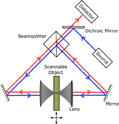
A 4Pi microscope is a laser scanning fluorescence microscope with an improved axial resolution. With it the typical range of the axial resolution of 500–700 nm can be improved to 100–150 nm, which corresponds to an almost spherical focal spot with 5–7 times less volume than that of standard confocal microscopy. Working principle The improvement in resolution is achieved by using two opposing objective lenses, which both are focused to the same geometrical location. Also the difference in optical path length through each of the two objective lenses is carefully aligned to be minimal. By this method, molecules residing in the common focal area of both objectives can be illuminated coherently from both sides and the reflected or emitted light can also be collected coherently, i.e. coherent superposition of emitted light on the detector is possible. The solid angle \Omega that is used for illumination and detection is increased and approaches its maximum. In this case ... [...More Info...] [...Related Items...] OR: [Wikipedia] [Google] [Baidu] |
Abbe Limit
The resolution of an optical imaging system a microscope, telescope, or camera can be limited by factors such as imperfections in the lenses or misalignment. However, there is a principal limit to the resolution of any optical system, due to the physics of diffraction. An optical system with resolution performance at the instrument's theoretical limit is said to be diffraction-limited. The diffraction-limited angular resolution of a telescopic instrument is inversely proportional to the wavelength of the light being observed, and proportional to the diameter of its objective's entrance aperture. For telescopes with circular apertures, the size of the smallest feature in an image that is diffraction limited is the size of the Airy disk. As one decreases the size of the aperture of a telescopic lens, diffraction proportionately increases. At small apertures, such as f/22, most modern lenses are limited only by diffraction and not by aberrations or other imperfections in the co ... [...More Info...] [...Related Items...] OR: [Wikipedia] [Google] [Baidu] |
The New York Times
''The New York Times'' (''the Times'', ''NYT'', or the Gray Lady) is a daily newspaper based in New York City with a worldwide readership reported in 2020 to comprise a declining 840,000 paid print subscribers, and a growing 6 million paid digital subscribers. It also is a producer of popular podcasts such as '' The Daily''. Founded in 1851 by Henry Jarvis Raymond and George Jones, it was initially published by Raymond, Jones & Company. The ''Times'' has won 132 Pulitzer Prizes, the most of any newspaper, and has long been regarded as a national "newspaper of record". For print it is ranked 18th in the world by circulation and 3rd in the U.S. The paper is owned by the New York Times Company, which is publicly traded. It has been governed by the Sulzberger family since 1896, through a dual-class share structure after its shares became publicly traded. A. G. Sulzberger, the paper's publisher and the company's chairman, is the fifth generation of the family to head the p ... [...More Info...] [...Related Items...] OR: [Wikipedia] [Google] [Baidu] |
Nanoscopic Scale
The nanoscopic scale (or nanoscale) usually refers to structures with a length scale applicable to nanotechnology, usually cited as 1–100 nanometers (nm). A nanometer is a billionth of a meter. The nanoscopic scale is (roughly speaking) a lower bound to the mesoscopic scale for most solids. For technical purposes, the nanoscopic scale is the size at which fluctuations in the averaged properties (due to the motion and behavior of individual particles) begin to have a significant effect (often a few percent) on the behavior of a system, and must be taken into account in its analysis. The nanoscopic scale is sometimes marked as the point where the properties of a material change; above this point, the properties of a material are caused by 'bulk' or 'volume' effects, namely which atoms are present, how they are bonded, and in what ratios. Below this point, the properties of a material change, and while the type of atoms present and their relative orientations are still importa ... [...More Info...] [...Related Items...] OR: [Wikipedia] [Google] [Baidu] |
Optical Microscopy
Optics is the branch of physics that studies the behaviour and properties of light, including its interactions with matter and the construction of instruments that use or detect it. Optics usually describes the behaviour of visible, ultraviolet, and infrared light. Because light is an electromagnetic wave, other forms of electromagnetic radiation such as X-rays, microwaves, and radio waves exhibit similar properties. Most optical phenomena can be accounted for by using the classical electromagnetic description of light. Complete electromagnetic descriptions of light are, however, often difficult to apply in practice. Practical optics is usually done using simplified models. The most common of these, geometric optics, treats light as a collection of rays that travel in straight lines and bend when they pass through or reflect from surfaces. Physical optics is a more comprehensive model of light, which includes wave effects such as diffraction and interference that cannot b ... [...More Info...] [...Related Items...] OR: [Wikipedia] [Google] [Baidu] |
Fluorescence Microscopy
A fluorescence microscope is an optical microscope that uses fluorescence instead of, or in addition to, scattering, reflection, and attenuation or absorption, to study the properties of organic or inorganic substances. "Fluorescence microscope" refers to any microscope that uses fluorescence to generate an image, whether it is a simple set up like an epifluorescence microscope or a more complicated design such as a confocal microscope, which uses optical sectioning to get better resolution of the fluorescence image. Principle The specimen is illuminated with light of a specific wavelength (or wavelengths) which is absorbed by the fluorophores, causing them to emit light of longer wavelengths (i.e., of a different color than the absorbed light). The illumination light is separated from the much weaker emitted fluorescence through the use of a spectral emission filter. Typical components of a fluorescence microscope are a light source ( xenon arc lamp or mercury-vapor ... [...More Info...] [...Related Items...] OR: [Wikipedia] [Google] [Baidu] |


.png)
