|
Subparietal Sulcus
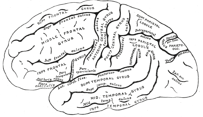
In neuroanatomy, the subparietal sulcus () or suprasplenial sulcus is a sulcus, or crevice, on the medial surface of each cerebral hemisphere, above the splenium of the corpus callosum. It separates the precuneus from the posterior part of the cingulate gyrus. It is the posterior continuation of the cingulate sulcus. The cingulate sulcus actually "terminates" as the marginal sulcus of the cingulate sulcus (margin of cingulate gyrus). It extends posteriorly toward the calcarine sulcus. The precuneus is bordered anteriorly by the marginal branch of the cingulate sulcus (margin of cingulate sulcus), posteriorly by the parieto-occipital sulcus In neuroanatomy, the parieto-occipital sulcus (also called the parieto-occipital fissure) is a deep sulcus in the cerebral cortex that marks the boundary between the cuneus and precuneus, and also between the parietal and occipital lobes. Only ..., and inferiorly by the subparietal sulcus. Additional images References * Michio On ... [...More Info...] [...Related Items...] OR: [Wikipedia] [Google] [Baidu] |
Cerebral Hemisphere
The vertebrate cerebrum (brain) is formed by two cerebral hemispheres that are separated by a groove, the longitudinal fissure. The brain can thus be described as being divided into left and right cerebral hemispheres. Each of these hemispheres has an outer layer of grey matter, the cerebral cortex, that is supported by an inner layer of white matter. In eutherian (placental) mammals, the hemispheres are linked by the corpus callosum, a very large bundle of nerve fibers. Smaller commissures, including the anterior commissure, the posterior commissure and the fornix, also join the hemispheres and these are also present in other vertebrates. These commissures transfer information between the two hemispheres to coordinate localized functions. There are three known poles of the cerebral hemispheres: the ''occipital pole'', the ''frontal pole'', and the ''temporal pole''. The central sulcus is a prominent fissure which separates the parietal lobe from the frontal lobe and the prim ... [...More Info...] [...Related Items...] OR: [Wikipedia] [Google] [Baidu] |
Neuroanatomy
Neuroanatomy is the study of the structure and organization of the nervous system. In contrast to animals with radial symmetry, whose nervous system consists of a distributed network of cells, animals with bilateral symmetry have segregated, defined nervous systems. Their neuroanatomy is therefore better understood. In vertebrates, the nervous system is segregated into the internal structure of the brain and spinal cord (together called the central nervous system, or CNS) and the routes of the nerves that connect to the rest of the body (known as the peripheral nervous system, or PNS). The delineation of distinct structures and regions of the nervous system has been critical in investigating how it works. For example, much of what neuroscientists have learned comes from observing how damage or "lesions" to specific brain areas affects behavior or other neural functions. For information about the composition of non-human animal nervous systems, see nervous system. For information ab ... [...More Info...] [...Related Items...] OR: [Wikipedia] [Google] [Baidu] |
Sulcus (neuroanatomy)
In neuroanatomy, a sulcus (Latin: "furrow", pl. ''sulci'') is a depression or groove in the cerebral cortex. It surrounds a gyrus (pl. gyri), creating the characteristic folded appearance of the brain in humans and other mammals. The larger sulci are usually called fissures. Structure Sulci, the grooves, and gyri, the folds or ridges, make up the folded surface of the cerebral cortex. Larger or deeper sulci are termed fissures, and in many cases the two terms are interchangeable. The folded cortex creates a larger surface area for the brain in humans and other mammals. When looking at the human brain, two-thirds of the surface are hidden in the grooves. The sulci and fissures are both grooves in the cortex, but they are differentiated by size. A sulcus is a shallower groove that surrounds a gyrus. A fissure is a large furrow that divides the brain into lobes and also into the two hemispheres as the longitudinal fissure. Importance of expanded surface area As the surfac ... [...More Info...] [...Related Items...] OR: [Wikipedia] [Google] [Baidu] |
Splenium
The corpus callosum (Latin for "tough body"), also callosal commissure, is a wide, thick nerve tract, consisting of a flat bundle of commissural fibers, beneath the cerebral cortex in the brain. The corpus callosum is only found in placental mammals. It spans part of the longitudinal fissure, connecting the left and right cerebral hemispheres, enabling communication between them. It is the largest white matter structure in the human brain, about in length and consisting of 200–300 million axonal projections. A number of separate nerve tracts, classed as subregions of the corpus callosum, connect different parts of the hemispheres. The main ones are known as the genu, the rostrum, the trunk or body, and the splenium. Structure The corpus callosum forms the floor of the longitudinal fissure that separates the two cerebral hemispheres. Part of the corpus callosum forms the roof of the lateral ventricles. The corpus callosum has four main parts – individual nerve tracts ... [...More Info...] [...Related Items...] OR: [Wikipedia] [Google] [Baidu] |
Corpus Callosum
The corpus callosum (Latin for "tough body"), also callosal commissure, is a wide, thick nerve tract, consisting of a flat bundle of commissural fibers, beneath the cerebral cortex in the brain. The corpus callosum is only found in placental mammals. It spans part of the longitudinal fissure, connecting the left and right cerebral hemispheres, enabling communication between them. It is the largest white matter structure in the human brain, about in length and consisting of 200–300 million axonal projections. A number of separate nerve tracts, classed as subregions of the corpus callosum, connect different parts of the hemispheres. The main ones are known as the genu, the rostrum, the trunk or body, and the splenium. Structure The corpus callosum forms the floor of the longitudinal fissure that separates the two cerebral hemispheres. Part of the corpus callosum forms the roof of the lateral ventricles. The corpus callosum has four main parts – individual nerve tracts ... [...More Info...] [...Related Items...] OR: [Wikipedia] [Google] [Baidu] |
Precuneus
In neuroanatomy, the precuneus is the portion of the superior parietal lobule on the medial surface of each brain hemisphere. It is located in front of the cuneus (the upper portion of the occipital lobe). The precuneus is bounded in front by the marginal branch of the cingulate sulcus, at the rear by the parieto-occipital sulcus, and underneath by the subparietal sulcus. It is involved with episodic memory, visuospatial processing, reflections upon self, and aspects of consciousness. The location of the precuneus makes it difficult to study. Furthermore, it is rarely subject to isolated injury due to strokes, or trauma such as gunshot wounds. This has resulted in it being "one of the less accurately mapped areas of the whole cortical surface". While originally described as homogeneous by Korbinian Brodmann, it is now appreciated to contain three subdivisions. It is also known after Achille-Louis Foville as the ''quadrate lobule of Foville''. The Latin form of was first used ... [...More Info...] [...Related Items...] OR: [Wikipedia] [Google] [Baidu] |
Cingulate Gyrus
The cingulate cortex is a part of the brain situated in the medial aspect of the cerebral cortex. The cingulate cortex includes the entire cingulate gyrus, which lies immediately above the corpus callosum, and the continuation of this in the cingulate sulcus. The cingulate cortex is usually considered part of the limbic lobe. It receives inputs from the thalamus and the neocortex, and projects to the entorhinal cortex via the cingulum. It is an integral part of the limbic system, which is involved with emotion formation and processing, learning, and memory. The combination of these three functions makes the cingulate gyrus highly influential in linking motivational outcomes to behavior (e.g. a certain action induced a positive emotional response, which results in learning). This role makes the cingulate cortex highly important in disorders such as depression and schizophrenia. It also plays a role in executive function and respiratory control. Etymology The term ''cingul ... [...More Info...] [...Related Items...] OR: [Wikipedia] [Google] [Baidu] |
Cingulate Sulcus
The cingulate sulcus is a sulcus (brain fold) on the cingulate cortex in the medial wall of the cerebral cortex. The frontal and parietal lobes are separated from the cingulate gyrus by the cingulate sulcus. It terminates as the marginal sulcus of the cingulate sulcus. It sends a ramus to separate the paracentral lobule from the frontal gyri, the paracentral sulcus. Additional images File:Cingulate sulcus animation small.gif, Position of cingulate sulcus (shown in red). File:LobesCaptsMedial1.png, Medial surface of right cerebral hemisphere. Cingulate sulcus (labeled as sulcus cinguli) and brain lobes. File:Slide2ZEN.JPG, Medial surface of cerebral hemisphere.Medial view.Deep dissection. File:Slide3ZEN.JPG, Medial surface of cerebral hemisphere.Medial view.Deep dissection. File:Slide4ZE.JPG, Medial surface of cerebral hemisphere.Medial view.Deep dissection. External links * NIF Search - Cingulate Sulcusvia the Neuroscience Information Framework The Neuroscience Information ... [...More Info...] [...Related Items...] OR: [Wikipedia] [Google] [Baidu] |
Marginal Sulcus
In neuroanatomy, the marginal sulcus (margin of the cingulate sulcus) is a Sulcus (neuroanatomy), sulcus (crevice) that may be considered the termination of the cingulate sulcus. It separates the paracentral lobule anteriorly and the precuneus posteriorly. Additional images File:Marginal sulcus animation small.gif, Position of marginal sulcus (shown in red). File:Cingulate sulcus of Vervet Monkey.png, Transverse plane, Transverse sections of brains of vervet monkey. It showing difference of the relative position of the left and right ascending ramus of the cingulate sulcus. References External links * https://web.archive.org/web/20070416083824/http://www.med.harvard.edu/AANLIB/cases/case3/mr1/033.html Parietal lobe Sulci (neuroanatomy) {{neuroanatomy-stub ... [...More Info...] [...Related Items...] OR: [Wikipedia] [Google] [Baidu] |
Calcarine Sulcus
The calcarine sulcus (or calcarine fissure) is an anatomical landmark located at the caudal end of the medial surface of the brain of humans and other primates. Its name comes from the Latin "calcar" meaning "spur". It is very deep, and known as a complete sulcus. Structure The calcarine sulcus begins near the occipital pole in two converging rami. It runs forward to a point a little below the splenium of the corpus callosum. Here, it is joined at an acute angle by the medial part of the parieto-occipital sulcus. The anterior part of this sulcus gives rise to the prominence of the calcar avis in the posterior cornu of the lateral ventricle. The cuneus is above the calcarine sulcus, while the lingual gyrus is below it. Development In humans, the calcarine sulcus usually becomes visible between 20 weeks and 28 weeks of gestation. Function The calcarine sulcus is associated with visual cortex. It is where the primary visual cortex (V1) is concentrated. The central visua ... [...More Info...] [...Related Items...] OR: [Wikipedia] [Google] [Baidu] |
Parieto-occipital Sulcus
In neuroanatomy, the parieto-occipital sulcus (also called the parieto-occipital fissure) is a deep sulcus in the cerebral cortex that marks the boundary between the cuneus and precuneus, and also between the parietal and occipital lobes. Only a small part can be seen on the lateral surface of the hemisphere, its chief part being on the medial surface. The lateral part of the parieto-occipital sulcus (Fig. 726) is situated about 5 cm in front of the occipital pole of the hemisphere, and measures about 1.25 cm. in length. The medial part of the parieto-occipital sulcus (Fig. 727) runs downward and forward as a deep cleft on the medial surface of the hemisphere, and joins the calcarine fissure below and behind the posterior end of the corpus callosum. In most cases, it contains a submerged gyrus. Function The parieto-occipital lobe has been found in various neuroimaging studies, including PET (positron-emission-tomography) studies, and SPECT (single-photon emission computed ... [...More Info...] [...Related Items...] OR: [Wikipedia] [Google] [Baidu] |
Precuneus
In neuroanatomy, the precuneus is the portion of the superior parietal lobule on the medial surface of each brain hemisphere. It is located in front of the cuneus (the upper portion of the occipital lobe). The precuneus is bounded in front by the marginal branch of the cingulate sulcus, at the rear by the parieto-occipital sulcus, and underneath by the subparietal sulcus. It is involved with episodic memory, visuospatial processing, reflections upon self, and aspects of consciousness. The location of the precuneus makes it difficult to study. Furthermore, it is rarely subject to isolated injury due to strokes, or trauma such as gunshot wounds. This has resulted in it being "one of the less accurately mapped areas of the whole cortical surface". While originally described as homogeneous by Korbinian Brodmann, it is now appreciated to contain three subdivisions. It is also known after Achille-Louis Foville as the ''quadrate lobule of Foville''. The Latin form of was first used ... [...More Info...] [...Related Items...] OR: [Wikipedia] [Google] [Baidu] |
_-_inferiror_view.png)


