|
Suboccipital Venous Plexus
The suboccipital venous plexus drains deoxygenated blood from the back of the head. It communicates with the external vertebral venous plexuses. The external vertebral venous plexuses travel inferiorly from this suboccipital region to drain into the brachiocephalic vein. The occipital vein joins in the formation of the plexus deep to the musculature of the back and from here drains into the external jugular vein. The plexus surrounds segments of the vertebral artery The vertebral arteries are major arteries of the neck. Typically, the vertebral arteries originate from the subclavian arteries. Each vessel courses superiorly along each side of the neck, merging within the skull to form the single, midline .... Veins of the head and neck {{circulatory-stub ... [...More Info...] [...Related Items...] OR: [Wikipedia] [Google] [Baidu] |
Occipital Vein
The occipital vein is a vein of the scalp. It originates from a plexus around the external occipital protuberance and superior nuchal line to the back part of the vertex of the skull. It usually drains into the internal jugular vein, but may also drain into the posterior auricular vein (which joins the external jugular vein). It drains part of the scalp. Structure The occipital vein is part of the scalp. It begins as a plexus at the posterior aspect of the scalp from the external occipital protuberance and superior nuchal line to the back part of the vertex of the skull. It pierces the cranial attachment of the trapezius and, dipping into the venous plexus of the suboccipital triangle, joins the deep cervical vein and the vertebral vein. Occasionally it follows the course of the occipital artery, and ends in the internal jugular vein. Alternatively, it joins the posterior auricular vein, and ends in the external jugular vein. The parietal emissary vein connects it with th ... [...More Info...] [...Related Items...] OR: [Wikipedia] [Google] [Baidu] |
Vertebral Vein
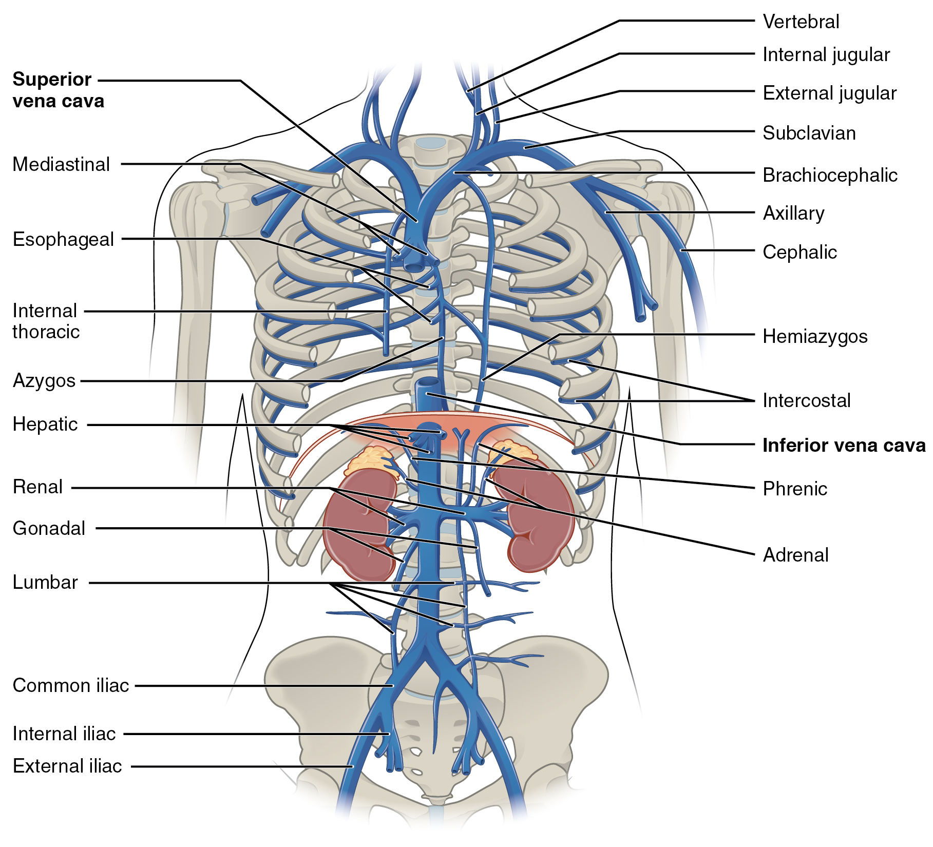
The vertebral vein is formed in the suboccipital triangle, from numerous small tributaries which spring from the internal vertebral venous plexuses and issue from the vertebral canal above the posterior arch of the atlas. They unite with small veins from the deep muscles at the upper part of the back of the neck, and form a vessel which enters the foramen in the transverse process of the atlas, and descends, forming a dense plexus around the vertebral artery, in the canal formed by the transverse foramina of the upper six cervical vertebrae. This plexus ends in a single trunk, which emerges from the transverse foramina of the sixth cervical vertebra, and opens at the root of the neck into the back part of the innominate vein near its origin, its mouth being guarded by a pair of valves. On the right side, it crosses the first part of the subclavian artery In human anatomy, the subclavian arteries are paired major arteries of the upper thorax, below the clavicle. They receive ... [...More Info...] [...Related Items...] OR: [Wikipedia] [Google] [Baidu] |
External Vertebral Venous Plexuses
External may refer to: * External (mathematics), a concept in abstract algebra * Externality, in economics, the cost or benefit that affects a party who did not choose to incur that cost or benefit * Externals, a fictional group of X-Men antagonists See also * * Internal (other) {{disambig ... [...More Info...] [...Related Items...] OR: [Wikipedia] [Google] [Baidu] |
Suboccipital Triangle
The suboccipital triangle is a region of the neck bounded by the following three muscles of the suboccipital group of muscles: * Rectus capitis posterior major - above and medially * Obliquus capitis superior - above and laterally * Obliquus capitis inferior - below and laterally (Rectus capitis posterior minor is also in this region but does not form part of the triangle) It is covered by a layer of dense fibro-fatty tissue, situated beneath the semispinalis capitis. The floor is formed by the posterior atlantooccipital membrane, and the posterior arch of the atlas. In the deep groove on the upper surface of the posterior arch of the atlas are the vertebral artery and the first cervical or suboccipital nerve. In the past, the vertebral artery was accessed here in order to conduct angiography of the circle of Willis. Presently, formal angiography of the circle of Willis is performed via catheter angiography, with access usually being acquired at the common femoral artery. A ... [...More Info...] [...Related Items...] OR: [Wikipedia] [Google] [Baidu] |
Brachiocephalic Vein
The left and right brachiocephalic veins (previously called innominate veins) are major veins in the upper chest, formed by the union of each corresponding internal jugular vein and subclavian vein. This is at the level of the sternoclavicular joint. The left brachiocephalic vein is nearly always longer than the right. These veins merge to form the superior vena cava, a great vessel, posterior to the junction of the first costal cartilage with the manubrium of the sternum. The brachiocephalic veins are the major veins returning blood to the superior vena cava. Tributaries The brachiocephalic vein is formed by the confluence of the subclavian and internal jugular veins. In addition it receives drainage from: * Left and right internal thoracic vein (Also called internal mammary veins): drain into the inferior border of their corresponding vein * Left and right inferior thyroid veins: drain into the superior aspect of their corresponding veins near the confluence * Left and rig ... [...More Info...] [...Related Items...] OR: [Wikipedia] [Google] [Baidu] |
Vertebral Artery
The vertebral arteries are major arteries of the neck. Typically, the vertebral arteries originate from the subclavian arteries. Each vessel courses superiorly along each side of the neck, merging within the skull to form the single, midline basilar artery. As the supplying component of the ''vertebrobasilar vascular system'', the vertebral arteries supply blood to the upper spinal cord, brainstem, cerebellum, and posterior part of brain. Structure The vertebral arteries usually arise from the posterosuperior aspect of the central subclavian arteries on each side of the body, then enter deep to the transverse process at the level of the 6th cervical vertebrae (C6), or occasionally (in 7.5% of cases) at the level of C7. They then proceed superiorly, in the transverse foramen of each cervical vertebra. Once they have passed through the transverse foramen of C1 (also known as the atlas), the vertebral arteries travel across the posterior arch of C1 and through the suboccipi ... [...More Info...] [...Related Items...] OR: [Wikipedia] [Google] [Baidu] |

