|
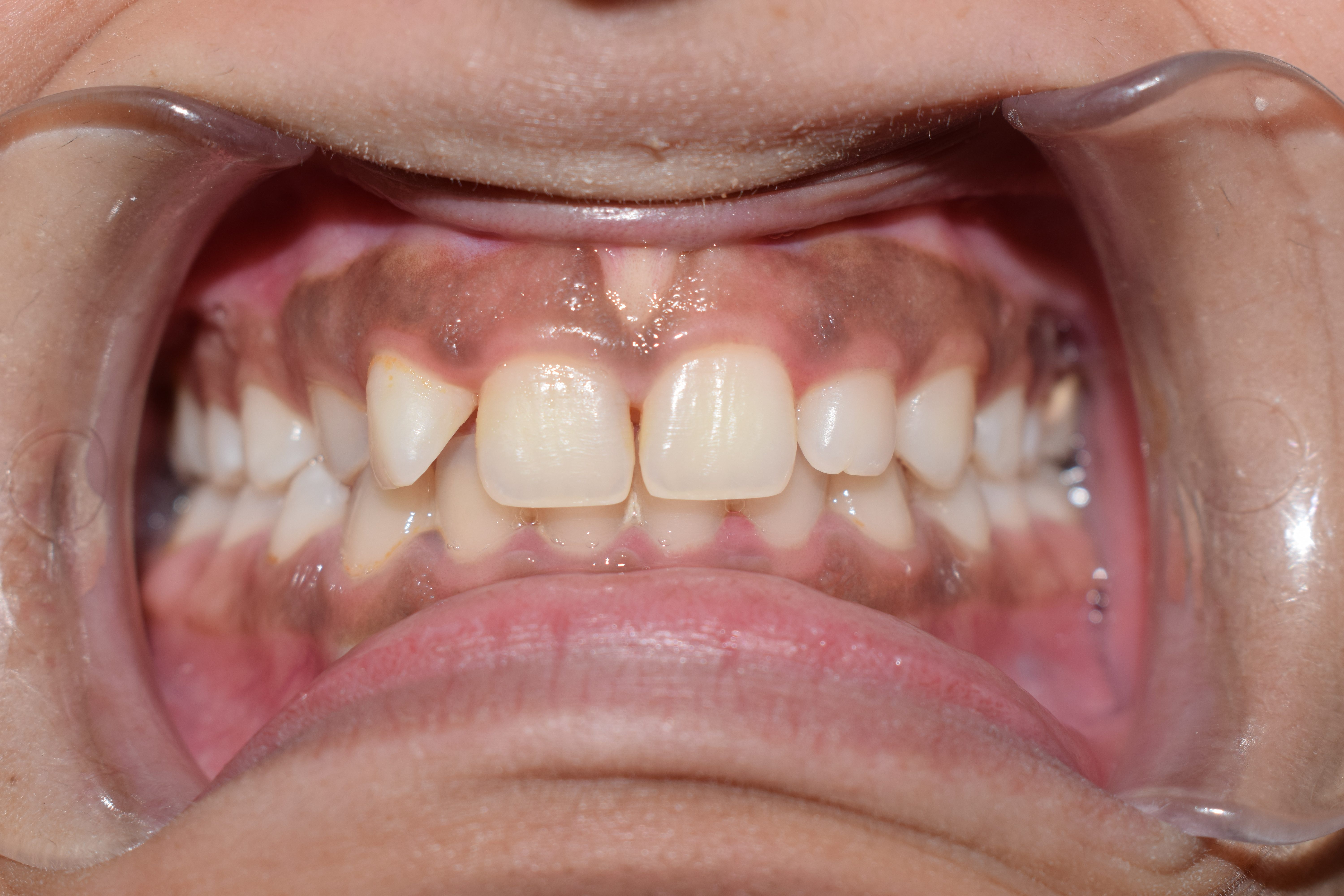
Stippling (dentistry)
The gingiva often possess a textured surface that is referred to as being stippled (engraved points). Stippling only presents on the attached gingiva bound to underlying alveolar bone, not the freely moveable alveolar mucosa or free gingiva. Stippling used to be thought to indicate health, but it has since been shown that smooth gingiva is not an indication of disease, unless it is smooth ''due to a loss'' of previously existing stippling. Stippling is a consequence of the microscopic elevations and depressions of the surface of the gingival tissue due to the connective tissue projections within the tissue. The degree of keratinization and the prominence of stippling appear to be related. To be more specific, stippling occurs at sites of fusion of the epithelial ridges (also known as rete pegs Rete pegs (also known as rete processes or rete ridges) are the epithelial extensions that project into the underlying connective tissue in both skin and mucous membranes. In the epithelium ... [...More Info...] [...Related Items...] OR: [Wikipedia] [Google] [Baidu] |
Stippling SSF
Stippling is the creation of a pattern simulating varying Grayscale, degrees of solidity or shading by using small dots. Such a pattern may occur in nature and these effects are frequently emulated by artists. Art In printmaking, stipple engraving is a technique using flicks of the burin to build up the image in short lines or dots, often combined with conventional linear engraving. In engraved glass a similar stipple technique has often been popular. In a drawing or painting, the dots are made of pigment of a single colour, applied with a pen or brush; the denser the dots, the darker the apparent shade—or lighter, if the pigment is lighter than the surface. This is similar to—but distinct from—pointillism, which uses dots of different colours to simulate blended colours. Botany In description of flora species, a stippling is a kind of pattern, especially in the case of flowering plants, produced in nature that occur on flower petals and sepals. These are similar to ... [...More Info...] [...Related Items...] OR: [Wikipedia] [Google] [Baidu] |
Gingiva
The gums or gingiva (plural: ''gingivae'') consist of the mucosal tissue that lies over the mandible and maxilla inside the mouth. Gum health and disease can have an effect on general health. Structure The gums are part of the soft tissue lining of the mouth. They surround the teeth and provide a seal around them. Unlike the soft tissue linings of the lips and cheeks, most of the gums are tightly bound to the underlying bone which helps resist the friction of food passing over them. Thus when healthy, it presents an effective barrier to the barrage of periodontal insults to deeper tissue. Healthy gums are usually coral pink in light skinned people, and may be naturally darker with melanin pigmentation. Changes in color, particularly increased redness, together with swelling and an increased tendency to bleed, suggest an inflammation that is possibly due to the accumulation of bacterial plaque. Overall, the clinical appearance of the tissue reflects the underlying histology, b ... [...More Info...] [...Related Items...] OR: [Wikipedia] [Google] [Baidu] |
Alveolar Bone
The alveolar process () or alveolar bone is the thickened ridge of bone that contains the tooth sockets on the jaw bones (in humans, the maxilla and the mandible). The structures are covered by gums as part of the oral cavity. The synonymous terms ''alveolar ridge'' and ''alveolar margin'' are also sometimes used more specifically to refer to the ridges on the inside of the mouth which can be felt with the tongue, either on roof of the mouth between the upper teeth and the hard palate or on the bottom of the mouth behind the lower teeth. Terminology The term ''alveolar'' () ('hollow') refers to the cavities of the tooth sockets, known as dental alveoli. The alveolar process is also called the ''alveolar bone'' or ''alveolar ridge''. The curved portion is referred to as the alveolar arch. The alveolar bone proper, also called bundle bone, directly surrounds the teeth. The term alveolar crest describes the extreme rim of the bone nearest to the crowns of the teeth. The portion of ... [...More Info...] [...Related Items...] OR: [Wikipedia] [Google] [Baidu] |
Alveolar Mucosa
The oral mucosa is the mucous membrane lining the inside of the mouth. It comprises stratified squamous epithelium, termed "oral epithelium", and an underlying connective tissue termed ''lamina propria''. The oral cavity has sometimes been described as a mirror that reflects the health of the individual. Changes indicative of disease are seen as alterations in the oral mucosa lining the mouth, which can reveal systemic conditions, such as diabetes or vitamin deficiency, or the local effects of chronic tobacco or alcohol use. The oral mucosa tends to heal faster and with less scar formation compared to the skin. The underlying mechanism remains unknown, but research suggests that extracellular vesicles might be involved. Classification Oral mucosa can be divided into three main categories based on function and histology: *Lining mucosa, nonkeratinized stratified squamous epithelium, found almost everywhere else in the oral cavity, including the: **Alveolar mucosa, the lining between ... [...More Info...] [...Related Items...] OR: [Wikipedia] [Google] [Baidu] |
Keratinization
Keratin () is one of a family of structural fibrous proteins also known as ''scleroproteins''. Alpha-keratin (α-keratin) is a type of keratin found in vertebrates. It is the key structural material making up scales, hair, nails, feathers, horns, claws, hooves, and the outer layer of skin among vertebrates. Keratin also protects epithelial cells from damage or stress. Keratin is extremely insoluble in water and organic solvents. Keratin monomers assemble into bundles to form intermediate filaments, which are tough and form strong unmineralized epidermal appendages found in reptiles, birds, amphibians, and mammals. Excessive keratinization participate in fortification of certain tissues such as in horns of cattle and rhinos, and armadillos' osteoderm. The only other biological matter known to approximate the toughness of keratinized tissue is chitin. Keratin comes in two types, the primitive, softer forms found in all vertebrates and harder, derived forms found only amon ... [...More Info...] [...Related Items...] OR: [Wikipedia] [Google] [Baidu] |
Rete Pegs
Rete pegs (also known as rete processes or rete ridges) are the epithelial extensions that project into the underlying connective tissue in both skin and mucous membranes. In the epithelium of the mouth, the attached gingiva exhibit rete pegs, while the sulcularItoiz, ME; Carranza, FA: The Gingiva. In Newman, MG; Takei, HH; Carranza, FA; editors: ''Carranza’s Clinical Periodontology'', 9th Edition. Philadelphia: W.B. Saunders Company, 2002. pages 23. and junctional epithelia do not.Page, RC; Schroeder, HE. "Pathogenesis of Inflammatory Periodontal Disease: A Summary of Current Work." ''Lab Invest'' 1976;34(3):235-249 Scar tissue lacks rete pegs and scars tend to shear off more easily than normal tissue as a result. Also known as ''papillae'', they are downward thickenings of the epidermis between the dermal papillae The dermis or corium is a layer of skin between the epidermis (with which it makes up the cutis) and subcutaneous tissues, that primarily consists of dense irr ... [...More Info...] [...Related Items...] OR: [Wikipedia] [Google] [Baidu] |
.jpg)

.jpg)

