|
Stab Wounds
A stab wound is a specific form of penetrating trauma to the skin that results from a knife or a similar pointed object. While stab wounds are typically known to be caused by knives, they can also occur from a variety of implements, including broken bottles and ice picks. Most stabbings occur because of intentional violence or through self-infliction. The treatment is dependent on many different variables such as the anatomical location and the severity of the injury. Even though stab wounds are inflicted at a much greater rate than gunshot wounds, they account for less than 10% of all penetrating trauma deaths. Management Stab wounds can cause various internal and external injuries. They are generally caused by low-velocity weapons, meaning the injuries inflicted on a person are typically confined to the path it took internally, instead of causing damage to surrounding tissue, which is common of gunshot wounds. The abdomen is the most commonly injured area from a stab wound. Int ... [...More Info...] [...Related Items...] OR: [Wikipedia] [Google] [Baidu] |
Intravenous
Intravenous therapy (abbreviated as IV therapy) is a medical technique that administers fluids, medications and nutrients directly into a person's vein. The intravenous route of administration is commonly used for rehydration or to provide nutrients for those who cannot, or will not—due to reduced mental states or otherwise—consume food or water by mouth. It may also be used to administer medications or other medical therapy such as blood products or electrolytes to correct electrolyte imbalances. Attempts at providing intravenous therapy have been recorded as early as the 1400s, but the practice did not become widespread until the 1900s after the development of techniques for safe, effective use. The intravenous route is the fastest way to deliver medications and fluid replacement throughout the body as they are introduced directly into the circulatory system and thus quickly distributed. For this reason, the intravenous route of administration is also used for the consu ... [...More Info...] [...Related Items...] OR: [Wikipedia] [Google] [Baidu] |
Liver Function Tests
Liver function tests (LFTs or LFs), also referred to as a hepatic panel, are groups of blood tests that provide information about the state of a patient's liver. These tests include prothrombin time (PT/INR), activated partial thromboplastin time (aPTT), albumin, bilirubin (direct and indirect), and others. The liver transaminases aspartate transaminase (AST or SGOT) and alanine transaminase (ALT or SGPT) are useful biomarkers of liver injury in a patient with some degree of intact liver function. Most liver diseases cause only mild symptoms initially, but these diseases must be detected early. Hepatic (liver) involvement in some diseases can be of crucial importance. This testing is performed on a patient's blood sample. Some tests are associated with functionality (e.g., albumin), some with cellular integrity (e.g., transaminase), and some with conditions linked to the biliary tract ( gamma-glutamyl transferase and alkaline phosphatase). Because some of these tests do no ... [...More Info...] [...Related Items...] OR: [Wikipedia] [Google] [Baidu] |
White Blood Cell Count
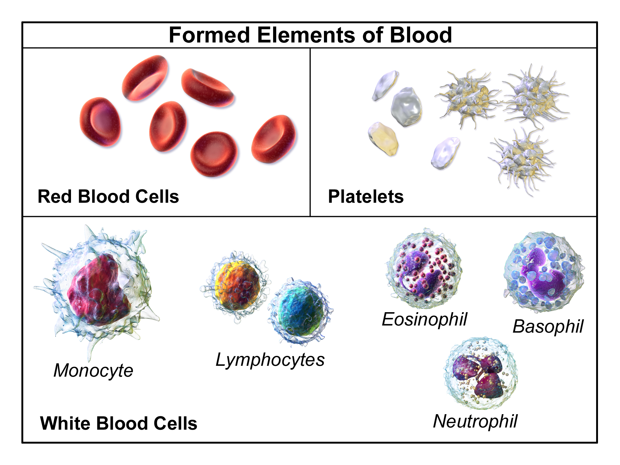
A complete blood count (CBC), also known as a full blood count (FBC), is a set of medical laboratory tests that provide information about the cells in a person's blood. The CBC indicates the counts of white blood cells, red blood cells and platelets, the concentration of hemoglobin, and the hematocrit (the volume percentage of red blood cells). The red blood cell indices, which indicate the average size and hemoglobin content of red blood cells, are also reported, and a white blood cell differential, which counts the different types of white blood cells, may be included. The CBC is often carried out as part of a medical assessment and can be used to monitor health or diagnose diseases. The results are interpreted by comparing them to reference ranges, which vary with sex and age. Conditions like anemia and thrombocytopenia are defined by abnormal complete blood count results. The red blood cell indices can provide information about the cause of a person's anemia such as iron d ... [...More Info...] [...Related Items...] OR: [Wikipedia] [Google] [Baidu] |
Hematocrit
The hematocrit () (Ht or HCT), also known by several other names, is the volume percentage (vol%) of red blood cells (RBCs) in blood, measured as part of a blood test. The measurement depends on the number and size of red blood cells. It is normally 40.7–50.3% for males and 36.1–44.3% for females. It is a part of a person's complete blood count results, along with hemoglobin concentration, white blood cell count and platelet count. Because the purpose of red blood cells is to transfer oxygen from the lungs to body tissues, a blood sample's hematocrit—the red blood cell volume percentage—can become a point of reference of its capability of delivering oxygen. Hematocrit levels that are too high or too low can indicate a blood disorder, dehydration, or other medical conditions. An abnormally low hematocrit may suggest anemia, a decrease in the total amount of red blood cells, while an abnormally high hematocrit is called polycythemia. Both are potentially life-threate ... [...More Info...] [...Related Items...] OR: [Wikipedia] [Google] [Baidu] |
Hypovolemia
Hypovolemia, also known as volume depletion or volume contraction, is a state of abnormally low extracellular fluid in the body. This may be due to either a loss of both salt and water or a decrease in blood volume. Hypovolemia refers to the loss of extracellular fluid and should not be confused with dehydration. Hypovolemia is caused by a variety of events, but these can be simplified into two categories: those that are associated with kidney function and those that are not. The signs and symptoms of hypovolemia worsen as the amount of fluid lost increases. Immediately or shortly after mild fluid loss (from blood donation, diarrhea, vomiting, bleeding from trauma, etc.), one may experience headache, fatigue, weakness, dizziness, or thirst. Untreated hypovolemia or excessive and rapid losses of volume may lead to hypovolemic shock. Signs and symptoms of hypovolemic shock include increased heart rate, low blood pressure, pale or cold skin, and altered mental status. When ... [...More Info...] [...Related Items...] OR: [Wikipedia] [Google] [Baidu] |
Contrast Medium
A contrast agent (or contrast medium) is a substance used to increase the contrast of structures or fluids within the body in medical imaging. Contrast agents absorb or alter external electromagnetism or ultrasound, which is different from radiopharmaceuticals, which emit radiation themselves. In x-ray imaging, contrast agents enhance the radiodensity in a target tissue or structure. In magnetic resonance imaging, contrast agents shorten (or in some instances increase) the relaxation times of nuclei within body tissues in order to alter the contrast in the image. Contrast agents are commonly used to improve the visibility of blood vessels and the gastrointestinal tract. The types of contrast agent are classified according to their intended imaging modalities. Radiocontrast media For radiography, which is based on X-rays, iodine and barium are the most common types of contrast agent. Various sorts of iodinated contrast agents exist, with variations occurring between the o ... [...More Info...] [...Related Items...] OR: [Wikipedia] [Google] [Baidu] |
Computed Tomography
A computed tomography scan (CT scan; formerly called computed axial tomography scan or CAT scan) is a medical imaging technique used to obtain detailed internal images of the body. The personnel that perform CT scans are called radiographers or radiology technologists. CT scanners use a rotating X-ray tube and a row of detectors placed in a gantry to measure X-ray attenuations by different tissues inside the body. The multiple X-ray measurements taken from different angles are then processed on a computer using tomographic reconstruction algorithms to produce tomographic (cross-sectional) images (virtual "slices") of a body. CT scans can be used in patients with metallic implants or pacemakers, for whom magnetic resonance imaging (MRI) is contraindicated. Since its development in the 1970s, CT scanning has proven to be a versatile imaging technique. While CT is most prominently used in medical diagnosis, it can also be used to form images of non-living objects. The 1979 N ... [...More Info...] [...Related Items...] OR: [Wikipedia] [Google] [Baidu] |
Diagnostic Peritoneal Lavage
Diagnostic peritoneal lavage (DPL) or diagnostic peritoneal aspiration (DPA) is a surgical diagnostic procedure to determine if there is free floating fluid (most often blood) in the abdominal cavity. Indications This procedure is performed when intra-abdominal bleeding ( hemoperitoneum), usually secondary to trauma, is suspected. In a hemodynamically unstable patient with high-risk mechanism of injury, peritoneal lavage is a means of rapidly diagnosing intra-abdominal injury requiring laparotomy, but has largely been replaced in trauma care by the use of a focused assessment with sonography for trauma (FAST scan) due to its repeatability, non-invasiveness and non-interference with subsequent computed tomography (CT scan). Abdominal CT and contrast duodenography may complement lavage in stable patients, but in an unstable or uncooperative persons, these studies are too time-consuming or require ill-advised sedation. Magnetic resonance imaging is extremely accurate for the anatom ... [...More Info...] [...Related Items...] OR: [Wikipedia] [Google] [Baidu] |
Focused Assessment With Sonography For Trauma
Focused assessment with sonography in trauma (commonly abbreviated as FAST) is a rapid bedside ultrasound examination performed by surgeons, emergency physicians, and paramedics as a screening test for blood around the heart (pericardial effusion) or abdominal organs ( hemoperitoneum) after trauma. There is also the extended FAST (eFAST) which includes some additional ultrasound views to assess for pneumothorax. The four classic areas that are examined for free fluid are the perihepatic space (including Morison's pouch or the hepatorenal recess), peri splenic space, pericardium, and the pelvis. With this technique it is possible to identify the presence of intraperitoneal or pericardial free fluid. In the context of traumatic injury, this fluid will usually be due to bleeding. Indications Reasons a FAST or eFAST would be performed would be: #Blunt abdominal trauma #Penetrating abdominal trauma #Blunt thoracic trauma #Penetrating thoracic trauma #Undifferentiated shock (lo ... [...More Info...] [...Related Items...] OR: [Wikipedia] [Google] [Baidu] |
Trauma Center
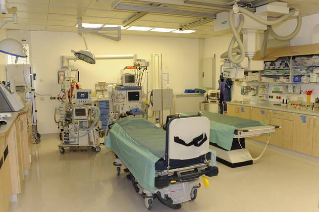
A trauma center (or trauma centre) is a hospital equipped and staffed to provide care for patients suffering from major traumatic injuries such as falls, motor vehicle collisions, or gunshot wounds. A trauma center may also refer to an emergency department (also known as a "casualty department" or "accident and emergency") without the presence of specialized services to care for victims of major trauma. In the United States, a hospital can receive trauma center status by meeting specific criteria established by the American College of Surgeons (ACS) and passing a site review by the Verification Review Committee. Official designation as a trauma center is determined by individual state law provisions. Trauma centers vary in their specific capabilities and are identified by "Level" designation: Level I (Level-1) being the highest and Level III (Level-3) being the lowest (some states have five designated levels, in which case Level V (Level-5) is the lowest). The highest lev ... [...More Info...] [...Related Items...] OR: [Wikipedia] [Google] [Baidu] |
Shock (circulatory)
Shock is the state of insufficient blood flow to the tissues of the body as a result of problems with the circulatory system. Initial symptoms of shock may include weakness, fast heart rate, fast breathing, sweating, anxiety, and increased thirst. This may be followed by confusion, unconsciousness, or cardiac arrest, as complications worsen. Shock is divided into four main types based on the underlying cause: low volume, cardiogenic, obstructive, and distributive shock. Low volume shock, also known as hypovolemic shock, may be from bleeding, diarrhea, or vomiting. Cardiogenic shock may be due to a heart attack or cardiac contusion. Obstructive shock may be due to cardiac tamponade or a tension pneumothorax. Distributive shock may be due to sepsis, anaphylaxis, injury to the upper spinal cord, or certain overdoses. The diagnosis is generally based on a combination of symptoms, physical examination, and laboratory tests. A decreased pulse pressure ( systolic blood ... [...More Info...] [...Related Items...] OR: [Wikipedia] [Google] [Baidu] |


