|
Sphenoparietal Suture
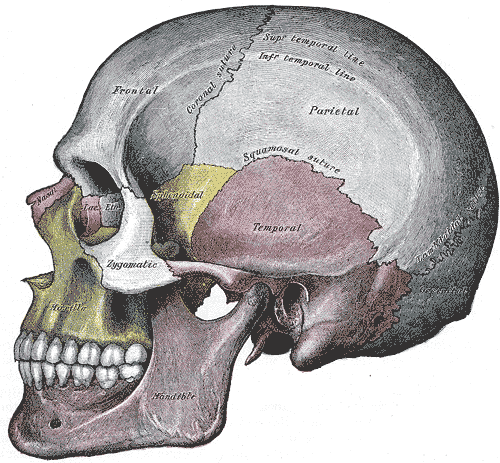
The sphenoparietal suture is the cranial suture between the sphenoid bone and the parietal bone. It is one of the sutures that comprises the pterion. Additional images File:Sphenoparietal suture.gif, Position of sphenoparietal suture (shown in red). Animation. File:Sphenoparietal suture4.gif, Parietal bones (above) and sphenoid bone (below) File:Gray188-Sphenoparietal suture.png, Side view of the skull. Sphenoparietal suture indicated by the arrow. File:Gray164 - Sphenoparietal suture.png, Left zygomatic bone In the human skull, the zygomatic bone (from grc, ζῠγόν, zugón, yoke), also called cheekbone or malar bone, is a paired irregular bone which articulates with the maxilla, the temporal bone, the sphenoid bone and the frontal bone. It is si ... in situ. (Sphenoparietal suture visible at upper right in blue.) References External links Bones of the head and neck Cranial sutures Human head and neck Joints Joints of the head and neck Skeletal system Skul ... [...More Info...] [...Related Items...] OR: [Wikipedia] [Google] [Baidu] |
Parietal Bone
The parietal bones () are two bones in the Human skull, skull which, when joined at a fibrous joint, form the sides and roof of the Human skull, cranium. In humans, each bone is roughly quadrilateral in form, and has two surfaces, four borders, and four angles. It is named from the Latin ''paries'' (''-ietis''), wall. Surfaces External The external surface [Fig. 1] is convex, smooth, and marked near the center by an eminence, the parietal eminence (''tuber parietale''), which indicates the point where ossification commenced. Crossing the middle of the bone in an arched direction are two curved lines, the superior and inferior temporal lines; the former gives attachment to the temporal fascia, and the latter indicates the upper limit of the muscular origin of the temporal muscle. Above these lines the bone is covered by a tough layer of fibrous tissue – the epicranial aponeurosis; below them it forms part of the temporal fossa, and affords attachment to the temporal muscle. ... [...More Info...] [...Related Items...] OR: [Wikipedia] [Google] [Baidu] |
Sphenoid Bone
The sphenoid bone is an unpaired bone of the neurocranium. It is situated in the middle of the skull towards the front, in front of the basilar part of occipital bone, basilar part of the occipital bone. The sphenoid bone is one of the seven bones that articulate to form the orbit (anatomy), orbit. Its shape somewhat resembles that of a butterfly or bat with its wings extended. Structure It is divided into the following parts: * a median portion, known as the body of sphenoid bone, containing the sella turcica, which houses the pituitary gland as well as the paired paranasal sinuses, the sphenoidal sinuses * two Greater wing of sphenoid bone, greater wings on the lateral side of the body and two Lesser wing of sphenoid bone, lesser wings from the anterior side. * Pterygoid processes of the sphenoides, directed downwards from the junction of the body and the greater wings. Two sphenoidal conchae are situated at the anterior and inferior part of the body. Intrinsic ligaments of ... [...More Info...] [...Related Items...] OR: [Wikipedia] [Google] [Baidu] |
Cranial Suture
In anatomy, fibrous joints are joints connected by Fibrous connective tissue, fibrous tissue, consisting mainly of collagen. These are fixed joints where bones are united by a layer of white fibrous tissue of varying thickness. In the skull the joints between the bones are called Suture (anatomy), sutures. Such immovable joints are also referred to as synarthrosis, synarthroses. Types Most fibrous joints are also called "fixed" or "immovable". These joints have no joint cavity and are connected via fibrous connective tissue. The skull bones are connected by fibrous joints called ''#Sutures, sutures''. In fetus, fetal skulls the sutures are wide to allow slight movement during birth. They later become rigid (synarthrosis, synarthrodial). Some of the long bones in the body such as the radius (bone), radius and ulna in the forearm are joined by a ''#Syndesmosis, syndesmosis'' (along the interosseous membrane of forearm, interosseous membrane). Syndemoses are slightly moveable (amph ... [...More Info...] [...Related Items...] OR: [Wikipedia] [Google] [Baidu] |
Sphenoid Bone
The sphenoid bone is an unpaired bone of the neurocranium. It is situated in the middle of the skull towards the front, in front of the basilar part of occipital bone, basilar part of the occipital bone. The sphenoid bone is one of the seven bones that articulate to form the orbit (anatomy), orbit. Its shape somewhat resembles that of a butterfly or bat with its wings extended. Structure It is divided into the following parts: * a median portion, known as the body of sphenoid bone, containing the sella turcica, which houses the pituitary gland as well as the paired paranasal sinuses, the sphenoidal sinuses * two Greater wing of sphenoid bone, greater wings on the lateral side of the body and two Lesser wing of sphenoid bone, lesser wings from the anterior side. * Pterygoid processes of the sphenoides, directed downwards from the junction of the body and the greater wings. Two sphenoidal conchae are situated at the anterior and inferior part of the body. Intrinsic ligaments of ... [...More Info...] [...Related Items...] OR: [Wikipedia] [Google] [Baidu] |
Parietal Bone
The parietal bones () are two bones in the Human skull, skull which, when joined at a fibrous joint, form the sides and roof of the Human skull, cranium. In humans, each bone is roughly quadrilateral in form, and has two surfaces, four borders, and four angles. It is named from the Latin ''paries'' (''-ietis''), wall. Surfaces External The external surface [Fig. 1] is convex, smooth, and marked near the center by an eminence, the parietal eminence (''tuber parietale''), which indicates the point where ossification commenced. Crossing the middle of the bone in an arched direction are two curved lines, the superior and inferior temporal lines; the former gives attachment to the temporal fascia, and the latter indicates the upper limit of the muscular origin of the temporal muscle. Above these lines the bone is covered by a tough layer of fibrous tissue – the epicranial aponeurosis; below them it forms part of the temporal fossa, and affords attachment to the temporal muscle. ... [...More Info...] [...Related Items...] OR: [Wikipedia] [Google] [Baidu] |
Pterion
The pterion is the region where the frontal, parietal, temporal, and sphenoid bones join. It is located on the side of the skull, just behind the temple. Structure The pterion is located in the temporal fossa, approximately 2.6 cm behind and 1.3 cm above the posterolateral margin of the frontozygomatic suture. It is the junction between four bones: * the parietal bone. * the squamous part of temporal bone. * the greater wing of sphenoid bone. * the frontal bone. These bones are typically joined by five cranial sutures: * the sphenoparietal suture joins the sphenoid and parietal bones. * the coronal suture joins the frontal bone to the sphenoid and parietal bones. * the squamous suture joins the temporal bone to the sphenoid and parietal bones. * the sphenofrontal suture joins the sphenoid and frontal bones. * the sphenosquamosal suture joins the sphenoid and temporal bones. Clinical significance Haematoma The pterion is known as the weakest part of the sk ... [...More Info...] [...Related Items...] OR: [Wikipedia] [Google] [Baidu] |
Zygomatic Bone
In the human skull, the zygomatic bone (from grc, ζῠγόν, zugón, yoke), also called cheekbone or malar bone, is a paired irregular bone which articulates with the maxilla, the temporal bone, the sphenoid bone and the frontal bone. It is situated at the upper and lateral part of the face and forms the prominence of the cheek, part of the lateral wall and floor of the orbit, and parts of the temporal fossa and the infratemporal fossa. It presents a malar and a temporal surface; four processes (the frontosphenoidal, orbital, maxillary, and temporal), and four borders. Etymology The term ''zygomatic'' derives from the Ancient Greek , ''zygoma'', meaning "yoke". The zygomatic bone is occasionally referred to as the zygoma, but this term may also refer to the zygomatic arch. Structure Surfaces The ''malar surface'' is convex and perforated near its center by a small aperture, the zygomaticofacial foramen, for the passage of the zygomaticofacial nerve and vessels; below ... [...More Info...] [...Related Items...] OR: [Wikipedia] [Google] [Baidu] |
Bones Of The Head And Neck
A bone is a rigid organ that constitutes part of the skeleton in most vertebrate animals. Bones protect the various other organs of the body, produce red and white blood cells, store minerals, provide structure and support for the body, and enable mobility. Bones come in a variety of shapes and sizes and have complex internal and external structures. They are lightweight yet strong and hard and serve multiple functions. Bone tissue (osseous tissue), which is also called bone in the uncountable sense of that word, is hard tissue, a type of specialized connective tissue. It has a honeycomb-like matrix internally, which helps to give the bone rigidity. Bone tissue is made up of different types of bone cells. Osteoblasts and osteocytes are involved in the formation and mineralization of bone; osteoclasts are involved in the resorption of bone tissue. Modified (flattened) osteoblasts become the lining cells that form a protective layer on the bone surface. The mineralized matrix ... [...More Info...] [...Related Items...] OR: [Wikipedia] [Google] [Baidu] |
Cranial Sutures
In anatomy, fibrous joints are joints connected by fibrous tissue, consisting mainly of collagen Collagen () is the main structural protein in the extracellular matrix found in the body's various connective tissues. As the main component of connective tissue, it is the most abundant protein in mammals, making up from 25% to 35% of the whole .... These are fixed joints where bones are united by a layer of white fibrous tissue of varying thickness. In the skull the joints between the bones are called Suture (anatomy), sutures. Such immovable joints are also referred to as synarthrosis, synarthroses. Types Most fibrous joints are also called "fixed" or "immovable". These joints have no joint cavity and are connected via fibrous connective tissue. The skull bones are connected by fibrous joints called ''#Sutures, sutures''. In fetus, fetal skulls the sutures are wide to allow slight movement during birth. They later become rigid (synarthrosis, synarthrodial). Some of the long bo ... [...More Info...] [...Related Items...] OR: [Wikipedia] [Google] [Baidu] |
Human Head And Neck
Humans (''Homo sapiens'') are the most abundant and widespread species of primate, characterized by bipedalism and exceptional cognitive skills due to a large and complex brain. This has enabled the development of advanced tools, culture, and language. Humans are highly social and tend to live in complex social structures composed of many cooperating and competing groups, from families and kinship networks to political states. Social interactions between humans have established a wide variety of values, social norms, and rituals, which bolster human society. Its intelligence and its desire to understand and influence the environment and to explain and manipulate phenomena have motivated humanity's development of science, philosophy, mythology, religion, and other fields of study. Although some scientists equate the term ''humans'' with all members of the genus ''Homo'', in common usage, it generally refers to ''Homo sapiens'', the only extant member. Anatomically mode ... [...More Info...] [...Related Items...] OR: [Wikipedia] [Google] [Baidu] |
Joints
A joint or articulation (or articular surface) is the connection made between bones, ossicles, or other hard structures in the body which link an animal's skeletal system into a functional whole.Saladin, Ken. Anatomy & Physiology. 7th ed. McGraw-Hill Connect. Webp.274/ref> They are constructed to allow for different degrees and types of movement. Some joints, such as the knee, elbow, and shoulder, are self-lubricating, almost frictionless, and are able to withstand compression and maintain heavy loads while still executing smooth and precise movements. Other joints such as suture (joint), sutures between the bones of the skull permit very little movement (only during birth) in order to protect the brain and the sense organs. The connection between a tooth and the jawbone is also called a joint, and is described as a fibrous joint known as a gomphosis. Joints are classified both structurally and functionally. Classification The number of joints depends on if Sesamoid bone, sesamoi ... [...More Info...] [...Related Items...] OR: [Wikipedia] [Google] [Baidu] |
Joints Of The Head And Neck
A joint or articulation (or articular surface) is the connection made between bones, ossicles, or other hard structures in the body which link an animal's skeletal system into a functional whole.Saladin, Ken. Anatomy & Physiology. 7th ed. McGraw-Hill Connect. Webp.274/ref> They are constructed to allow for different degrees and types of movement. Some joints, such as the knee, elbow, and shoulder, are self-lubricating, almost frictionless, and are able to withstand compression and maintain heavy loads while still executing smooth and precise movements. Other joints such as sutures between the bones of the skull permit very little movement (only during birth) in order to protect the brain and the sense organs. The connection between a tooth and the jawbone is also called a joint, and is described as a fibrous joint known as a gomphosis. Joints are classified both structurally and functionally. Classification The number of joints depends on if sesamoids are included, age of the ... [...More Info...] [...Related Items...] OR: [Wikipedia] [Google] [Baidu] |


