|
Sphenoidal
The sphenoid bone is an unpaired bone of the neurocranium. It is situated in the middle of the skull towards the front, in front of the basilar part of the occipital bone. The sphenoid bone is one of the seven bones that articulate to form the orbit. Its shape somewhat resembles that of a butterfly or bat with its wings extended. Structure It is divided into the following parts: * a median portion, known as the body of sphenoid bone, containing the sella turcica, which houses the pituitary gland as well as the paired paranasal sinuses, the sphenoidal sinuses * two greater wings on the lateral side of the body and two lesser wings from the anterior side. * Pterygoid processes of the sphenoides, directed downwards from the junction of the body and the greater wings. Two sphenoidal conchae are situated at the anterior and inferior part of the body. Intrinsic ligaments of the sphenoid The more important of these are: * the pterygospinous, stretching between the spina angulari ... [...More Info...] [...Related Items...] OR: [Wikipedia] [Google] [Baidu] |
Sphenoidal Sinuses
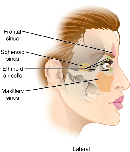
The sphenoid sinus is a paired paranasal sinus occurring within the within the body of the sphenoid bone. It represents one pair of the four paired paranasal sinuses.Illustrated Anatomy of the Head and Neck, Fehrenbach and Herring, Elsevier, 2012, page 64 The pair of sphenoid sinuses are separated in the middle by a septum of sphenoid sinuses. Each sphenoid sinus communicates with the nasal cavity via the opening of sphenoidal sinus. The two sphenoid sinuses vary in size and shape, and are usually asymmetrical. Anatomy On average, a sphenoid sinus measures 2.2 cm vertical height, 2 cm in transverse breadth; and 2.2 cm antero-posterior depth. Each spehoid sinus is contained within the body of sphenoid bone, being situated just inferior to the sella turcica. The two sphenoid sinuses are separated medially by the septum of sphenoidal sinuses (which is usually asymmetrical). An opening of sphenoidal sinus forms a passage between each sphenoidal sinus, and the nasal ca ... [...More Info...] [...Related Items...] OR: [Wikipedia] [Google] [Baidu] |
Body Of Sphenoid Bone
The body of the sphenoid bone, more or less cubical in shape, is hollowed out in its interior to form two large cavities, the sphenoidal sinuses, which are separated from each other by a septum. Superior surface The superior surface of the body ig. 1presents in front a prominent spine, the ethmoidal spine, for articulation with the cribriform plate of the ethmoid bone; behind this is a smooth surface slightly raised in the middle line, and grooved on either side for the olfactory lobes of the brain. This surface is bounded behind by a ridge, which forms the anterior border of a narrow, transverse groove, the prechiasmatic groove, above and behind which lies the optic chiasma; the groove ends on either side in the optic foramen, which transmits the optic nerve and ophthalmic artery into the orbital cavity. Behind the chiasmatic groove is an elevation, the tuberculum sellae; and behind this is a deep depression, the saddle-shaped sella turcica (Turkish seat), the deepest part o ... [...More Info...] [...Related Items...] OR: [Wikipedia] [Google] [Baidu] |
Sphenoidal Conchae
The sphenoidal conchae (sphenoidal turbinated processes) are two thin, curved plates, situated at the anterior and lower part of the body of the sphenoid. An aperture of variable size exists in the anterior wall of each, and through this the sphenoidal sinus opens into the nasal cavity. ''General Anatomy and Osteology of Head and Neck'' (I. K. International Pvt Ltd, 2009; by Mahdi Hasan)- Retrieved 2018-08-29 Each is irregular in form, and tapers to a point behind, being broader and thinner in front. Its upper surface is concave, and looks toward the cavity of the sinus; its under surface is convex, and forms part of the roof of the corresponding nasal cavity. Each bone ... [...More Info...] [...Related Items...] OR: [Wikipedia] [Google] [Baidu] |
Greater Wing Of Sphenoid Bone
The greater wing of the sphenoid bone, or alisphenoid, is a bony process of the sphenoid bone; there is one on each side, extending from the side of the body of the sphenoid and curving upward, laterally, and backward. Structure The greater wings of the sphenoid are two strong processes of bone, which arise from the sides of the body, and are curved upward, laterally, and backward; the posterior part of each projects as a triangular process that fits into the angle between the squamous and the petrous part of the temporal bone and presents at its apex a downward-directed process, the spine of sphenoid bone. Cerebral surface The superior or cerebral surface of each greater wing ig. 1forms part of the middle cranial fossa; it is deeply concave, and presents depressions for the convolutions of the temporal lobe of the brain. It has a number of foramina (holes) in it: * The foramen rotundum is a circular aperture at its anterior and medial part; it transmits the maxillary nerve. ... [...More Info...] [...Related Items...] OR: [Wikipedia] [Google] [Baidu] |
Spina Angularis
The sphenoidal spine (Latin: "''spina angularis''") is a downwardly directed process at the apex of the great wings of the sphenoid bone that serves as the origin of the sphenomandibular ligament. Additional images File:Spine of sphenoid bone.png, Base of skull The base of skull, also known as the cranial base or the cranial floor, is the most inferior area of the skull. It is composed of the endocranium and the lower parts of the calvaria. Structure Structures found at the base of the skull are for .... Inferior surface. Spine of sphenoid bone marked with black circle References External links * - "Schematic view of key landmarks of the infratemporal fossa." * Bones of the head and neck {{musculoskeletal-stub ... [...More Info...] [...Related Items...] OR: [Wikipedia] [Google] [Baidu] |
Lateral Pterygoid Plate
The pterygoid processes of the sphenoid (from Greek ''pteryx'', ''pterygos'', "wing"), one on either side, descend perpendicularly from the regions where the body and the greater wings of the sphenoid bone unite. Each process consists of a medial pterygoid plate and a lateral pterygoid plate, the latter of which serve as the origins of the medial and lateral pterygoid muscles. The medial pterygoid, along with the masseter allows the jaw to move in a vertical direction as it contracts and relaxes. The lateral pterygoid allows the jaw to move in a horizontal direction during mastication (chewing). Fracture of either plate are used in clinical medicine to distinguish the Le Fort fracture classification for high impact injuries to the sphenoid and maxillary bones. The superior portion of the pterygoid processes are fused anteriorly; a vertical groove, the pterygopalatine fossa, descends on the front of the line of fusion. The plates are separated below by an angular cleft, the pt ... [...More Info...] [...Related Items...] OR: [Wikipedia] [Google] [Baidu] |
Pterygoid Processes Of The Sphenoid
The pterygoid processes of the sphenoid (from Greek ''pteryx'', ''pterygos'', "wing"), one on either side, descend perpendicularly from the regions where the body and the greater wings of the sphenoid bone unite. Each process consists of a medial pterygoid plate and a lateral pterygoid plate, the latter of which serve as the origins of the medial and lateral pterygoid muscles. The medial pterygoid, along with the masseter allows the jaw to move in a vertical direction as it contracts and relaxes. The lateral pterygoid allows the jaw to move in a horizontal direction during mastication (chewing). Fracture of either plate are used in clinical medicine to distinguish the Le Fort fracture classification for high impact injuries to the sphenoid and maxillary bones. The superior portion of the pterygoid processes are fused anteriorly; a vertical groove, the pterygopalatine fossa, descends on the front of the line of fusion. The plates are separated below by an angular cleft, the pt ... [...More Info...] [...Related Items...] OR: [Wikipedia] [Google] [Baidu] |
Lesser Wing Of Sphenoid Bone
The lesser wings of the sphenoid or orbito-sphenoids are two thin triangular plates, which arise from the upper and anterior parts of the body, and, projecting lateralward, end in sharp points ig. 1 In some animals they remain as separate bones called orbitosphenoids. Structure The main features of the lesser wing are the optic canal, the anterior clinoid process, and the superior orbital fissure. Surfaces The superior surface of each is flat, and supports part of the frontal lobe of the brain. The inferior surface forms the back part of the roof of the orbit, and the upper boundary of the superior orbital fissure. This fissure is of a triangular form, and leads from the cavity of the cranium into that of the orbit: it is bounded medially by the body; above, by the small wing; below, by the medial margin of the orbital surface of the great wing; and is completed laterally by the frontal bone. It transmits the oculomotor nerve, the trochlear nerve, and the abducent nerve, the ... [...More Info...] [...Related Items...] OR: [Wikipedia] [Google] [Baidu] |
Ethmoid Bone
The ethmoid bone (; from grc, ἡθμός, hēthmós, sieve) is an unpaired bone in the skull that separates the nasal cavity from the brain. It is located at the roof of the nose, between the two orbits. The cubical bone is lightweight due to a spongy construction. The ethmoid bone is one of the bones that make up the orbit of the eye. Structure The ethmoid bone is an anterior cranial bone located between the eyes. It contributes to the medial wall of the orbit, the nasal cavity, and the nasal septum. The ethmoid has three parts: cribriform plate, ethmoidal labyrinth, and perpendicular plate. The cribriform plate forms the roof of the nasal cavity and also contributes to formation of the anterior cranial fossa, the ethmoidal labyrinth consists of a large mass on either side of the perpendicular plate, and the perpendicular plate forms the superior two-thirds of the nasal septum. Between the orbital plate and the nasal conchae are the ethmoidal sinuses or ethmoidal air cells, w ... [...More Info...] [...Related Items...] OR: [Wikipedia] [Google] [Baidu] |
Parietal Bone
The parietal bones () are two bones in the Human skull, skull which, when joined at a fibrous joint, form the sides and roof of the Human skull, cranium. In humans, each bone is roughly quadrilateral in form, and has two surfaces, four borders, and four angles. It is named from the Latin ''paries'' (''-ietis''), wall. Surfaces External The external surface [Fig. 1] is convex, smooth, and marked near the center by an eminence, the parietal eminence (''tuber parietale''), which indicates the point where ossification commenced. Crossing the middle of the bone in an arched direction are two curved lines, the superior and inferior temporal lines; the former gives attachment to the temporal fascia, and the latter indicates the upper limit of the muscular origin of the temporal muscle. Above these lines the bone is covered by a tough layer of fibrous tissue – the epicranial aponeurosis; below them it forms part of the temporal fossa, and affords attachment to the temporal muscle. ... [...More Info...] [...Related Items...] OR: [Wikipedia] [Google] [Baidu] |
Scaphoid Fossa
In the pterygoid processes of the sphenoid, above the pterygoid fossa is a small, oval, shallow depression, the scaphoid fossa, which gives origin to the Tensor veli palatini The tensor veli palatini muscle (tensor palati or tensor muscle of the velum palatinum) is a broad, thin, ribbon-like muscle in the head that tenses the soft palate. Structure The tensor veli palatini is found anterior-lateral to the levator ve .... It is not the same as and has to be distinguished from the scaphoid fossa of the external ear or pinna. References External links Diagram - look for #28(sourchere Bones of the head and neck {{musculoskeletal-stub ... [...More Info...] [...Related Items...] OR: [Wikipedia] [Google] [Baidu] |
Pterygoid Fossa
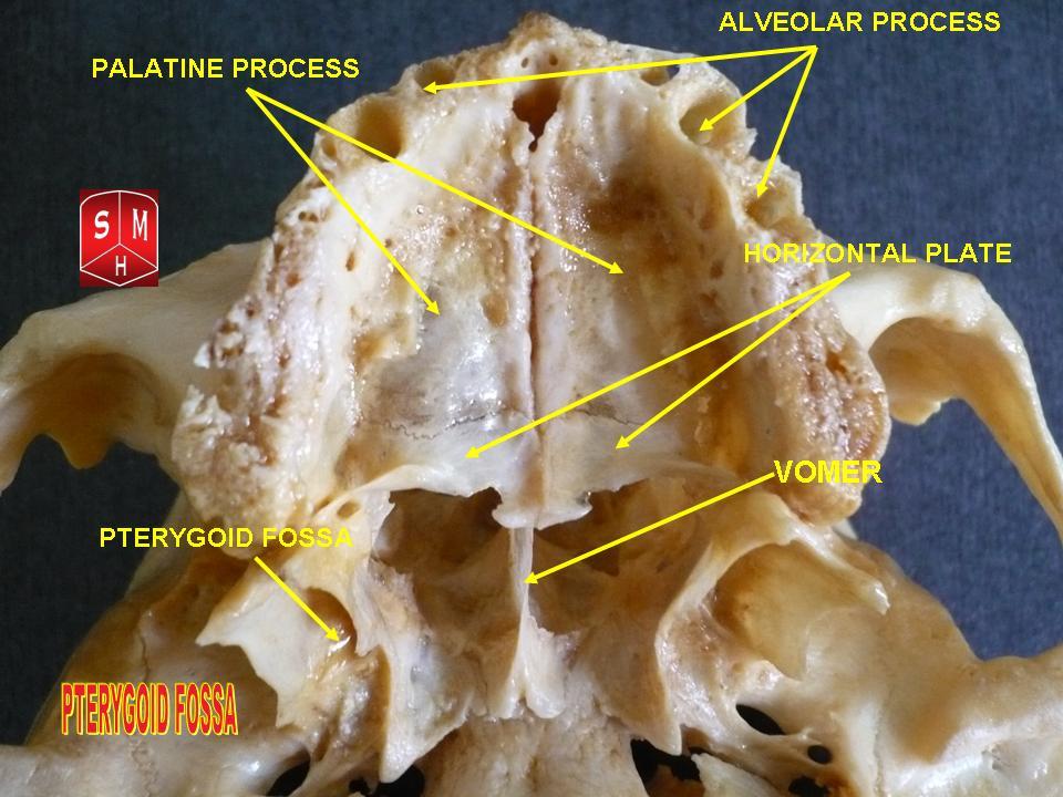
The pterygoid fossa is an anatomical term for the fossa formed by the divergence of the lateral pterygoid plate and the medial pterygoid plate of the sphenoid bone. Structure The lateral and medial pterygoid plates (of the pterygoid process of the sphenoid bone) diverge behind and enclose between them a V-shaped fossa, the pterygoid fossa. This fossa faces posteriorly, and contains the medial pterygoid muscle and the tensor veli palatini muscle. See also * Pterygoid fovea * Scaphoid fossa * Pterygoid process The pterygoid processes of the sphenoid (from Greek ''pteryx'', ''pterygos'', "wing"), one on either side, descend perpendicularly from the regions where the body and the greater wings of the sphenoid bone unite. Each process consists of a me ... References Bones of the head and neck {{musculoskeletal-stub ... [...More Info...] [...Related Items...] OR: [Wikipedia] [Google] [Baidu] |


