|
Small Supernumerary Marker Chromosome
A small supernumerary marker chromosome (sSMC) is an abnormal extra chromosome. It contains copies of parts of one or more normal chromosomes and like normal chromosomes is located in the cell's nucleus, is replicated and distributed into each daughter cell during cell division, and typically has genes which may be expressed. However, it may also be active in causing birth defects and neoplasms (e.g. tumors and cancers). The sSMC's small size makes it virtually undetectable using classical cytogenetic methods: the far larger DNA and gene content of the cell's normal chromosomes obscures those of the sSMC. Newer molecular techniques such as fluorescence in situ hybridization, next generation sequencing, comparative genomic hybridization, and highly specialized cytogenetic G banding analyses are required to study it. Using these methods, the DNA sequences and genes in sSMCs are identified and help define as well as explain any effect(s) it may have on individuals. Human cells t ... [...More Info...] [...Related Items...] OR: [Wikipedia] [Google] [Baidu] |
Marker Chromosome
A marker chromosome (mar) is a small fragment of a chromosome which generally cannot be identified without specialized genomic analysis due to the size of the fragment.Thompson & Thompson Genetics in Medicine, Chapter 5, 57-74 https://www.clinicalkey.com/#!/content/book/3-s2.0-B9781437706963000054?scrollTo=%23hl0000654 The significance of a marker is variable as it depends on what material is contained within the marker.Nelson Textbook of Pediatrics, Chapter 81, 604-627 https://www.clinicalkey.com/#!/content/book/3-s2.0-B9781455775668000818?scrollTo=%23hl0003126 The large majority of these marker chromosomes are smaller than one of the smaller human chromosomes, chromosome 20, and by definition are termed small supernumerary marker chromosomes. Marker chromosomes occur sporadically about 70% of the time, with the remainder being inherited from a parent. About 50% of cases involve mosaicism, which affects the severity of the condition. The frequency is approximately 3-4 per 10,000 pe ... [...More Info...] [...Related Items...] OR: [Wikipedia] [Google] [Baidu] |
Cytogenetic
Cytogenetics is essentially a branch of genetics, but is also a part of cell biology/cytology (a subdivision of human anatomy), that is concerned with how the chromosomes relate to cell behaviour, particularly to their behaviour during mitosis and meiosis. Techniques used include karyotyping, analysis of G-banded chromosomes, other cytogenetic banding techniques, as well as molecular cytogenetics such as fluorescent ''in situ'' hybridization (FISH) and comparative genomic hybridization (CGH). History Beginnings Chromosomes were first observed in plant cells by Carl Nägeli in 1842. Their behavior in animal (salamander) cells was described by Walther Flemming, the discoverer of mitosis, in 1882. The name was coined by another German anatomist, von Waldeyer in 1888. The next stage took place after the development of genetics in the early 20th century, when it was appreciated that the set of chromosomes (the karyotype) was the carrier of the genes. Levitsky seems to have been t ... [...More Info...] [...Related Items...] OR: [Wikipedia] [Google] [Baidu] |
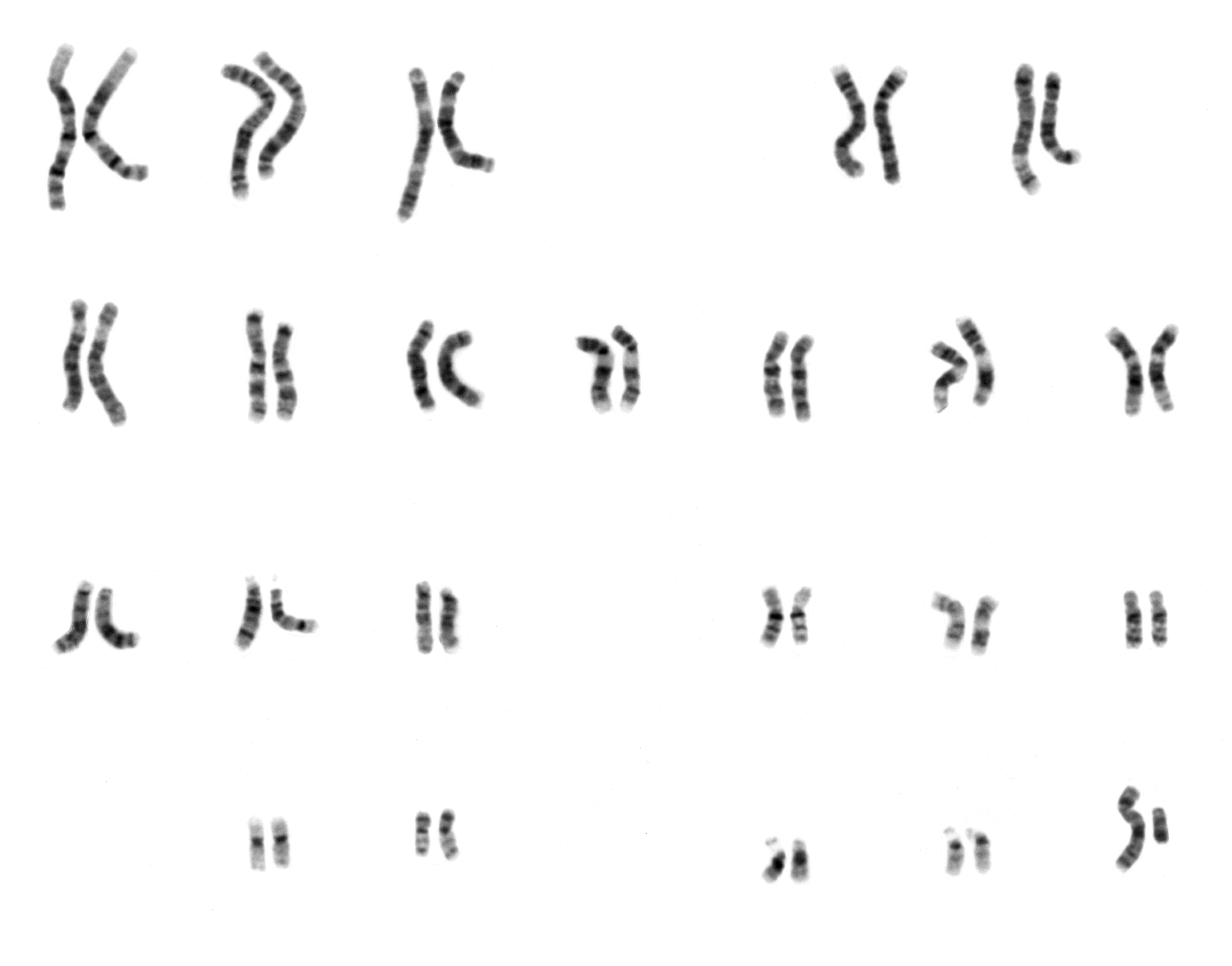
Karyotype
A karyotype is the general appearance of the complete set of metaphase chromosomes in the cells of a species or in an individual organism, mainly including their sizes, numbers, and shapes. Karyotyping is the process by which a karyotype is discerned by determining the chromosome complement of an individual, including the number of chromosomes and any abnormalities. A karyogram or idiogram is a graphical depiction of a karyotype, wherein chromosomes are organized in pairs, ordered by size and position of centromere for chromosomes of the same size. Karyotyping generally combines light microscopy and photography, and results in a photomicrographic (or simply micrographic) karyogram. In contrast, a schematic karyogram is a designed graphic representation of a karyotype. In schematic karyograms, just one of the sister chromatids of each chromosome is generally shown for brevity, and in reality they are generally so close together that they look as one on photomicrographs as well ... [...More Info...] [...Related Items...] OR: [Wikipedia] [Google] [Baidu] |
Centromere
The centromere links a pair of sister chromatids together during cell division. This constricted region of chromosome connects the sister chromatids, creating a short arm (p) and a long arm (q) on the chromatids. During mitosis, spindle fibers attach to the centromere via the kinetochore. The physical role of the centromere is to act as the site of assembly of the kinetochores – a highly complex multiprotein structure that is responsible for the actual events of chromosome segregation – i.e. binding microtubules and signaling to the cell cycle machinery when all chromosomes have adopted correct attachments to the spindle, so that it is safe for cell division to proceed to completion and for cells to enter anaphase. There are, broadly speaking, two types of centromeres. "Point centromeres" bind to specific proteins that recognize particular DNA sequences with high efficiency. Any piece of DNA with the point centromere DNA sequence on it will typically form a centromere if pr ... [...More Info...] [...Related Items...] OR: [Wikipedia] [Google] [Baidu] |
Chromosome 15
Chromosome 15 is one of the 23 pairs of chromosomes in humans. People normally have two copies of this chromosome. Chromosome 15 spans about 102 million base pairs (the building material of DNA) and represents between 3% and 3.5% of the total DNA in cells. Chromosome 15 is an acrocentric chromosome, with a very small short arm (the "p" arm, for "petite"), which contains few protein coding genes among its 19 million base pairs. It has a larger long arm (the "q" arm) that is gene rich, spanning about 83 million base pairs. The human leukocyte antigen gene for β2-microglobulin is found on chromosome 15, as well as the FBN1 gene, coding for both fibrillin-1 (a protein critical to the proper functioning of connective tissue), and aprosin (a small protein produced from part of the transcribed FBN1 gene mRNA), which is involved in fat metabolism. Genes Number of genes The following are some of the gene count estimates of human chromosome 15. Because researchers use different approac ... [...More Info...] [...Related Items...] OR: [Wikipedia] [Google] [Baidu] |
Chromosome 20
Chromosome 20 is one of the 23 pairs of chromosomes in humans. Chromosome 20 spans around 66 million base pairs (the building material of DNA) and represents between 2 and 2.5 percent of the total DNA in cells. Chromosome 20 was fully sequenced in 2001 and was reported to contain over 59 million base pairs. Since then, due to sequencing improvements and fixes, the length of chromosome 20 has been updated to just over 66 million base pairs. Genes Number of genes The following are some of the gene count estimates of human chromosome 20. Because researchers use different approaches to genome annotation their predictions of the number of genes on each chromosome varies (for technical details, see gene prediction). Among various projects, the collaborative consensus coding sequence project ( CCDS) takes an extremely conservative strategy. So CCDS's gene number prediction represents a lower bound on the total number of human protein-coding genes. Gene list The following is a ... [...More Info...] [...Related Items...] OR: [Wikipedia] [Google] [Baidu] |
Y Chromosome
The Y chromosome is one of two sex chromosomes (allosomes) in therian mammals, including humans, and many other animals. The other is the X chromosome. Y is normally the sex-determining chromosome in many species, since it is the presence or absence of Y that determines the male or female sex of offspring produced in sexual reproduction. In mammals, the Y chromosome contains the gene SRY, which triggers male development. The DNA in the human Y chromosome is composed of about 59 million base pairs, making it similar in size to chromosome 19. The Y chromosome is passed only from father to son. With a 30% difference between humans and chimpanzees, the Y chromosome is one of the fastest-evolving parts of the human genome. The human Y chromosome carries an estimated 100–200 genes, with between 45 and 73 of these being protein-coding. All single-copy Y-linked genes are hemizygous (present on only one chromosome) except in cases of aneuploidy such as XYY syndrome or XXYY syndrome. ... [...More Info...] [...Related Items...] OR: [Wikipedia] [Google] [Baidu] |
X Chromosome
The X chromosome is one of the two sex-determining chromosomes (allosomes) in many organisms, including mammals (the other is the Y chromosome), and is found in both males and females. It is a part of the XY sex-determination system and XO sex-determination system. The X chromosome was named for its unique properties by early researchers, which resulted in the naming of its counterpart Y chromosome, for the next letter in the alphabet, following its subsequent discovery. Discovery It was first noted that the X chromosome was special in 1890 by Hermann Henking in Leipzig. Henking was studying the testicles of ''Pyrrhocoris'' and noticed that one chromosome did not take part in meiosis. Chromosomes are so named because of their ability to take up staining (''chroma'' in Greek means ''color''). Although the X chromosome could be stained just as well as the others, Henking was unsure whether it was a different class of object and consequently named it ''X element'', which later be ... [...More Info...] [...Related Items...] OR: [Wikipedia] [Google] [Baidu] |
Sex Chromosome
A sex chromosome (also referred to as an allosome, heterotypical chromosome, gonosome, heterochromosome, or idiochromosome) is a chromosome that differs from an ordinary autosome in form, size, and behavior. The human sex chromosomes, a typical pair of mammal allosomes, determine the sex of an individual created in sexual reproduction. Autosomes differ from allosomes because autosomes appear in pairs whose members have the same form but differ from other pairs in a diploid cell, whereas members of an allosome pair may differ from one another and thereby determine sex. Nettie Stevens and Edmund Beecher Wilson both independently discovered sex chromosomes in 1905. However, Stevens is credited for discovering them earlier than Wilson. Differentiation In humans, each cell nucleus contains 23 pairs of chromosomes, a total of 46 chromosomes. The first 22 pairs are called autosomes. Autosomes are homologous chromosomes i.e. chromosomes which contain the same genes (regions of DNA) i ... [...More Info...] [...Related Items...] OR: [Wikipedia] [Google] [Baidu] |
Autosome
An autosome is any chromosome that is not a sex chromosome. The members of an autosome pair in a diploid cell have the same morphology, unlike those in allosome, allosomal (sex chromosome) pairs, which may have different structures. The DNA in autosomes is collectively known as atDNA or auDNA. For example, humans have a diploid human genome, genome that usually contains 22 pairs of autosomes and one allosome pair (46 chromosomes total). The autosome pairs are labeled with numbers (1–22 in humans) roughly in order of their sizes in base pairs, while allosomes are labelled with their letters. By contrast, the allosome pair consists of two X chromosomes in females or one X and one Y chromosome in males. Unusual combinations of XYY syndrome, XYY, Klinefelter syndrome, XXY, Triple X syndrome, XXX, XXXX syndrome, XXXX, XXXXX syndrome, XXXXX or XXYY syndrome, XXYY, among Aneuploidy, other Salome combinations, are known to occur and usually cause developmental abnormalities. Autosomes ... [...More Info...] [...Related Items...] OR: [Wikipedia] [Google] [Baidu] |
Cell (biology)
The cell is the basic structural and functional unit of life forms. Every cell consists of a cytoplasm enclosed within a membrane, and contains many biomolecules such as proteins, DNA and RNA, as well as many small molecules of nutrients and metabolites.Cell Movements and the Shaping of the Vertebrate Body in Chapter 21 of Molecular Biology of the Cell '' fourth edition, edited by Bruce Alberts (2002) published by Garland Science. The Alberts text discusses how the "cellular building blocks" move to shape developing embryos. It is also common to describe small molecules such as ... [...More Info...] [...Related Items...] OR: [Wikipedia] [Google] [Baidu] |
Cytogenetics
Cytogenetics is essentially a branch of genetics, but is also a part of cell biology/cytology (a subdivision of human anatomy), that is concerned with how the chromosomes relate to cell behaviour, particularly to their behaviour during mitosis and meiosis. Techniques used include karyotyping, analysis of G-banded chromosomes, other cytogenetic banding techniques, as well as molecular cytogenetics such as fluorescent ''in situ'' hybridization (FISH) and comparative genomic hybridization (CGH). History Beginnings Chromosomes were first observed in plant cells by Carl Nägeli in 1842. Their behavior in animal (salamander) cells was described by Walther Flemming, the discoverer of mitosis, in 1882. The name was coined by another German anatomist, von Waldeyer in 1888. The next stage took place after the development of genetics in the early 20th century, when it was appreciated that the set of chromosomes (the karyotype) was the carrier of the genes. Levitsky seems to have been t ... [...More Info...] [...Related Items...] OR: [Wikipedia] [Google] [Baidu] |


