|
Slow-wave Potential
A slow-wave potential is a rhythmic electrophysiological event in the gastrointestinal tract. The normal conduction of slow waves is one of the key regulators of gastrointestinal motility. Slow waves are generated and propagated by a class of pacemaker cells called the interstitial cells of Cajal, which also act as intermediates between nerves and smooth muscle cells. Slow waves generated in interstitial cells of Cajal spread to the surrounding smooth muscle cells and control motility. Description In the human enteric nervous system, the slow-wave threshold is the slow-wave potential which must be reached before a slow wave can be propagated in gut wall smooth muscle. Slow waves themselves seldom cause any smooth muscle contraction (Except for, probably in the stomach). When the amplitude of slow waves in smooth muscle cells reaches the slow-wave threshold — the L-type Ca2+ channels are activated, resulting in calcium influx and initiation of motility. Slow waves are generated ... [...More Info...] [...Related Items...] OR: [Wikipedia] [Google] [Baidu] |
Gastrointestinal Tract
The gastrointestinal tract (GI tract, digestive tract, alimentary canal) is the tract or passageway of the digestive system that leads from the mouth to the anus. The GI tract contains all the major organ (biology), organs of the digestive system, in humans and other animals, including the esophagus, stomach, and intestines. Food taken in through the mouth is digestion, digested to extract nutrients and absorb energy, and the waste expelled at the anus as feces. ''Gastrointestinal'' is an adjective meaning of or pertaining to the stomach and intestines. Nephrozoa, Most animals have a "through-gut" or complete digestive tract. Exceptions are more primitive ones: sponges have small pores (ostium (sponges), ostia) throughout their body for digestion and a larger dorsal pore (osculum) for excretion, comb jellies have both a ventral mouth and dorsal anal pores, while cnidarians and acoels have a single pore for both digestion and excretion. The human gastrointestinal tract consists o ... [...More Info...] [...Related Items...] OR: [Wikipedia] [Google] [Baidu] |
Interstitial Cell Of Cajal
Interstitial cells of Cajal (ICC) are interstitial cells found in the gastrointestinal tract. There are different types of ICC with different functions. ICC and another type of interstitial cell, known as platelet-derived growth factor receptor alpha (PDGFRα) cells, are electrically coupled to smooth muscle cells via gap junctions, that work together as an SIP functional syncytium. Myenteric interstitial cells of Cajal (ICC-MY) serve as pacemaker cells that generate the bioelectrical events known as slow waves. Slow waves conduct to smooth muscle cells and cause phasic contractions. The picture to the right shows an isolated Interstitial cell of Cajal from the Myenteric plexus of the mouse small intestine grown in a primary cell culture. This cell type can be characterized morphologically as having a small cell body often triangular or stellate-shaped with several long processes branching out into secondary and tertiary extensions - these processes often contact smooth muscl ... [...More Info...] [...Related Items...] OR: [Wikipedia] [Google] [Baidu] |
Nerve
A nerve is an enclosed, cable-like bundle of nerve fibers (called axons) in the peripheral nervous system. A nerve transmits electrical impulses. It is the basic unit of the peripheral nervous system. A nerve provides a common pathway for the electrochemical nerve impulses called action potentials that are transmitted along each of the axons to peripheral organs or, in the case of sensory nerves, from the periphery back to the central nervous system. Each axon, within the nerve, is an extension of an individual neuron, along with other supportive cells such as some Schwann cells that coat the axons in myelin. Within a nerve, each axon is surrounded by a layer of connective tissue called the endoneurium. The axons are bundled together into groups called fascicles, and each fascicle is wrapped in a layer of connective tissue called the perineurium. Finally, the entire nerve is wrapped in a layer of connective tissue called the epineurium. Nerve cells (often called neurons) are f ... [...More Info...] [...Related Items...] OR: [Wikipedia] [Google] [Baidu] |
Smooth Muscle
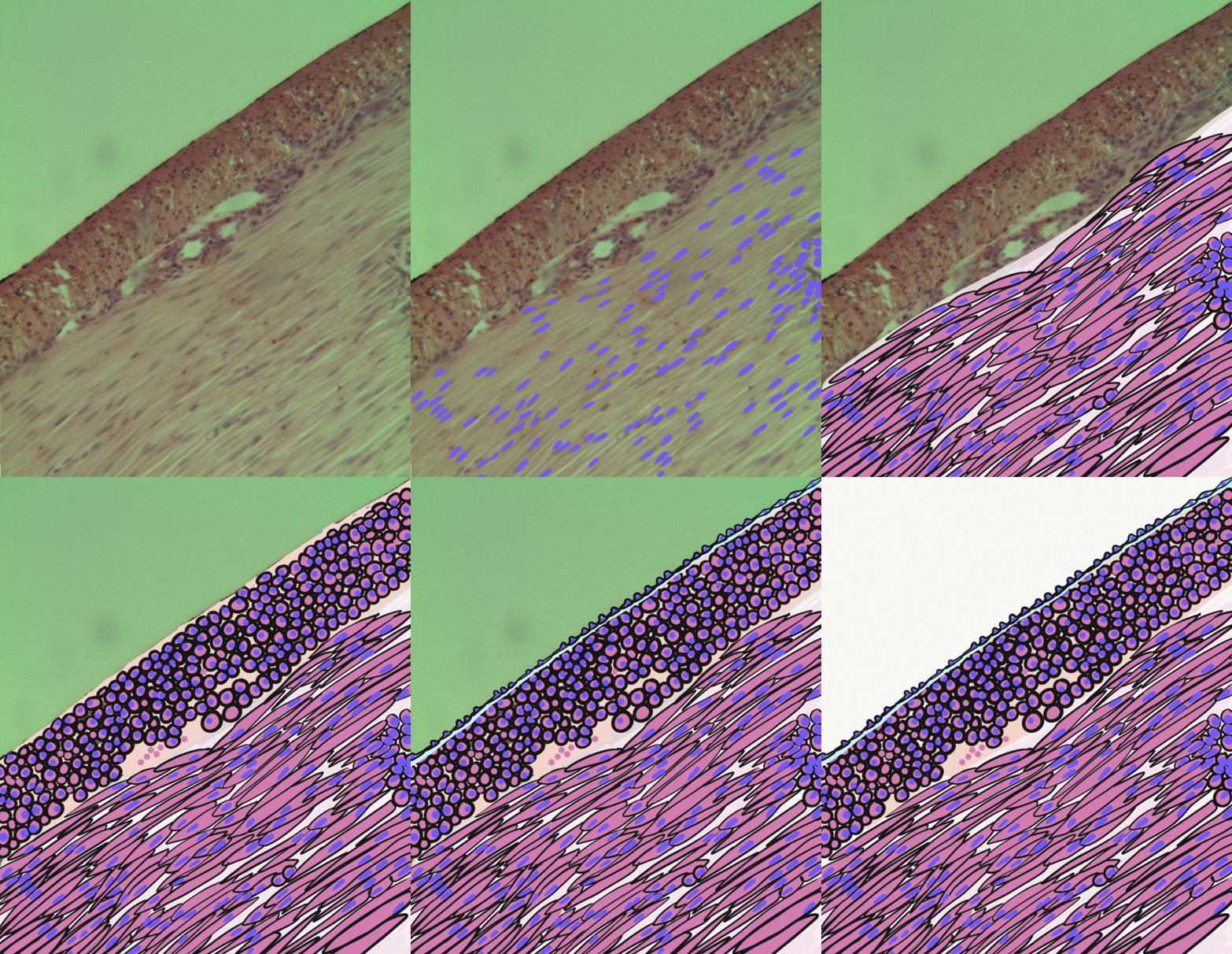
Smooth muscle is an involuntary non-striated muscle, so-called because it has no sarcomeres and therefore no striations (''bands'' or ''stripes''). It is divided into two subgroups, single-unit and multiunit smooth muscle. Within single-unit muscle, the whole bundle or sheet of smooth muscle cells contracts as a syncytium. Smooth muscle is found in the walls of hollow organs, including the stomach, intestines, bladder and uterus; in the walls of passageways, such as blood, and lymph vessels, and in the tracts of the respiratory, urinary, and reproductive systems. In the eyes, the ciliary muscles, a type of smooth muscle, dilate and contract the iris and alter the shape of the lens. In the skin, smooth muscle cells such as those of the arrector pili cause hair to stand erect in response to cold temperature or fear. Structure Gross anatomy Smooth muscle is grouped into two types: single-unit smooth muscle, also known as visceral smooth muscle, and multiunit smooth muscle. ... [...More Info...] [...Related Items...] OR: [Wikipedia] [Google] [Baidu] |
Enteric Nervous System
The enteric nervous system (ENS) or intrinsic nervous system is one of the main divisions of the autonomic nervous system (ANS) and consists of a mesh-like system of neurons that governs the function of the gastrointestinal tract. It is capable of acting independently of the sympathetic and parasympathetic nervous systems, although it may be influenced by them. The ENS is nicknamed the "second brain". It is derived from neural crest cells. The enteric nervous system is capable of operating independently of the brain and spinal cord, but does rely on innervation from the vagus nerve and prevertebral ganglia in healthy subjects. However, studies have shown that the system is operable with a severed vagus nerve. The neurons of the enteric nervous system control the motor functions of the system, in addition to the secretion of gastrointestinal enzymes. These neurons communicate through many neurotransmitters similar to the CNS, including acetylcholine, dopamine, and serotonin. Th ... [...More Info...] [...Related Items...] OR: [Wikipedia] [Google] [Baidu] |
Smooth Muscle
Smooth muscle is an involuntary non-striated muscle, so-called because it has no sarcomeres and therefore no striations (''bands'' or ''stripes''). It is divided into two subgroups, single-unit and multiunit smooth muscle. Within single-unit muscle, the whole bundle or sheet of smooth muscle cells contracts as a syncytium. Smooth muscle is found in the walls of hollow organs, including the stomach, intestines, bladder and uterus; in the walls of passageways, such as blood, and lymph vessels, and in the tracts of the respiratory, urinary, and reproductive systems. In the eyes, the ciliary muscles, a type of smooth muscle, dilate and contract the iris and alter the shape of the lens. In the skin, smooth muscle cells such as those of the arrector pili cause hair to stand erect in response to cold temperature or fear. Structure Gross anatomy Smooth muscle is grouped into two types: single-unit smooth muscle, also known as visceral smooth muscle, and multiunit smooth muscle. ... [...More Info...] [...Related Items...] OR: [Wikipedia] [Google] [Baidu] |
Entrainment (physics)
Injection locking and injection pulling are the frequency effects that can occur when a harmonic oscillator is disturbed by a second oscillator operating at a nearby frequency. When the coupling is strong enough and the frequencies near enough, the second oscillator can capture the first oscillator, causing it to have essentially identical frequency as the second. This is injection locking. When the second oscillator merely disturbs the first but does not capture it, the effect is called injection pulling. Injection locking and pulling effects are observed in numerous types of physical systems, however the terms are most often associated with electronic oscillators or laser resonators. Injection locking has been used in beneficial and clever ways in the design of early television sets and oscilloscopes, allowing the equipment to be synchronized to external signals at a relatively low cost. Injection locking has also been used in high performance frequency doubling circuits. How ... [...More Info...] [...Related Items...] OR: [Wikipedia] [Google] [Baidu] |
Uterine Smooth Muscle
The myometrium is the middle layer of the uterine wall, consisting mainly of uterine smooth muscle cells (also called uterine myocytes) but also of supporting stromal and vascular tissue. Its main function is to induce uterine contractions. Structure The myometrium is located between the endometrium (the inner layer of the uterine wall) and the serosa or perimetrium (the outer uterine layer). The inner one-third of the myometrium (termed the ''junctional'' or ''sub-endometrial'' layer) appears to be derived from the Müllerian duct, while the outer, more predominant layer of the myometrium appears to originate from non-Müllerian tissue and is the major contractile tissue during parturition and abortion. The junctional layer appears to function like a circular muscle layer, capable of peristaltic and anti-peristaltic activity, equivalent to the muscular layer of the intestines. Muscular structure The molecular structure of the smooth muscle of myometrium is very similar to ... [...More Info...] [...Related Items...] OR: [Wikipedia] [Google] [Baidu] |
Acetylcholine
Acetylcholine (ACh) is an organic chemical that functions in the brain and body of many types of animals (including humans) as a neurotransmitter. Its name is derived from its chemical structure: it is an ester of acetic acid and choline. Parts in the body that use or are affected by acetylcholine are referred to as cholinergic. Substances that increase or decrease the overall activity of the cholinergic system are called cholinergics and anticholinergics, respectively. Acetylcholine is the neurotransmitter used at the neuromuscular junction—in other words, it is the chemical that motor neurons of the nervous system release in order to activate muscles. This property means that drugs that affect cholinergic systems can have very dangerous effects ranging from paralysis to convulsions. Acetylcholine is also a neurotransmitter in the autonomic nervous system, both as an internal transmitter for the sympathetic nervous system and as the final product released by the parasymp ... [...More Info...] [...Related Items...] OR: [Wikipedia] [Google] [Baidu] |
Substance P
Substance P (SP) is an undecapeptide (a peptide composed of a chain of 11 amino acid residues) and a member of the tachykinin neuropeptide family. It is a neuropeptide, acting as a neurotransmitter and as a neuromodulator. Substance P and its closely related neurokinin A (NKA) are produced from a polyprotein precursor after differential splicing of the preprotachykinin A gene. The deduced amino acid sequence of substance P is as follows: * Arg Pro Lys Pro Gln Gln Phe Phe Gly Leu Met (RPKPQQFFGLM) with an amidation at the C-terminus. Substance P is released from the terminals of specific sensory nerves. It is found in the brain and spinal cord and is associated with inflammatory processes and pain. Discovery The original discovery of Substance P (SP) was in 1931 by Ulf von Euler and John H. Gaddum as a tissue extract that caused intestinal contraction ''in vitro''. Its tissue distribution and biologic actions were further investigated over the following decades. The ele ... [...More Info...] [...Related Items...] OR: [Wikipedia] [Google] [Baidu] |
Vasoactive Intestinal Peptide
Vasoactive intestinal peptide, also known as vasoactive intestinal polypeptide or VIP, is a peptide hormone that is vasoactive in the intestine. VIP is a peptide of 28 amino acid residues that belongs to a glucagon/secretin superfamily, the ligand of class II G protein–coupled receptors. VIP is produced in many tissues of vertebrates including the gut, pancreas, and suprachiasmatic nuclei of the hypothalamus in the brain. VIP stimulates contractility in the heart, causes vasodilation, increases glycogenolysis, lowers arterial blood pressure and relaxes the smooth muscle of trachea, stomach and gallbladder. In humans, the vasoactive intestinal peptide is encoded by the ''VIP'' gene. VIP has a half-life (t½) in the blood of about two minutes. Function In the body VIP has an effect on several tissues: In the digestive system, VIP seems to induce smooth muscle relaxation (lower esophageal sphincter, stomach, gallbladder), stimulate secretion of water into pancreatic juice a ... [...More Info...] [...Related Items...] OR: [Wikipedia] [Google] [Baidu] |


