|
Sinoatrial
The sinoatrial node (also known as the sinuatrial node, SA node or sinus node) is an oval shaped region of special cardiac muscle in the upper back wall of the right atrium made up of cells known as pacemaker cells. The sinus node is approximately fifteen mm long, three mm wide, and one mm thick, located directly below and to the side of the superior vena cava. These cells can produce an electrical impulse an action potential known as a cardiac action potential that travels through the electrical conduction system of the heart, causing it to contract. In a healthy heart, the SA node continuously produces action potentials, setting the rhythm of the heart (sinus rhythm), and so is known as the heart's natural pacemaker. The rate of action potentials produced (and therefore the heart rate) is influenced by the nerves that supply it. Structure The sinoatrial node is a oval-shaped structure that is approximately fifteen mm long, three mm wide, and one mm thick, located directly b ... [...More Info...] [...Related Items...] OR: [Wikipedia] [Google] [Baidu] |
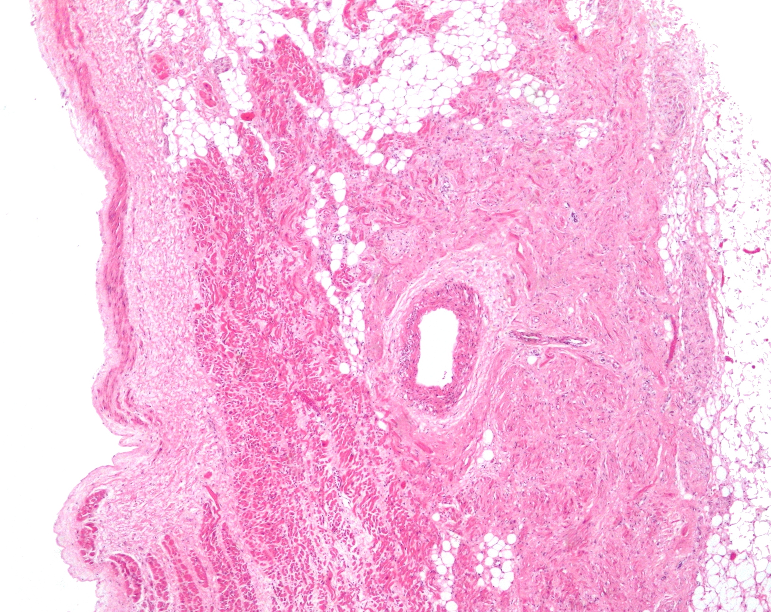
Sinoatrial Node 2 Low Mag
The sinoatrial node (also known as the sinuatrial node, SA node or sinus node) is an oval shaped region of special cardiac muscle in the upper back wall of the right atrium made up of cells known as pacemaker cells. The sinus node is approximately fifteen mm long, three mm wide, and one mm thick, located directly below and to the side of the superior vena cava. These cells can produce an electrical impulse an action potential known as a cardiac action potential that travels through the electrical conduction system of the heart, causing it to contract. In a healthy heart, the SA node continuously produces action potentials, setting the rhythm of the heart (sinus rhythm), and so is known as the heart's natural pacemaker. The rate of action potentials produced (and therefore the heart rate) is influenced by the nerves that supply it. Structure The sinoatrial node is a oval-shaped structure that is approximately fifteen mm long, three mm wide, and one mm thick, located directly b ... [...More Info...] [...Related Items...] OR: [Wikipedia] [Google] [Baidu] |
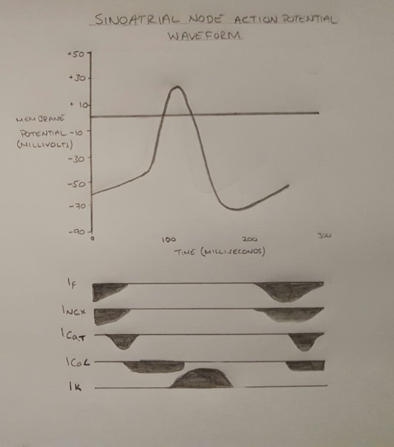
Cardiac Action Potential
The cardiac action potential is a brief change in voltage ( membrane potential) across the cell membrane of heart cells. This is caused by the movement of charged atoms (called ions) between the inside and outside of the cell, through proteins called ion channels. The cardiac action potential differs from action potentials found in other types of electrically excitable cells, such as nerves. Action potentials also vary within the heart; this is due to the presence of different ion channels in different cells. Unlike the action potential in skeletal muscle cells, the cardiac action potential is not initiated by nervous activity. Instead, it arises from a group of specialized cells known as pacemaker cells, that have automatic action potential generation capability. In healthy hearts, these cells form the cardiac pacemaker and are found in the sinoatrial node in the right atrium. They produce roughly 60–100 action potentials every minute. The action potential passes along t ... [...More Info...] [...Related Items...] OR: [Wikipedia] [Google] [Baidu] |
Heart
The heart is a muscular organ in most animals. This organ pumps blood through the blood vessels of the circulatory system. The pumped blood carries oxygen and nutrients to the body, while carrying metabolic waste such as carbon dioxide to the lungs. In humans, the heart is approximately the size of a closed fist and is located between the lungs, in the middle compartment of the chest. In humans, other mammals, and birds, the heart is divided into four chambers: upper left and right atria and lower left and right ventricles. Commonly the right atrium and ventricle are referred together as the right heart and their left counterparts as the left heart. Fish, in contrast, have two chambers, an atrium and a ventricle, while most reptiles have three chambers. In a healthy heart blood flows one way through the heart due to heart valves, which prevent backflow. The heart is enclosed in a protective sac, the pericardium, which also contains a small amount of fluid. The wall ... [...More Info...] [...Related Items...] OR: [Wikipedia] [Google] [Baidu] |
Sinoatrial Nodal Artery
The sinoatrial nodal artery (or sinuatrial nodal artery or sinoatrial artery) is an artery of the heart which supplies the sinoatrial node, the natural pacemaker center of the heart, and arises from the right coronary artery in around 60% of people. In about 40% of cases, the sinoatrial artery is a branch of the left circumflex coronary artery. In less than 1% of humans, the artery has an anomalous origin directly from the coronary sinus, descending aorta, or distal right coronary artery. In more than 50% of human hearts, the artery actually courses close to the superior posterior aspect of the interatrial septum. The origin of the sinoatrial node artery is not related to coronary artery dominance, which means the side (right or left) that provides the circulation to the back of the heart. In contrast, the atrioventricular nodal branch, that is the artery that brings blood to the atrioventricular node, depends on coronary artery dominance. The sinoatrial node surrounds the sinoatri ... [...More Info...] [...Related Items...] OR: [Wikipedia] [Google] [Baidu] |
Heart Rate
Heart rate (or pulse rate) is the frequency of the heartbeat measured by the number of contractions (beats) of the heart per minute (bpm). The heart rate can vary according to the body's physical needs, including the need to absorb oxygen and excrete carbon dioxide, but is also modulated by numerous factors, including, but not limited to, genetics, physical fitness, stress or psychological status, diet, drugs, hormonal status, environment, and disease/illness as well as the interaction between and among these factors. It is usually equal or close to the pulse measured at any peripheral point. The American Heart Association states the normal resting adult human heart rate is 60–100 bpm. Tachycardia is a high heart rate, defined as above 100 bpm at rest. Bradycardia is a low heart rate, defined as below 60 bpm at rest. When a human sleeps, a heartbeat with rates around 40–50 bpm is common and is considered normal. When the heart is not beating in a regular pattern, this is ref ... [...More Info...] [...Related Items...] OR: [Wikipedia] [Google] [Baidu] |
Pacemaker Cell
350px, Image showing the cardiac pacemaker or SA node, the primary pacemaker within the electrical_conduction_system_of_the_heart">SA_node,_the_primary_pacemaker_within_the_electrical_conduction_system_of_the_heart. The_muscle_contraction.html" "title="electrical conduction system of the heart.">electrical conduction system of the heart">SA node, the primary pacemaker within the electrical conduction system of the heart. The muscle contraction">contraction of cardiac muscle (heart muscle) in all animals is initiated by electrical impulses known as action potentials that in the heart are known as cardiac action potentials. The rate at which these impulses fire controls the rate of cardiac contraction, that is, the heart rate. The cells that create these rhythmic impulses, setting the pace for blood pumping, are called pacemaker cells, and they directly control the heart rate. They make up the cardiac pacemaker, that is, the natural pacemaker of the heart. In most humans, the h ... [...More Info...] [...Related Items...] OR: [Wikipedia] [Google] [Baidu] |
Cardiac Pacemaker
350px, Image showing the cardiac pacemaker or SA node, the primary pacemaker within the electrical_conduction_system_of_the_heart">SA_node,_the_primary_pacemaker_within_the_electrical_conduction_system_of_the_heart. The_muscle_contraction.html" "title="electrical conduction system of the heart.">electrical conduction system of the heart">SA node, the primary pacemaker within the electrical conduction system of the heart. The muscle contraction">contraction of cardiac muscle (heart muscle) in all animals is initiated by electrical impulses known as action potentials that in the heart are known as cardiac action potentials. The rate at which these impulses fire controls the rate of cardiac contraction, that is, the heart rate. The cells that create these rhythmic impulses, setting the pace for blood pumping, are called pacemaker cells, and they directly control the heart rate. They make up the cardiac pacemaker, that is, the natural pacemaker of the heart. In most humans, the h ... [...More Info...] [...Related Items...] OR: [Wikipedia] [Google] [Baidu] |
Electrical Conduction System Of The Heart
The cardiac conduction system (CCS) (also called the electrical conduction system of the heart) transmits the signals generated by the sinoatrial node – the heart's pacemaker, to cause the heart muscle to contract, and pump blood through the body's circulatory system. The pacemaking signal travels through the right atrium to the atrioventricular node, along the bundle of His, and through the bundle branches to Purkinje fibers in the walls of the ventricles. The Purkinje fibers transmit the signals more rapidly to stimulate contraction of the ventricles. The conduction system consists of specialized heart muscle cells, situated within the myocardium. There is a skeleton of fibrous tissue that surrounds the conduction system which can be seen on an ECG. Dysfunction of the conduction system can cause irregular heart rhythms including rhythms that are too fast or too slow. Structure Electrical signals arising in the SA node (located in the right atrium) stimulate the atr ... [...More Info...] [...Related Items...] OR: [Wikipedia] [Google] [Baidu] |
Atrium (heart)
The atrium ( la, ātrium, , entry hall) is one of two upper chambers in the heart that receives blood from the circulatory system. The blood in the atria is pumped into the heart ventricles through the atrioventricular valves. There are two atria in the human heart – the left atrium receives blood from the pulmonary circulation, and the right atrium receives blood from the venae cavae of the systemic circulation. During the cardiac cycle the atria receive blood while relaxed in diastole, then contract in systole to move blood to the ventricles. Each atrium is roughly cube-shaped except for an ear-shaped projection called an atrial appendage, sometimes known as an auricle. All animals with a closed circulatory system have at least one atrium. The atrium was formerly called the 'auricle'. That term is still used to describe this chamber in some other animals, such as the ''Mollusca''. They have thicker muscular walls than the atria do. Structure Humans have a four-chambered ... [...More Info...] [...Related Items...] OR: [Wikipedia] [Google] [Baidu] |
Atrial
The atrium ( la, ātrium, , entry hall) is one of two upper chambers in the heart that receives blood from the circulatory system. The blood in the atria is pumped into the heart ventricles through the atrioventricular valves. There are two atria in the human heart – the left atrium receives blood from the pulmonary circulation, and the right atrium receives blood from the venae cavae of the systemic circulation. During the cardiac cycle the atria receive blood while relaxed in diastole, then contract in systole to move blood to the ventricles. Each atrium is roughly cube-shaped except for an ear-shaped projection called an atrial appendage, sometimes known as an auricle. All animals with a closed circulatory system have at least one atrium. The atrium was formerly called the 'auricle'. That term is still used to describe this chamber in some other animals, such as the ''Mollusca''. They have thicker muscular walls than the atria do. Structure Humans have a four-chambered h ... [...More Info...] [...Related Items...] OR: [Wikipedia] [Google] [Baidu] |
Electrical Conduction System Of The Heart
The cardiac conduction system (CCS) (also called the electrical conduction system of the heart) transmits the signals generated by the sinoatrial node – the heart's pacemaker, to cause the heart muscle to contract, and pump blood through the body's circulatory system. The pacemaking signal travels through the right atrium to the atrioventricular node, along the bundle of His, and through the bundle branches to Purkinje fibers in the walls of the ventricles. The Purkinje fibers transmit the signals more rapidly to stimulate contraction of the ventricles. The conduction system consists of specialized heart muscle cells, situated within the myocardium. There is a skeleton of fibrous tissue that surrounds the conduction system which can be seen on an ECG. Dysfunction of the conduction system can cause irregular heart rhythms including rhythms that are too fast or too slow. Structure Electrical signals arising in the SA node (located in the right atrium) stimulate the atr ... [...More Info...] [...Related Items...] OR: [Wikipedia] [Google] [Baidu] |
Action Potential
An action potential occurs when the membrane potential of a specific cell location rapidly rises and falls. This depolarization then causes adjacent locations to similarly depolarize. Action potentials occur in several types of animal cells, called excitable cells, which include neurons, muscle cells, and in some plant cells. Certain endocrine cells such as pancreatic beta cells, and certain cells of the anterior pituitary gland are also excitable cells. In neurons, action potentials play a central role in cell-cell communication by providing for—or with regard to saltatory conduction, assisting—the propagation of signals along the neuron's axon toward synaptic boutons situated at the ends of an axon; these signals can then connect with other neurons at synapses, or to motor cells or glands. In other types of cells, their main function is to activate intracellular processes. In muscle cells, for example, an action potential is the first step in the chain of events l ... [...More Info...] [...Related Items...] OR: [Wikipedia] [Google] [Baidu] |


