|
Seminal Colliculus
The seminal colliculus (Latin ''colliculus seminalis''), or verumontanum, of the prostatic urethra is a landmark distal to the entrance of the ejaculatory ducts (on both sides, corresponding vas deferens and seminal vesicle feed into corresponding ejaculatory duct). ''Verumontanum'' is translated from Latin to mean 'mountain ridge', a reference to the distinctive median elevation of urothelium that characterizes the landmark on magnified views. Embryologically, it is derived from the uterovaginal primordium. The landmark is important in classification of several urethral developmental disorders. The margins of seminal colliculus are the following: * the orifices of the prostatic utricle * the slit-like openings of the ejaculatory ducts. * the openings of the prostatic ducts Posterior urethral valves The verumontanum is an important anatomic landmark for pathology in a congenital anomaly known as posterior urethral valves, in which there is a developmental obstruction of th ... [...More Info...] [...Related Items...] OR: [Wikipedia] [Google] [Baidu] |
Seminal Vesicle
The seminal vesicles (also called vesicular glands, or seminal glands) are a pair of two convoluted tubular glands that lie behind the urinary bladder of some male mammals. They secrete fluid that partly composes the semen. The vesicles are 5–10 cm in size, 3–5 cm in diameter, and are located between the bladder and the rectum. They have multiple outpouchings which contain secretory glands, which join together with the vas deferens at the ejaculatory duct. They receive blood from the vesiculodeferential artery, and drain into the vesiculodeferential veins. The glands are lined with column-shaped and cuboidal cells. The vesicles are present in many groups of mammals, but not marsupials, monotremes or carnivores. Inflammation of the seminal vesicles is called seminal vesiculitis, most often is due to bacterial infection as a result of a sexually transmitted disease or following a surgical procedure. Seminal vesiculitis can cause pain in the lower abdomen, scrot ... [...More Info...] [...Related Items...] OR: [Wikipedia] [Google] [Baidu] |
Prostatic Utricle
The prostatic utricle (Latin for "small pouch of the prostate") is a small indentation in the prostatic urethra, at the apex of the urethral crest, on the seminal colliculus (''verumontanum''), laterally flanked by openings of the ejaculatory ducts. It is also known as the ''vagina masculina'' or ''uterus masculinus'' or (in older literature) ''vesicula prostatica''. Structure It is often described as "blind", meaning that it is a duct that does not lead to any other structures. It tends to be about one cm in length. It can sometimes be enlarged. The utricle is deemed enlarged if it allows insertion of a cystoscope at least 2 cm deep. This is often associated with Hypospadias. Function The prostatic utricle is the homologue of the uterus and vagina, usually described as derived from the paramesonephric duct, although this is occasionally disputed. In 1905 Robert William Taylor described the function of the utricle: "In coitus it so contracts that it draws upon the openings of ... [...More Info...] [...Related Items...] OR: [Wikipedia] [Google] [Baidu] |
Hypospadias
Hypospadias is a common variation in fetal development of the penis in which the urethra does not open from its usual location in the head of the penis. It is the second-most common birth abnormality of the male reproductive system, affecting about one of every 250 males at birth. Roughly 90% of cases are the less serious distal hypospadias, in which the urethral opening (the meatus) is on or near the head of the penis (glans). The remainder have proximal hypospadias, in which the meatus is all the way back on the shaft of the penis, near or within the scrotum. Shiny tissue that should have made the urethra extends from the meatus to the tip of the glans; this tissue is called the urethral plate. In most cases, the foreskin is less developed and does not wrap completely around the penis, leaving the underside of the glans uncovered. Also, a downward bending of the penis, commonly referred to as chordee, may occur. Chordee is found in 10% of distal hypospadias and 50% of proximal ... [...More Info...] [...Related Items...] OR: [Wikipedia] [Google] [Baidu] |
Urogenital Sinus
The urogenital sinus is a part of the human body only present in the development of the urinary and reproductive organs. It is the ventral part of the cloaca, formed after the cloaca separates from the anal canal during the fourth to seventh weeks of development. In males, the UG sinus is divided into three regions: upper, pelvic, and phallic. The upper part gives rise to the urinary bladder and the pelvic part gives rise to the prostatic and membranous parts of the urethra, the prostate and the bulbourethral gland (Cowper's). The phallic portion gives rise to the spongy (bulbar) part of the urethra and the urethral glands (Littre's). Note that the penile part of the urethra originates from urogenital fold. In females, the pelvic part of the UG sinus gives rise to the sinovaginal bulbs, structures that will eventually form the inferior two thirds of the vagina. This process begins when the lower tip of the paramesonephric ducts, the structures that will eventually form the ... [...More Info...] [...Related Items...] OR: [Wikipedia] [Google] [Baidu] |
Hypospadia
Hypospadias is a common variation in fetal development of the penis in which the urethra does not open from its usual location in the head of the penis. It is the second-most common birth abnormality of the male reproductive system, affecting about one of every 250 males at birth. Roughly 90% of cases are the less serious distal hypospadias, in which the urethral opening (the meatus) is on or near the head of the penis (glans). The remainder have proximal hypospadias, in which the meatus is all the way back on the shaft of the penis, near or within the scrotum. Shiny tissue that should have made the urethra extends from the meatus to the tip of the glans; this tissue is called the urethral plate. In most cases, the foreskin is less developed and does not wrap completely around the penis, leaving the underside of the glans uncovered. Also, a downward bending of the penis, commonly referred to as chordee, may occur. Chordee is found in 10% of distal hypospadias and 50% of proximal h ... [...More Info...] [...Related Items...] OR: [Wikipedia] [Google] [Baidu] |
Caudal (anatomical Term)
Standard anatomical terms of location are used to unambiguously describe the anatomy of animals, including humans. The terms, typically derived from Latin or Greek roots, describe something in its standard anatomical position. This position provides a definition of what is at the front ("anterior"), behind ("posterior") and so on. As part of defining and describing terms, the body is described through the use of anatomical planes and anatomical axes. The meaning of terms that are used can change depending on whether an organism is bipedal or quadrupedal. Additionally, for some animals such as invertebrates, some terms may not have any meaning at all; for example, an animal that is radially symmetrical will have no anterior surface, but can still have a description that a part is close to the middle ("proximal") or further from the middle ("distal"). International organisations have determined vocabularies that are often used as standard vocabularies for subdisciplines of ana ... [...More Info...] [...Related Items...] OR: [Wikipedia] [Google] [Baidu] |
Carcinoid
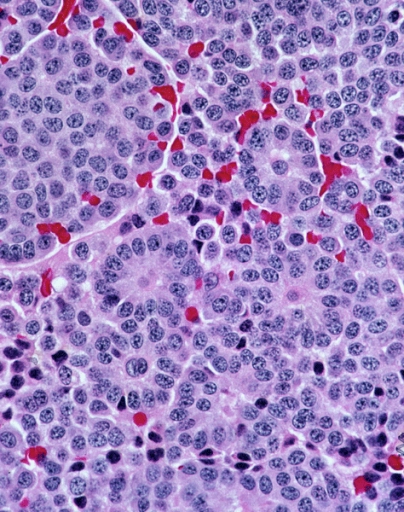
A carcinoid (also carcinoid tumor) is a slow-growing type of neuroendocrine tumor originating in the cells of the neuroendocrine system. In some cases, metastasis may occur. Carcinoid tumors of the midgut (jejunum, ileum, appendix, and cecum) are associated with carcinoid syndrome. Carcinoid tumors are the most common malignant tumor of the appendix, but they are most commonly associated with the small intestine, and they can also be found in the rectum and stomach. They are known to grow in the liver, but this finding is usually a manifestation of metastatic disease from a primary carcinoid occurring elsewhere in the body. They have a very slow growth rate compared to most malignant tumors. The median age at diagnosis for all patients with neuroendocrine tumors is 63 years. Signs and symptoms While most carcinoids are asymptomatic through the natural life and are discovered only upon surgery for unrelated reasons (so-called ''coincidental carcinoids''), all carcinoids are co ... [...More Info...] [...Related Items...] OR: [Wikipedia] [Google] [Baidu] |
EMedicine
eMedicine is an online clinical medical knowledge base founded in 1996 by doctors Scott Plantz and Jonathan Adler, and computer engineer Jeffrey Berezin. The eMedicine website consists of approximately 6,800 medical topic review articles, each of which is associated with a clinical subspecialty "textbook". The knowledge base includes over 25,000 clinically multimedia files. Each article is authored by board certified specialists in the subspecialty to which the article belongs and undergoes three levels of physician peer-review, plus review by a Doctor of Pharmacy. The article's authors are identified with their current faculty appointments. Each article is updated yearly, or more frequently as changes in practice occur, and the date is published on the article. eMedicine.com was sold to WebMD in January, 2006 and is available as the Medscape Reference. History Plantz, Adler and Berezin evolved the concept for eMedicine.com in 1996 and deployed the initial site via Boston Medi ... [...More Info...] [...Related Items...] OR: [Wikipedia] [Google] [Baidu] |
Posterior Urethral Valve
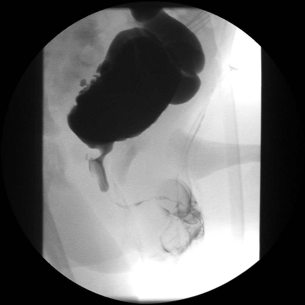
Posterior urethral valve (PUV) disorder is an obstructive developmental anomaly in the urethra and genitourinary system of male newborns. A posterior urethral valve is an obstructing membrane in the posterior male urethra as a result of abnormal '' in utero'' development. It is the most common cause of bladder outlet obstruction in male newborns. The disorder varies in degree, with mild cases presenting late due to milder symptoms. More severe cases can have renal and respiratory failure from lung underdevelopment as result of low amniotic fluid volumes, requiring intensive care and close monitoring. It occurs in about one in 8,000 babies. Presentation PUV can be diagnosed before birth, or even at birth when the ultrasound shows that the male baby has a hydronephrosis. Some babies may also have oligohydramnios due to the urinary obstruction. The later presentation can be a urinary tract infection, diurnal enuresis, or voiding pain. Complications * Incontinence * Urinary t ... [...More Info...] [...Related Items...] OR: [Wikipedia] [Google] [Baidu] |
Congenital
A birth defect, also known as a congenital disorder, is an abnormal condition that is present at birth regardless of its cause. Birth defects may result in disabilities that may be physical, intellectual, or developmental. The disabilities can range from mild to severe. Birth defects are divided into two main types: structural disorders in which problems are seen with the shape of a body part and functional disorders in which problems exist with how a body part works. Functional disorders include metabolic and degenerative disorders. Some birth defects include both structural and functional disorders. Birth defects may result from genetic or chromosomal disorders, exposure to certain medications or chemicals, or certain infections during pregnancy. Risk factors include folate deficiency, drinking alcohol or smoking during pregnancy, poorly controlled diabetes, and a mother over the age of 35 years old. Many are believed to involve multiple factors. Birth defects may be v ... [...More Info...] [...Related Items...] OR: [Wikipedia] [Google] [Baidu] |
Prostatic Urethra
The prostatic urethra, the widest and most dilatable part of the urethra canal, is about 3 cm long. It runs almost vertically through the prostate from its base to its apex, lying nearer its anterior than its posterior surface; the form of the canal is spindle-shaped, being wider in the middle than at either extremity, and narrowest below, where it joins the membranous portion. A transverse section of the canal as it lies in the prostate is horse-shoe-shaped, with the convexity directed forward. The keyhole sign, in ultrasound, is associated with a dilated bladder and prostatic urethra. Additional images File:Illu prostate lobes.jpg, Lobes of prostate File:Illu prostate zones.jpg, Zones of prostate File:Illu penis.jpg, Structure of the penis File:Gray1156.png, Vertical section of bladder The urinary bladder, or simply bladder, is a hollow organ in humans and other vertebrates that stores urine from the kidneys before disposal by urination. In humans the bladder ... [...More Info...] [...Related Items...] OR: [Wikipedia] [Google] [Baidu] |
Prostatic Ducts
The prostatic ducts (or prostatic ductules) open into the floor of the prostatic portion of the urethra, and are lined by two layers of epithelium, the inner layer consisting of columnar and the outer of small cubical cells. Small colloid masses, known as amyloid bodies are often found in the gland tubes. They open onto the prostatic sinus. See also * Prostate The prostate is both an accessory gland of the male reproductive system and a muscle-driven mechanical switch between urination and ejaculation. It is found only in some mammals. It differs between species anatomically, chemically, and phys ... References External links * - "The Male Pelvis: The Prostate Gland" Prostate {{genitourinary-stub ... [...More Info...] [...Related Items...] OR: [Wikipedia] [Google] [Baidu] |

