|
Second Intercostal Nerve
The intercostal nerves are part of the somatic nervous system, and arise from the anterior rami of the thoracic spinal nerves from T1 to T11. The intercostal nerves are distributed chiefly to the thoracic pleura and abdominal peritoneum, and differ from the anterior rami of the other spinal nerves in that each pursues an independent course without plexus formation. The first two nerves supply fibers to the upper limb and thorax; the next four distribute to the walls of the thorax; the lower five supply the walls of the thorax and abdomen. The 7th intercostal nerve end at the xyphoid process of the sternum. The 10th intercostal nerve terminates at the navel. The 12th ( subcostal) thoracic is distributed to the walls of the abdomen and groin. Each of these fibers contains around 1300 axons. Unlike the nerves from the autonomic nervous system that innervate the visceral pleura of the thoracic cavity, the intercostal nerves arise from the somatic nervous system. This enables them to c ... [...More Info...] [...Related Items...] OR: [Wikipedia] [Google] [Baidu] |
Intercostal Muscle
Intercostal muscles are many different groups of muscles that run between the ribs, and help form and move the chest wall. The intercostal muscles are mainly involved in the mechanical aspect of breathing by helping expand and shrink the size of the chest cavity. Structure There are three principal layers; # External intercostal muscles also known as intercostalis externus aid in quiet and forced inhalation. They originate on ribs 1–11 and have their insertion on ribs 2–12. The external intercostals are responsible for the elevation of the ribs and bending them more open, thus expanding the transverse dimensions of the thoracic cavity. The muscle fibers are directed downwards, forwards and medially in the anterior part. # Internal intercostal muscles also known as intercostalis internus aid in forced expiration (quiet expiration is a passive process). They originate on ribs 2–12 and have their insertions on ribs 1–11.Their fibers pass anterior and supe ... [...More Info...] [...Related Items...] OR: [Wikipedia] [Google] [Baidu] |
Parietal Pleura
The pulmonary pleurae (''sing.'' pleura) are the two opposing layers of serous membrane overlying the lungs and the inside of the surrounding chest walls. The inner pleura, called the visceral pleura, covers the surface of each lung and dips between the lobes of the lung as ''fissures'', and is formed by the invagination of lung buds into each thoracic sac during embryonic development. The outer layer, called the parietal pleura, lines the inner surfaces of the thoracic cavity on each side of the mediastinum, and can be subdivided into ''mediastinal'' (covering the side surfaces of the fibrous pericardium, oesophagus and thoracic aorta), ''diaphragmatic'' (covering the upper surface of the diaphragm), ''costal'' (covering the inside of rib cage) and cervical (covering the underside of the suprapleural membrane) pleurae. The visceral and the mediastinal parietal pleurae are connected at the root of the lung ("hilum") through a smooth fold known as ''pleural reflections'', and ... [...More Info...] [...Related Items...] OR: [Wikipedia] [Google] [Baidu] |
Lactation
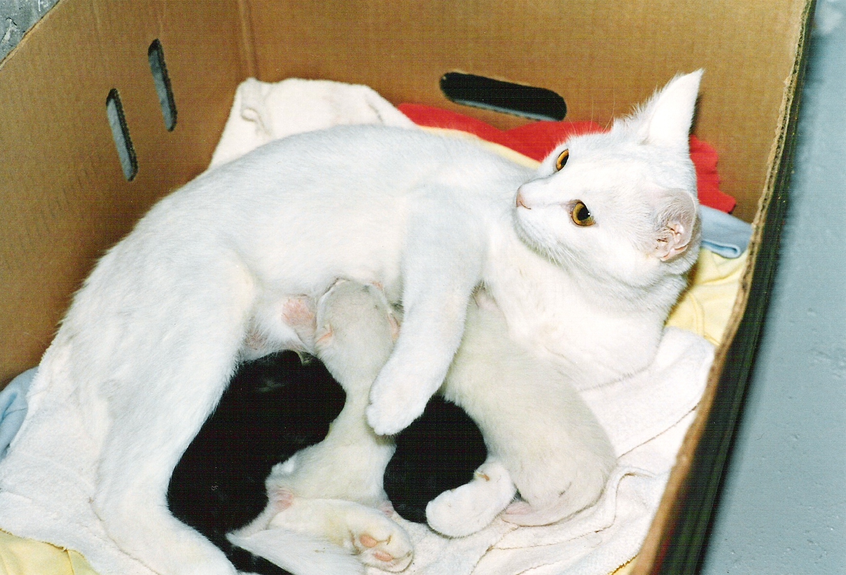
Lactation describes the secretion of milk from the mammary glands and the period of time that a mother lactates to feed her young. The process naturally occurs with all sexually mature female mammals, although it may predate mammals. The process of feeding milk in all animals (including humans) is called ''nursing'', and in humans it is also called ''breastfeeding''. Newborn infants often produce some milk from their own breast tissue, known colloquially as witch's milk. In most species, lactation is a sign that the female has been pregnant at some point in her life, although it can happen without pregnancy. Nearly every species of mammal has nipples; except for monotremes, egg-laying mammals, which instead release milk through ducts in the abdomen. In only one species of mammal, the Dayak fruit bat from Southeast Asia, is milk production a normal male function. ''Galactopoiesis'' is the maintenance of milk production. This stage requires prolactin. Oxytocin is critical for t ... [...More Info...] [...Related Items...] OR: [Wikipedia] [Google] [Baidu] |
Myoepithelial Cell
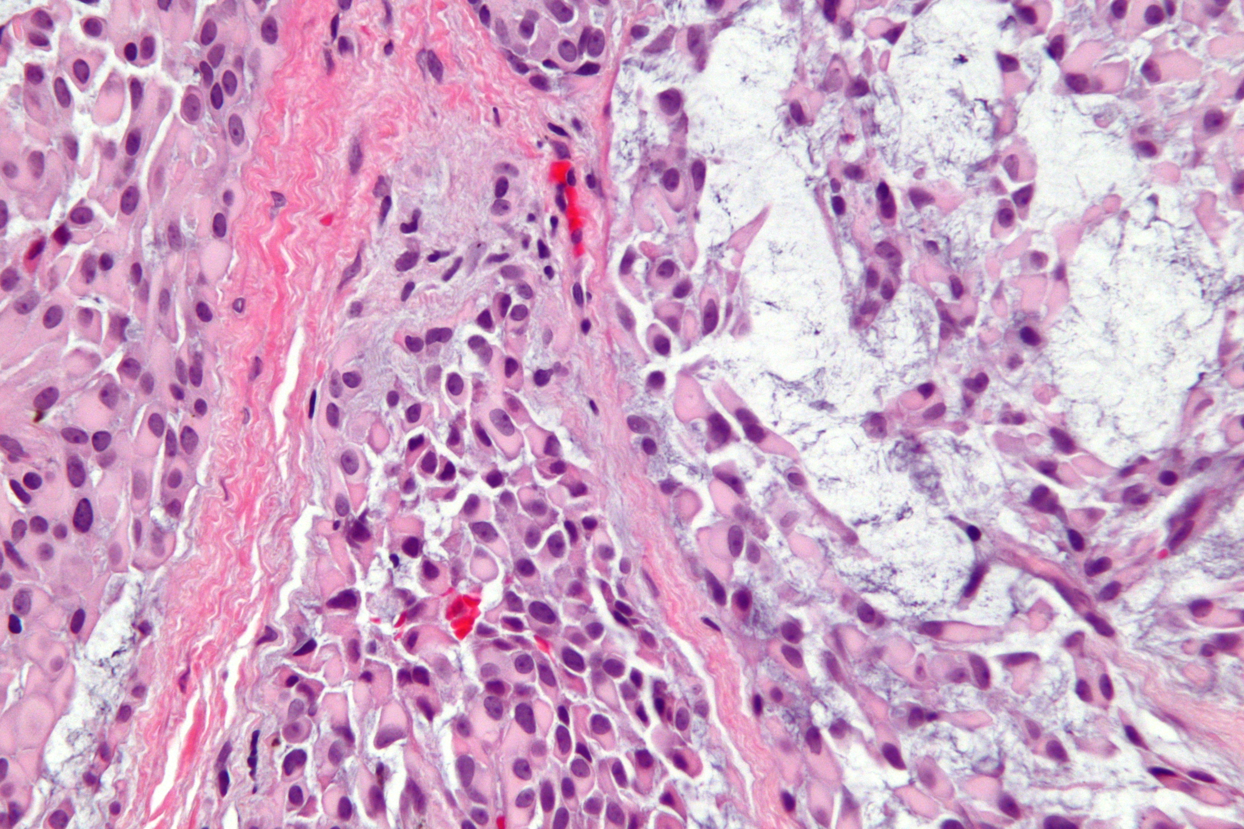
Myoepithelial cells (sometimes referred to as myoepithelium) are cells usually found in glandular epithelium as a thin layer above the basement membrane but generally beneath the luminal cells. These may be positive for alpha smooth muscle actin and can contract and expel the secretions of exocrine glands. They are found in the sweat glands, mammary glands, lacrimal glands, and salivary glands. Myoepithelial cells in these cases constitute the basal cell layer of an epithelium that harbors the epithelial progenitor. In the case of wound healing, myoepithelial cells reactively proliferate. Presence of myoepithelial cells in a hyperplastic tissue proves the benignity of the gland and, when absent, indicates cancer. Only rare cancers like adenoid cystic carcinomas contains myoepithelial cells as one of the malignant component. It can be found in endoderm or ectoderm. Markers Myoepithelial cells are true epithelial cells positive for keratins, not to be confused with myofibr ... [...More Info...] [...Related Items...] OR: [Wikipedia] [Google] [Baidu] |
Oxytocin
Oxytocin (Oxt or OT) is a peptide hormone and neuropeptide normally produced in the hypothalamus and released by the posterior pituitary. It plays a role in social bonding, reproduction, childbirth, and the period after childbirth. Oxytocin is released into the bloodstream as a hormone in response to sexual activity and during labour. It is also available in pharmaceutical form. In either form, oxytocin stimulates uterine contractions to speed up the process of childbirth. In its natural form, it also plays a role in bonding with the baby and milk production. Production and secretion of oxytocin is controlled by a positive feedback mechanism, where its initial release stimulates production and release of further oxytocin. For example, when oxytocin is released during a contraction of the uterus at the start of childbirth, this stimulates production and release of more oxytocin and an increase in the intensity and frequency of contractions. This process compounds in intensity ... [...More Info...] [...Related Items...] OR: [Wikipedia] [Google] [Baidu] |
Mechanoreceptor
A mechanoreceptor, also called mechanoceptor, is a sensory receptor that responds to mechanical pressure or distortion. Mechanoreceptors are innervated by sensory neurons that convert mechanical pressure into electrical signals that, in animals, are sent to the central nervous system. Vertebrate mechanoreceptors Cutaneous mechanoreceptors Cutaneous mechanoreceptors respond to mechanical stimuli that result from physical interaction, including pressure and vibration. They are located in the skin, like other cutaneous receptors. They are all innervated by Aβ fibers, except the mechanorecepting free nerve endings, which are innervated by Aδ fibers. Cutaneous mechanoreceptors can be categorized by what kind of sensation they perceive, by the rate of adaptation, and by morphology. Furthermore, each has a different receptive field. By sensation *The Slowly Adapting type 1 (SA1) mechanoreceptor, with the Merkel corpuscle end-organ (also known as Merkel discs) detect sustained p ... [...More Info...] [...Related Items...] OR: [Wikipedia] [Google] [Baidu] |
Cervical Plexus
The cervical plexus is a plexus of the anterior rami of the first four cervical spinal nerves which arise from C1 to C4 cervical segment in the neck. They are located laterally to the transverse processes between prevertebral muscles from the medial side and vertebral (m. scalenus, m. levator scapulae, m. splenius cervicis) from lateral side. There is anastomosis with accessory nerve, hypoglossal nerve and sympathetic trunk. It is located in the neck, deep to the sternocleidomastoid muscle. Nerves formed from the cervical plexus innervate the back of the head, as well as some neck muscles. The branches of the cervical plexus emerge from the posterior triangle at the nerve point, a point which lies midway on the posterior border of the sternocleidomastoid. Branches The cervical plexus has two types of branches: cutaneous and muscular. *Cutaneous (4 branches): **Lesser occipital nerve - innervates the skin and the scalp posterosuperior to the auricle (C2) **Great auricular nerve - ... [...More Info...] [...Related Items...] OR: [Wikipedia] [Google] [Baidu] |
Supraclavicular Nerve
The supraclavicular nerves (descending branches) arise from the third and fourth cervical nerves. They emerge beneath the posterior border of the sternocleidomastoideus (sternocleidomastoid muscle), and descend in the posterior triangle of the neck beneath the platysma muscle and the deep cervical fascia. Together, they innervate skin over the shoulder. The supraclavicular nerve can be blocked during shoulder surgery. Branches The supraclavicular nerves arise from C3 and C4 spinal nerve roots. Near the clavicle, the supraclavicular nerves perforate the fascia and the platysma muscle to become cutaneous. They are arranged, according to their position, into three groups—anterior, middle, and posterior. Medial supraclavicular nerve The medial supraclavicular nerves or ''anterior supraclavicular nerves'' (nn. supraclaviculares anteriores; suprasternal nerves) cross obliquely over the external jugular vein and the clavicular and sternal heads of the sternocleidomastoideus, and supp ... [...More Info...] [...Related Items...] OR: [Wikipedia] [Google] [Baidu] |
Pectoralis Major
The pectoralis major () is a thick, fan-shaped or triangular convergent muscle, situated at the chest of the human body. It makes up the bulk of the chest muscles and lies under the breast. Beneath the pectoralis major is the pectoralis minor, a thin, triangular muscle. The pectoralis major's primary functions are flexion, adduction, and internal rotation of the humerus. The pectoral major may colloquially be referred to as "pecs", "pectoral muscle", or "chest muscle", because it is the largest and most superficial muscle in the chest area. Structure It arises from the anterior surface of the sternal half of the clavicle from breadth of the half of the anterior surface of the sternum, as low down as the attachment of the cartilage of the sixth or seventh rib; from the cartilages of all the true ribs, with the exception, frequently, of the first or seventh, and from the aponeurosis of the abdominal external oblique muscle. From this extensive origin the fibers converge toward the ... [...More Info...] [...Related Items...] OR: [Wikipedia] [Google] [Baidu] |
Intercostales Interni
The internal intercostal muscles (intercostales interni) are a group of skeletal muscles located between the ribs. They are eleven in number on either side. They commence anteriorly at the sternum, in the intercostal spaces between the cartilages of the true ribs, and at the anterior extremities of the cartilages of the false ribs, and extend backward as far as the angles of the ribs, hence they are continued to the vertebral column by thin aponeuroses, the posterior intercostal membranes. They pull the sternum and ribs upward and inward. Structure Their fibers are also directed obliquely, but pass in a direction opposite to those of the external intercostal muscles. The internal intercostal muscles originate from the costal groove of the rib and insert into the superior aspect of the rib below in a direction perpendicular to the external intercostal muscles. It is this arrangement that allows these muscles to facilitate exhalation. For the most part, they are muscles of exhala ... [...More Info...] [...Related Items...] OR: [Wikipedia] [Google] [Baidu] |
Transversus Thoracis Muscle
The transversus thoracis muscle (), also known as triangularis sterni, lies internal to the thoracic cage, anteriorly. It is usually a thin plane of muscular and tendinous fibers, however on athletic individuals it can be a thick 'slab of meat', situated upon the inner surface of the front wall of the chest. It is in the same layer as the subcostal muscles and the innermost intercostal muscles. Structure It arises on either side from the lower third of the posterior surface of the body of the sternum, from the posterior surface of the xiphoid process, and from the sternal ends of the costal cartilages of the lower three or four true ribs. Its fibers diverge upward and lateralward, to be inserted by slips into the lower borders and inner surfaces of the costal cartilages of the second, third, fourth, fifth, and sixth ribs. The lowest fibers of this muscle are horizontal in their direction, and are continuous with those of the transversus abdominis; the intermediate fibers are ob ... [...More Info...] [...Related Items...] OR: [Wikipedia] [Google] [Baidu] |
Internal Mammary Artery
In human anatomy, the internal thoracic artery (ITA), previously commonly known as the internal mammary artery (a name still common among surgeons), is an artery that supplies the anterior chest wall and the breasts. It is a paired artery, with one running along each side of the sternum, to continue after its bifurcation as the superior epigastric and musculophrenic arteries. Structure The internal thoracic artery arises from the anterior surface of the subclavian artery near its origin. It has a width of between 1-2 mm. It travels downward on the inside of the rib cage, approximately 1 cm from the sides of the sternum, and thus medial to the nipple. It is accompanied by the internal thoracic vein. It runs deep to the abdominal external oblique muscle, but superficial to the vagus nerve. In adults, internal thoracic artery lies closest to the sternum at the first intercoastal space. The gap between the artery and lateral border of the sternum increases when going downwards, u ... [...More Info...] [...Related Items...] OR: [Wikipedia] [Google] [Baidu] |


