|
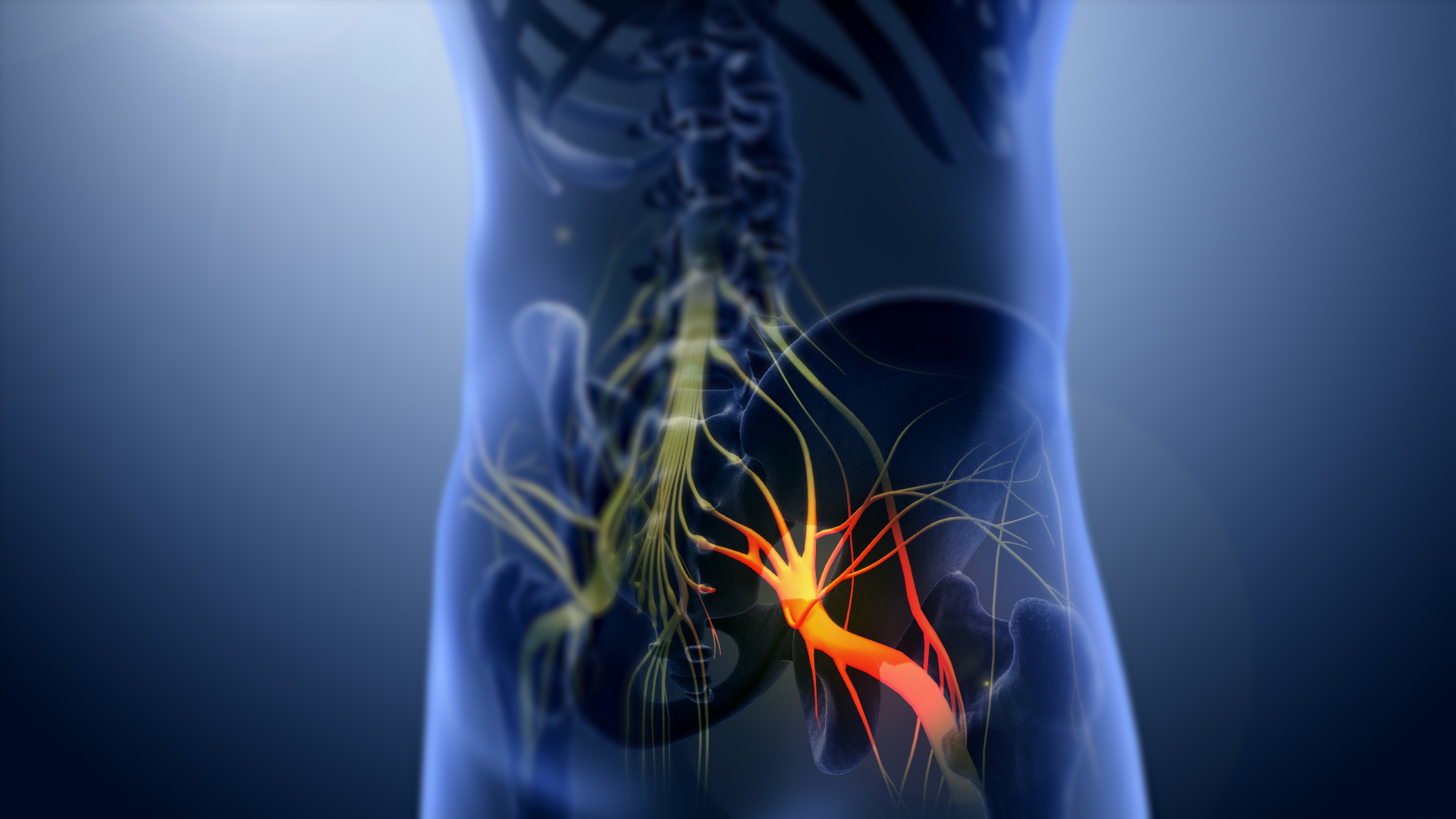
Sciatic Nerve Block
A sciatic nerve block is a nerve block that uses local anesthetic to achieve analgesia in the leg. The block works by affecting the sciatic nerve and is used for surgeries at or below the knee. The sciatic nerve is located in the gluteus maximus muscle, where the block is performed. The sciatic nerve can be blocked at different locations. At the popliteal fossa, the sciatic nerve divides into its two branches: The tibial and the common peroneal nerve. If surgery is performed on the ankle, achilles tendon or foot a popliteal block Popliteal refers to anatomical structures located in the back of the knee: *Popliteal artery *Popliteal vein *Popliteal fossa * Popliteal lymph nodes *Popliteus muscle *Popliteal nerves *Popliteal pterygium syndrome Popliteal pterygium syndrome (P ... can be performed, affecting the two branches of the sciatic nerve. It is done above the knee on the posterior leg where the sciatic nerve starts splitting into the common peroneal and tibial nerves. Refe ... [...More Info...] [...Related Items...] OR: [Wikipedia] [Google] [Baidu] |
Local Anesthetic Nerve Block
Local anesthetic nerve block (local anesthetic regional nerve blockade, or often simply nerve block) is a short-term nerve block involving the injection (medicine), injection of local anesthetic as close to the nerve as possible for analgesia, pain relief. The local anesthetic bathes the nerve and numbs the area of the body that is supplied by that nerve. The goal of the nerve block is to prevent pain by blocking the transmission of pain signals from the affected area. Nerve blocks have a number of uses including treating headache disorders and providing anesthesia during surgery. The analgesia, pain relief provided by the block is present during the surgery and continues to last after the procedure. This can lead to a reduction in the amount of opiates needed for pain control. The advantages of nerve blocks over General anaesthesia, general anesthesia include faster recovery, Anesthesia awareness#Conscious sedation and monitored anesthesia care, monitored anesthesia care vs. Trac ... [...More Info...] [...Related Items...] OR: [Wikipedia] [Google] [Baidu] |
Local Anesthetic
A local anesthetic (LA) is a medication that causes absence of pain sensation. In the context of surgery, a local anesthetic creates an absence of pain in a specific location of the body without a loss of consciousness, as opposed to a general anesthetic. When it is used on specific nerve pathways (local anesthetic nerve block), paralysis (loss of muscle power) also can be achieved. Examples Short Duration & Low Potency Procaine Chloroprocaine Medium Duration & Potency Lidocaine Prilocaine High Duration & Potency Tetracaine Bupivacaine Cinchocaine Ropivacaine Clinical LAs belong to one of two classes: aminoamide and aminoester local anesthetics. Synthetic LAs are structurally related to cocaine. They differ from cocaine mainly in that they have a very low abuse potential and do not produce hypertension or (with few exceptions) vasoconstriction. They are used in various techniques of local anesthesia such as: * Topical anesthesia (surface) * Topical administration ... [...More Info...] [...Related Items...] OR: [Wikipedia] [Google] [Baidu] |
Analgesia
Pain management is an aspect of medicine and health care involving relief of pain (pain relief, analgesia, pain control) in various dimensions, from acute and simple to chronic and challenging. Most physicians and other health professionals provide some pain control in the normal course of their practice, and for the more complex instances of pain, they also call on additional help from a specific medical specialty devoted to pain, which is called pain medicine. Pain management often uses a multidisciplinary approach for easing the suffering and improving the quality of life of anyone experiencing pain, whether acute pain or chronic pain. Relief of pain in general (analgesia) is often an acute affair, whereas managing chronic pain requires additional dimensions. The typical pain management team includes medical practitioners, pharmacists, clinical psychologists, physiotherapists, occupational therapists, recreational therapists, physician assistants, nurses, and dentist ... [...More Info...] [...Related Items...] OR: [Wikipedia] [Google] [Baidu] |
Sciatic Nerve
The sciatic nerve, also called the ischiadic nerve, is a large nerve in humans and other vertebrate animals which is the largest branch of the sacral plexus and runs alongside the hip joint and down the lower limb. It is the longest and widest single nerve in the human body, going from the top of the leg to the foot on the posterior aspect. The sciatic nerve has no cutaneous branches for the thigh. This nerve provides the connection to the nervous system for the skin of the lateral leg and the whole foot, the muscles of the back of the thigh, and those of the leg and foot. It is derived from spinal nerves L4 to S3. It contains fibers from both the anterior and posterior divisions of the lumbosacral plexus. Structure In humans, the sciatic nerve is formed from the L4 to S3 segments of the sacral plexus, a collection of nerve fibres that emerge from the sacral part of the spinal cord. The lumbosacral trunk from the L4 and L5 roots descends between the sacral promontory and ala and ... [...More Info...] [...Related Items...] OR: [Wikipedia] [Google] [Baidu] |
Gluteus Maximus Muscle
The gluteal muscles, often called glutes are a group of three muscles which make up the gluteal region commonly known as the buttocks: the gluteus maximus, gluteus medius and gluteus minimus. The three muscles originate from the ilium and sacrum and insert on the femur. The functions of the muscles include extension, abduction, external rotation, and internal rotation of the hip joint. Structure The gluteus maximus is the largest and most superficial of the three gluteal muscles. It makes up a large part of the shape and appearance of the hips. It is a narrow and thick fleshy mass of a quadrilateral shape, and forms the prominence of the buttocks. The gluteus medius is a broad, thick, radiating muscle, situated on the outer surface of the pelvis. It lies profound to the gluteus maximus and its posterior third is covered by the gluteus maximus, its anterior two-thirds by the gluteal aponeurosis, which separates it from the superficial fascia and skin. The gluteus minimus is t ... [...More Info...] [...Related Items...] OR: [Wikipedia] [Google] [Baidu] |
Tibial Nerve
The tibial nerve is a branch of the sciatic nerve. The tibial nerve passes through the popliteal fossa to pass below the arch of soleus. Structure Popliteal fossa The tibial nerve is the larger terminal branch of the sciatic nerve with root values of L4, L5, S1, S2, and S3. It lies superficial (or posterior) to the popliteal vessels, extending from the superior angle to the inferior angle of the popliteal fossa, crossing the popliteal vessels from lateral to medial side. It gives off branches as shown below: * Muscular branches - Muscular branches arise from the distal part of the popliteal fossa. It supplies the medial and lateral heads of gastrocnemius, soleus, plantaris and popliteus muscles. Nerve to popliteus crosses the popliteus muscle, runs downwards and laterally, winds around the lower border of the popliteus to supply the deep (or anterior) surface of the popliteus. This nerve also supplies the tibialis posterior muscle, superior tibiofibular joint, tibia bone, intero ... [...More Info...] [...Related Items...] OR: [Wikipedia] [Google] [Baidu] |
Common Peroneal Nerve
The common fibular nerve (also known as the common peroneal nerve, external popliteal nerve, or lateral popliteal nerve) is a nerve in the lower leg that provides sensation over the posterolateral part of the leg and the knee joint. It divides at the knee into two terminal branches: the superficial fibular nerve and deep fibular nerve, which innervate the muscles of the lateral and anterior compartments of the leg respectively. When the common fibular nerve is damaged or compressed, foot drop can ensue. Structure The common fibular nerve is the smaller terminal branch of the sciatic nerve. The common fibular nerve has root values of L4, L5, S1, and S2. It arises from the superior angle of the popliteal fossa and extends to the lateral angle of the popliteal fossa, along the medial border of the biceps femoris. It then winds around the neck of the fibula to pierce the fibularis longus and divides into terminal branches of the superficial fibular nerve and the deep fibular nerve. Bef ... [...More Info...] [...Related Items...] OR: [Wikipedia] [Google] [Baidu] |
Ankle
The ankle, or the talocrural region, or the jumping bone (informal) is the area where the foot and the leg meet. The ankle includes three joints: the ankle joint proper or talocrural joint, the subtalar joint, and the inferior tibiofibular joint. The movements produced at this joint are dorsiflexion and plantarflexion of the foot. In common usage, the term ankle refers exclusively to the ankle region. In medical terminology, "ankle" (without qualifiers) can refer broadly to the region or specifically to the talocrural joint. The main bones of the ankle region are the talus (in the foot), and the tibia and fibula (in the leg). The talocrural joint is a synovial hinge joint that connects the distal ends of the tibia and fibula in the lower limb with the proximal end of the talus. The articulation between the tibia and the talus bears more weight than that between the smaller fibula and the talus. Structure Region The ankle region is found at the junction of the leg and the f ... [...More Info...] [...Related Items...] OR: [Wikipedia] [Google] [Baidu] |
Achilles Tendon
The Achilles tendon or heel cord, also known as the calcaneal tendon, is a tendon at the back of the lower leg, and is the thickest in the human body. It serves to attach the plantaris, gastrocnemius (calf) and soleus muscles to the calcaneus (heel) bone. These muscles, acting via the tendon, cause plantar flexion of the foot at the ankle joint, and (except the soleus) flexion at the knee. Abnormalities of the Achilles tendon include inflammation ( Achilles tendinitis), degeneration, rupture, and becoming embedded with cholesterol deposits (xanthomas). The Achilles tendon was named in 1693 after the Greek hero Achilles. History The oldest-known written record of the tendon being named for Achilles is in 1693 by the Flemish/Dutch anatomist Philip Verheyen. In his widely used text he described the tendon's location and said that it was commonly called "the cord of Achilles." The tendon has been described as early as the time of Hippocrates, who described it as the "" (Latin f ... [...More Info...] [...Related Items...] OR: [Wikipedia] [Google] [Baidu] |
Foot
The foot ( : feet) is an anatomical structure found in many vertebrates. It is the terminal portion of a limb which bears weight and allows locomotion. In many animals with feet, the foot is a separate organ at the terminal part of the leg made up of one or more segments or bones, generally including claws or nails. Etymology The word "foot", in the sense of meaning the "terminal part of the leg of a vertebrate animal" comes from "Old English fot "foot," from Proto-Germanic *fot (source also of Old Frisian fot, Old Saxon fot, Old Norse fotr, Danish fod, Swedish fot, Dutch voet, Old High German fuoz, German Fuß, Gothic fotus "foot"), from PIE root *ped- "foot". The "plural form feet is an instance of i-mutation." Structure The human foot is a strong and complex mechanical structure containing 26 bones, 33 joints (20 of which are actively articulated), and more than a hundred muscles, tendons, and ligaments.Podiatry Channel, ''Anatomy of the foot and ankle'' The joints of the ... [...More Info...] [...Related Items...] OR: [Wikipedia] [Google] [Baidu] |
Popliteal Block
Popliteal refers to anatomical structures located in the back of the knee: *Popliteal artery *Popliteal vein *Popliteal fossa *Popliteal lymph nodes *Popliteus muscle *Popliteal nerves *Popliteal pterygium syndrome Popliteal pterygium syndrome (PPS) is an heredity, inherited condition affecting the face, Limb (anatomy), limbs, and genitalia. The syndrome goes by a number of names including the ''popliteal web syndrome'' and, more inclusively, the ''facio-geni ... {{disambig Lower limb anatomy ... [...More Info...] [...Related Items...] OR: [Wikipedia] [Google] [Baidu] |


.jpg)

