|
Synostotic
Synostosis (plural: synostoses) is fusion of two or more bones. It can be normal in puberty, fusion of the epiphyseal plate to become the epiphyseal line, or abnormal. When synostosis is abnormal it is a type of dysostosis. Examples of synostoses include: * craniosynostosis – an abnormal fusion of two or more cranial bones; * radioulnar synostosis – the abnormal fusion of the radius and ulna bones of the forearm; * tarsal coalition – a failure to separately form all seven bones of the tarsus (the hind part of the foot) resulting in an amalgamation of two bones; and * syndactyly – the abnormal fusion of neighboring digits. Synostosis within joints can cause ankylosis. __TOC__ Clinical significance Radioulnar synostosis is one of the more common failures of separation of parts of the upper limb. There are two general types: one is characterized by fusion of the radius and ulna at their proximal borders and the other is fused distal to the proximal radial epiphysis. Mos ... [...More Info...] [...Related Items...] OR: [Wikipedia] [Google] [Baidu] |
Plagiocephaly
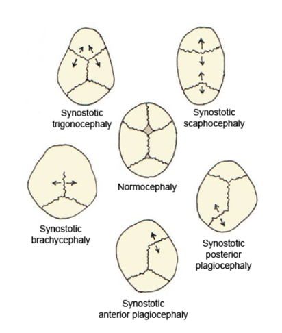
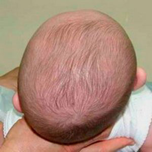
Plagiocephaly, also known as flat head syndrome, is a condition characterized by an asymmetrical distortion (flattening of one side) of the skull. A mild and widespread form is characterized by a flat spot on the back or one side of the head caused by remaining in a supine position for prolonged periods. Plagiocephaly is a diagonal asymmetry across the head shape. Often it is a flattening which is to one side at the back of the head and there is often some facial asymmetry. Depending on whether synostosis is involved, plagiocephaly divides into two groups: synostotic, with one or more fused cranial sutures, and non-synostotic (deformational). Surgical treatment of these groups includes the deference method; however, the treatment of deformational plagiocephaly is controversial. Brachycephaly describes a very wide head shape with a flattening across the whole back of the head. Causes Slight plagiocephaly is routinely diagnosed at birth and may be the result of a restrictive intrau ... [...More Info...] [...Related Items...] OR: [Wikipedia] [Google] [Baidu] |
Craniosynostosis
Craniosynostosis is a condition in which one or more of the fibrous sutures in a young infant's skull prematurely fuses by turning into bone (ossification), thereby changing the growth pattern of the skull. Because the skull cannot expand perpendicular to the fused suture, it compensates by growing more in the direction parallel to the closed sutures. Sometimes the resulting growth pattern provides the necessary space for the growing brain, but results in an abnormal head shape and abnormal facial features. In cases in which the compensation does not effectively provide enough space for the growing brain, craniosynostosis results in increased intracranial pressure leading possibly to visual impairment, sleeping impairment, eating difficulties, or an impairment of mental development combined with a significant reduction in IQ. Craniosynostosis occurs in one in 2000 births. Craniosynostosis is part of a syndrome in 15% to 40% of affected patients, but it usually occurs as an isol ... [...More Info...] [...Related Items...] OR: [Wikipedia] [Google] [Baidu] |
Epiphyseal Plate
The epiphyseal plate (or epiphysial plate, physis, or growth plate) is a hyaline cartilage plate in the metaphysis at each end of a long bone. It is the part of a long bone where new bone growth takes place; that is, the whole bone is alive, with maintenance remodeling throughout its existing bone tissue, but the growth plate is the place where the long bone grows longer (adds length). The plate is only found in children and adolescents; in adults, who have stopped growing, the plate is replaced by an epiphyseal line. This replacement is known as epiphyseal closure or growth plate fusion. Complete fusion can occur as early as 12 for girls (with the most common being 14-15 years for girls) and as early as 14 for boys (with the most common being 15–17 years for boys). Structure Development Endochondral ossification is responsible for the initial bone development from cartilage in utero and infants and the longitudinal growth of long bones in the epiphyseal plate. The plate' ... [...More Info...] [...Related Items...] OR: [Wikipedia] [Google] [Baidu] |
Antley–Bixler Syndrome
Antley–Bixler syndrome is a rare, severe autosomal recessive congenital disorder characterized by malformations and deformities affecting the majority of the skeleton and other areas of the body. Presentation Antley–Bixler syndrome presents itself at birth or prenatally. Features of the disorder include brachycephaly (flat forehead), craniosynostosis (complete skull-joint closure) of both coronal and lambdoid sutures, facial hypoplasia (underdevelopment); bowed ulna (forearm bone) and femur (thigh bone), synostosis of the radius (forearm bone), humerus (upper arm bone) and trapezoid (hand bone); camptodactyly (fused interphalangeal joints in the fingers), thin ilial wings (outer pelvic plate) and renal malformations. Other symptoms, such as cardiac malformations, proptotic exophthalmos (bulging eyes), arachnodactyly (spider-like fingers) as well as nasal, anal and vaginal atresia (occlusion) have been reported. Pathophysiology There are two distinct genetic mutations asso ... [...More Info...] [...Related Items...] OR: [Wikipedia] [Google] [Baidu] |
Anterior Plagiocephaly
Standard anatomical terms of location are used to unambiguously describe the anatomy of animals, including humans. The terms, typically derived from Latin or Greek roots, describe something in its standard anatomical position. This position provides a definition of what is at the front ("anterior"), behind ("posterior") and so on. As part of defining and describing terms, the body is described through the use of anatomical planes and anatomical axes. The meaning of terms that are used can change depending on whether an organism is bipedal or quadrupedal. Additionally, for some animals such as invertebrates, some terms may not have any meaning at all; for example, an animal that is radially symmetrical will have no anterior surface, but can still have a description that a part is close to the middle ("proximal") or further from the middle ("distal"). International organisations have determined vocabularies that are often used as standard vocabularies for subdisciplines o ... [...More Info...] [...Related Items...] OR: [Wikipedia] [Google] [Baidu] |
Trigonocephaly
Trigonocephaly is a congenital condition of premature fusion of the metopic suture (from the Greek , "forehead"), leading to a triangular forehead. The merging of the two frontal bones leads to transverse growth restriction and parallel growth expansion. It may occur syndromic, involving other abnormalities, or isolated. The term is from the Greek , "triangle", and , "head". Cause Trigonocephaly can either occur syndromatic or isolated. Trigonocephaly is associated with the following syndromes: Opitz syndrome, Muenke syndrome, Jacobsen syndrome, Baller–Gerold syndrome and Say–Meyer syndrome. The etiology of trigonocephaly is mostly unknown although there are three main theories. Trigonocephaly is probably a multifactorial congenital condition, but due to limited proof of these theories this cannot safely be concluded. Intrinsic bone malformation The first theory assumes that the origin of pathological synostosis lies within disturbed bone formation early on in the pregnan ... [...More Info...] [...Related Items...] OR: [Wikipedia] [Google] [Baidu] |
Scaphocephaly
Scaphocephaly is a type of cephalic disorder which occurs when there is a premature fusion of the sagittal suture. The sagittal suture joins together the two parietal bones of the skull. Scaphocephaly is the most common of the craniosynostosis conditions and is characterized by a long, narrow head. Classification Scaphocephaly is classified into 3 types, depending on morphology and position and suture closure: * Sphenocephaly ("wedge-shaped", most common) * Clinocephaly (camelback-shaped) * Leptocephaly ("thin head", least common); this occurs when the metopic suture is also fused Treatment This condition can be corrected by surgery if the child is young enough. The use of a cranial molding orthosis (a custom-made helmet) can also benefit the child if the child begins wearing it at an early age. Terminology The term is from Greek ''skaphe'' meaning 'light boat or skiff' and ''kephale'' meaning 'head') describes a specific shape of a long narrow head that resembles a boat. See a ... [...More Info...] [...Related Items...] OR: [Wikipedia] [Google] [Baidu] |
Bone
A bone is a Stiffness, rigid Organ (biology), organ that constitutes part of the skeleton in most vertebrate animals. Bones protect the various other organs of the body, produce red blood cell, red and white blood cells, store minerals, provide structure and support for the body, and enable animal locomotion, mobility. Bones come in a variety of shapes and sizes and have complex internal and external structures. They are lightweight yet strong and hard and serve multiple Function (biology), functions. Bone tissue (osseous tissue), which is also called bone in the mass noun, uncountable sense of that word, is hard tissue, a type of specialized connective tissue. It has a honeycomb-like matrix (biology), matrix internally, which helps to give the bone rigidity. Bone tissue is made up of different types of bone cells. Osteoblasts and osteocytes are involved in the formation and mineralization (biology), mineralization of bone; osteoclasts are involved in the bone resorption, resor ... [...More Info...] [...Related Items...] OR: [Wikipedia] [Google] [Baidu] |
Nievergelt Syndrome
Nievergelt is a Swiss surname. It may refer to the following: * Ernst Nievergelt Ernst Nievergelt (23 March 1910 – 1 July 1999) was a cyclist from Switzerland. He was born in Affoltern, Zurich, Switzerland, and died in Kappel am Albis. In 1935 Nievergelt won the amateur standings in the Championship of Zurich. He ... (1910–1999), Swiss cyclist * Erwin Nievergelt (1929–2019), Swiss chess player {{disambig German-language surnames ... [...More Info...] [...Related Items...] OR: [Wikipedia] [Google] [Baidu] |
Limb-body Wall Complex
Limb body wall complex (LBWC) is a rare fetal malformation of unknown origins. Traditionally diagnosis has been based on the Van Allen et al., criteria, i.e. the presence of two out of three of the following anomalies: # Exencephaly or encephalocele Encephalocele is a neural tube defect characterized by sac-like protrusions of the brain and the membranes that cover it through openings in the skull. These defects are caused by failure of the neural tube to close completely during fetal develop ... with facial clefts # Thoraco and or abdominoschisis and # Limb defects. LBWC occurs in approximately 0.32 in 100,000 births. At this time, there is no known cause of Limb Body Wall Complex. However, there have been tentative links made between a diagnosis of LBWC and cocaine use. In addition, current research has shown that there may be a genetic cause for a small limited number of LBWC cases. Limb Body Wall Complex is a lethal birth defect. There are only anecdotal stories of surviv ... [...More Info...] [...Related Items...] OR: [Wikipedia] [Google] [Baidu] |


