|
Superior Pubic Ramus
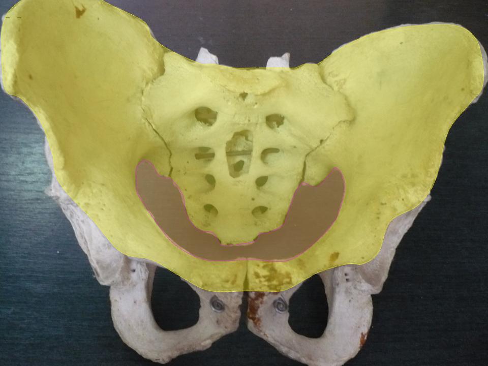
In vertebrates, the pubic region ( la, pubis) is the most forward-facing (ventral and anterior) of the three main regions making up the coxal bone. The left and right pubic regions are each made up of three sections, a superior ramus, inferior ramus, and a body. Structure The pubic region is made up of a ''body'', ''superior ramus'', and ''inferior ramus'' (). The left and right coxal bones join at the pubic symphysis. It is covered by a layer of fat, which is covered by the mons pubis. The pubis is the lower limit of the suprapubic region. In the female, the pubic region is anterior to the urethral sponge. Body The body forms the wide, strong, middle and flat part of the pubic region. The bodies of the left and right pubic regions join at the pubic symphysis. The rough upper edge is the pubic crest, ending laterally in the pubic tubercle. This tubercle, found roughly 3 cm from the pubic symphysis, is a distinctive feature on the lower part of the abdominal wall; important ... [...More Info...] [...Related Items...] OR: [Wikipedia] [Google] [Baidu] |
Pelvis
The pelvis (plural pelves or pelvises) is the lower part of the trunk, between the abdomen and the thighs (sometimes also called pelvic region), together with its embedded skeleton (sometimes also called bony pelvis, or pelvic skeleton). The pelvic region of the trunk includes the bony pelvis, the pelvic cavity (the space enclosed by the bony pelvis), the pelvic floor, below the pelvic cavity, and the perineum, below the pelvic floor. The pelvic skeleton is formed in the area of the back, by the sacrum and the coccyx and anteriorly and to the left and right sides, by a pair of hip bones. The two hip bones connect the spine with the lower limbs. They are attached to the sacrum posteriorly, connected to each other anteriorly, and joined with the two femurs at the hip joints. The gap enclosed by the bony pelvis, called the pelvic cavity, is the section of the body underneath the abdomen and mainly consists of the reproductive organs (sex organs) and the rectum, while the pelvic f ... [...More Info...] [...Related Items...] OR: [Wikipedia] [Google] [Baidu] |
Lesser Pelvis
The pelvic cavity is a body cavity that is bounded by the bones of the pelvis. Its oblique roof is the pelvic inlet (the superior opening of the pelvis). Its lower boundary is the pelvic floor. The pelvic cavity primarily contains the reproductive organs, urinary bladder, distal ureters, proximal urethra, terminal sigmoid colon, rectum, and anal canal. In females, the uterus, Fallopian tubes, ovaries and upper vagina occupy the area between the other viscera. The rectum is located at the back of the pelvis, in the curve of the sacrum and coccyx; the bladder is in front, behind the pubic symphysis. The pelvic cavity also contains major arteries, veins, muscles, and nerves. These structures coexist in a crowded space, and disorders of one pelvic component may impact upon another; for example, constipation may overload the rectum and compress the urinary bladder, or childbirth might damage the pudendal nerves and later lead to anal weakness. Structure The pelvis has an antero ... [...More Info...] [...Related Items...] OR: [Wikipedia] [Google] [Baidu] |
Pectineal Line (pubis)
The pectineal line of the pubis (also pecten pubis) is a ridge on the superior ramus of the pubic bone. It forms part of the pelvic brim. Lying across from the pectineal line are fibers of the pectineal ligament, and the proximal origin of the pectineus muscle. In combination with the arcuate line, it makes the iliopectineal line The iliopectineal line is the border of the iliopubic eminence. It can be defined as a compound structure of the arcuate line (from the ilium) and pectineal line (from the pubis). With the sacral promontory, it makes up the linea terminalis The .... References External links * () {{Authority control Bones of the pelvis Pubis (bone) ... [...More Info...] [...Related Items...] OR: [Wikipedia] [Google] [Baidu] |
Inguinal Ligament
The inguinal ligament (), also known as Poupart's ligament or groin ligament, is a band running from the pubic tubercle to the anterior superior iliac spine. It forms the base of the inguinal canal through which an indirect inguinal hernia may develop. Structure The inguinal ligament runs from the anterior superior iliac crest of the ilium to the pubic tubercle of the pubic bone. It is formed by the external abdominal oblique aponeurosis and is continuous with the fascia lata of the thigh. There is some dispute over the attachments. Structures that pass deep to the inguinal ligament include: *Psoas major, iliacus, pectineus *Femoral nerve, artery, and vein *Lateral cutaneous nerve of thigh *Lymphatics Function The ligament serves to contain soft tissues as they course anteriorly from the trunk to the lower extremity. This structure demarcates the superior border of the femoral triangle. It demarcates the inferior border of the inguinal triangle. The midpoint of the ingui ... [...More Info...] [...Related Items...] OR: [Wikipedia] [Google] [Baidu] |
Subcutaneous Inguinal Ring
The inguinal canals are the two passages in the anterior abdominal wall of humans and animals which in males convey the spermatic cords and in females the round ligament of the uterus. The inguinal canals are larger and more prominent in males. There is one inguinal canal on each side of the midline. Structure The inguinal canals are situated just above the medial half of the inguinal ligament. In both sexes the canals transmit the ilioinguinal nerves. The canals are approximately 3.75 to 4 cm long. , angled anteroinferiorly and medially. In males, its diameter is normally 2 cm (±1 cm in standard deviation) at the deep inguinal ring.The diameter has been estimated to be ±2.2cm ±1.08cm in Africans, and 2.1 cm ±0.41cm in Europeans. A first-order approximation is to visualize each canal as a cylinder. Walls To help define the boundaries, these canals are often further approximated as boxes with six sides. Not including the two rings, the remaining four sides are usually ca ... [...More Info...] [...Related Items...] OR: [Wikipedia] [Google] [Baidu] |
Inferior Crus
The superficial inguinal ring is bounded below by the crest of the pubis; on either side by the margins of the opening in the aponeurosis, which are called the crura of the ring; and above, by a series of curved intercrural fibers. * The inferior crus (or lateral, or external pillar) is the stronger and is formed by that portion of the inguinal ligament which is inserted into the pubic tubercle; it is curved so as to form a kind of groove, upon which, in the male, the spermatic cord rests. * The superior crus (or medial, or internal pillar) is a broad, thin, flat band, attached to the front of the pubic symphysis The pubic symphysis is a secondary cartilaginous joint between the left and right superior rami of the pubis of the hip bones. It is in front of and below the urinary bladder. In males, the suspensory ligament of the penis attaches to the pubi ... and interlacing with its fellow of the opposite side. See also * Wiktionary: crus References External links * ... [...More Info...] [...Related Items...] OR: [Wikipedia] [Google] [Baidu] |
Pubic Spine
The pubic tubercle is a prominent tubercle on the superior ramus of the pubis bone of the pelvis. Structure The pubic tubercle is a prominent forward-projecting tubercle on the upper border of the medial portion of the superior ramus of the pubis bone. The inguinal ligament attaches to it. Part of the abdominal external oblique muscle inserts onto it. The inferior epigastric artery passes between the pubic tubercle and the anterior superior iliac spine. The pubic spine is a rough ridge that extends from the pubic tubercle to the upper border of the pubic symphysis. Clinical significance The pubic tubercle may be palpated. It serves as a landmark for local anaesthetic of the genital branch of the genitofemoral nerve, which lies slightly lateral to the pubic tubercle. This may also be used for the obturator nerve The obturator nerve in human anatomy arises from the ventral divisions of the second, third, and fourth lumbar nerves in the lumbar plexus; the branch from t ... [...More Info...] [...Related Items...] OR: [Wikipedia] [Google] [Baidu] |
Puboprostatic Ligament
The pubovesical ligament is a ligament that extends from the neck of the urinary bladder to the inferior aspect of the pubis (bone), pubis bones. Structure The pubovesical ligament is the continuation of the detrusor muscle and the adventitia surrounding the urinary bladder. It connects the urinary bladder to the Pubis (bone), pubis and to the Tendinous arch of pelvic fascia, tendinous arch of the pelvic fascia. It may also integrate fibres from the proximal side of the prostate in Man, men. Variation In the female it is divided into two branches, the lateral pubovesical ligament and the medial pubovesical ligament. The lateral branch extends from the neck of the bladder to the tendinous arch of pelvic fascia, tendinous arch of the pelvic fascia. The medial pubovesical ligament arises from the neck of the bladder and is a forward continuation of the tendinous arch to the pubis. In the male the pubovesical ligament is parallel and medial to the puboprostatic ligament. The pubo ... [...More Info...] [...Related Items...] OR: [Wikipedia] [Google] [Baidu] |
Levator Ani
The levator ani is a broad, thin muscle group, situated on either side of the pelvis. It is formed from three muscle components: the pubococcygeus, the iliococcygeus, and the puborectalis. It is attached to the inner surface of each side of the lesser pelvis, and these unite to form the greater part of the pelvic floor. The coccygeus muscle completes the pelvic floor, which is also called the ''pelvic diaphragm''. It supports the viscera in the pelvic cavity, and surrounds the various structures that pass through it. The levator ani is the main pelvic floor muscle and painfully contracts during vaginismus. It also contracts rhythmically during orgasm. Structure The levator ani is made up of 3 parts: * Iliococcygeus muscle * Pubococcygeus muscle * Puborectalis muscle The iliococcygeus arises from the inner side of the ischium (the lower and back part of the hip bone) and from the posterior part of the tendinous arch of the obturator fascia, and is attached to the coccyx and a ... [...More Info...] [...Related Items...] OR: [Wikipedia] [Google] [Baidu] |
Gracilis Muscle
The gracilis muscle (; Latin for "slender") is the most superficial muscle on the medial side of the thigh. It is thin and flattened, broad above, narrow and tapering below. Structure It arises by a thin aponeurosis from the anterior margins of the lower half of the symphysis pubis and the upper half of the pubic arch. The muscle's fibers run vertically downward, ending in a rounded tendon. This tendon passes behind the medial condyle of the femur, curves around the medial condyle of the tibia where it becomes flattened, and inserts into the upper part of the medial surface of the body of the tibia, below the condyle. For this reason, the muscle is a lower limb adductor. At its insertion the tendon is situated immediately above that of the semitendinosus muscle, and its upper edge is overlapped by the tendon of the sartorius muscle, which it joins to form the pes anserinus. The pes anserinus is separated from the medial collateral ligament of the knee-joint by a bursa. A ... [...More Info...] [...Related Items...] OR: [Wikipedia] [Google] [Baidu] |
Adductor Brevis
The adductor brevis is a muscle in the thigh situated immediately deep to the pectineus and adductor longus. It belongs to the adductor muscle group. The main function of the adductor brevis is to pull the thigh medially. The adductor brevis and the rest of the adductor muscle group is also used to stabilize left to right movements of the trunk, when standing on both feet, or to balance when standing on a moving surface. The adductor muscle group is used pressing the thighs together to ride a horse, and kicking with the inside of the foot in soccer or swimming. Last, they contribute to flexion of the thigh when running or against resistance (squats, jumping, etc.). Structure It is somewhat triangular in form, and arises by a narrow origin from the outer surfaces of the body of the pubis and inferior ramus of the pubis, between the gracilis and obturator externus. The Adductor brevis muscle widens in triangular fashion to be inserted into the upper part of the linea aspe ... [...More Info...] [...Related Items...] OR: [Wikipedia] [Google] [Baidu] |
Obturator Externus
The external obturator muscle, obturator externus muscle (; OE) is a flat, triangular muscle, which covers the outer surface of the anterior wall of the pelvis. It is sometimes considered part of the medial compartment of thigh, and sometimes considered part of the gluteal region. Structure It arises from the margin of bone immediately around the medial side of the obturator membrane and surrounding bone, viz., from the inferior pubic ramus, and the ramus of the ischium; it also arises from the medial two-thirds of the outer surface of the obturator membrane, and from the tendinous arch which completes the canal for the passage of the obturator vessels and nerves. The fibers springing from the pubic arch extend on to the inner surface of the bone, where they obtain a narrow origin between the margin of the foramen and the attachment of the obturator membrane. The fibers converge and pass posterolateral and upward, and end in a tendon which runs across the back of the neck of ... [...More Info...] [...Related Items...] OR: [Wikipedia] [Google] [Baidu] |


