|
Submucosal Gland
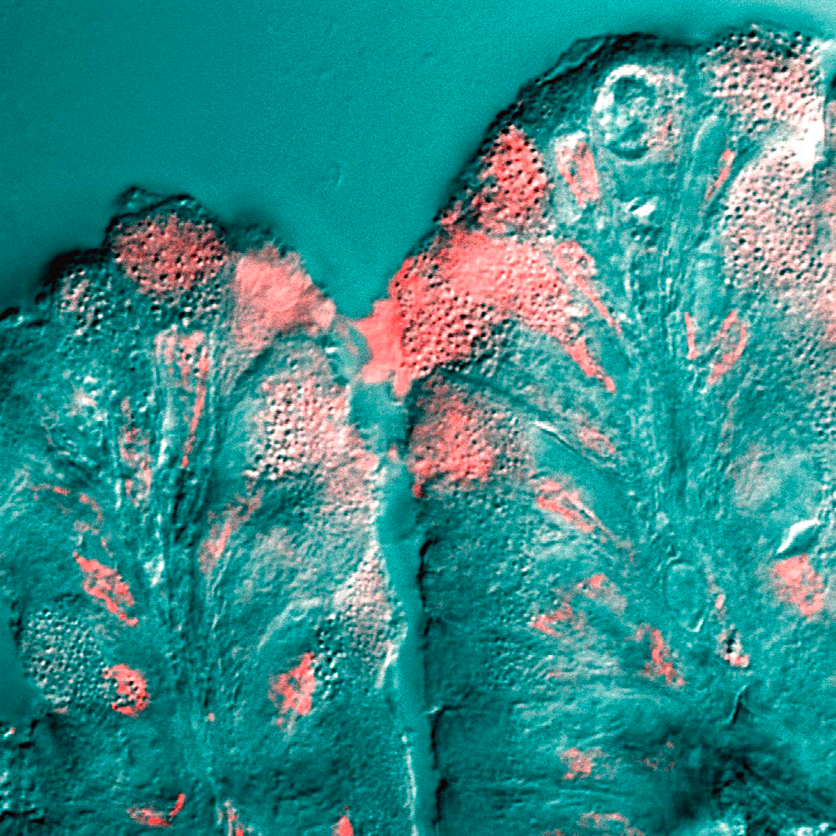
Submucosal glands can refer to various racemose exocrine glands of the mucous type. These glands secrete mucus to facilitate the movement of particles along the body's various tubes, such as the throat and intestines. The mucosa is the lining of the tubes, like a kind of ''skin''. ''Submucosal'' means that the actual gland resides in the connecting tissue below the mucosa. The submucosa is the tissue that connects the mucosa to the muscle outside the tube. The glands themselves are quite complex. The mucus factory is at the bottom, in the submucosa, it is composed of many little sacs (acini) where the mucus originates. Each sac (acinus) has one end that can open and close (dilate) to allow the mucus out. The acini empty into little tubes (tubules) that lead to a reservoir (collecting duct) that has a portal through the ''skin'' (mucosa) that can open and close allowing the mucus into the main tube. The submucosal glands are a companion to goblet cells which also produce mucus, an ... [...More Info...] [...Related Items...] OR: [Wikipedia] [Google] [Baidu] |
Racemic Mixture
In chemistry, a racemic mixture, or racemate (), is one that has equal amounts of left- and right-handed enantiomers of a chiral molecule or salt. Racemic mixtures are rare in nature, but many compounds are produced industrially as racemates. History The first known racemic mixture was racemic acid, which Louis Pasteur found to be a mixture of the two enantiomeric isomers of tartaric acid. He manually separated the crystals of a mixture by hand, starting from an aqueous solution of the sodium ammonium salt of racemate tartaric acid. Pasteur benefited from the fact that ammonium tartrate salt that gives enantiomeric crystals with distinct crystal forms (at 77 °F). Reasoning from the macroscopic scale down to the molecular, he reckoned that the molecules had to have non-superimposable mirror images. A sample with only a single enantiomer is an ''enantiomerically pure'' or ''enantiopure'' compound. Etymology From racemic acid found in grapes; from Latin ''racemus'', meani ... [...More Info...] [...Related Items...] OR: [Wikipedia] [Google] [Baidu] |
Exocrine Glands
Exocrine glands are glands that secrete substances on to an epithelial surface by way of a duct. Examples of exocrine glands include sweat, salivary, mammary, ceruminous, lacrimal, sebaceous, prostate and mucous. Exocrine glands are one of two types of glands in the human body, the other being endocrine glands, which secrete their products directly into the bloodstream. The liver and pancreas are both exocrine and endocrine glands; they are exocrine glands because they secrete products—bile and pancreatic juice—into the gastrointestinal tract through a series of ducts, and endocrine because they secrete other substances directly into the bloodstream. Exocrine sweat glands are part of the integumentary system; they have eccrine and apocrine types. Classification Structure Exocrine glands contain a glandular portion and a duct portion, the structures of which can be used to classify the gland. * The duct portion may be branched (called compound) or unbranched (called simple) ... [...More Info...] [...Related Items...] OR: [Wikipedia] [Google] [Baidu] |
Mucus
Mucus ( ) is a slippery aqueous secretion produced by, and covering, mucous membranes. It is typically produced from cells found in mucous glands, although it may also originate from mixed glands, which contain both serous and mucous cells. It is a viscous colloid containing inorganic salts, antimicrobial enzymes (such as lysozymes), immunoglobulins (especially IgA), and glycoproteins such as lactoferrin and mucins, which are produced by goblet cells in the mucous membranes and submucosal glands. Mucus serves to protect epithelial cells in the linings of the respiratory, digestive, and urogenital systems, and structures in the visual and auditory systems from pathogenic fungi, bacteria and viruses. Most of the mucus in the body is produced in the gastrointestinal tract. Amphibians, fish, snails, slugs, and some other invertebrates also produce external mucus from their epidermis as protection against pathogens, and to help in movement and is also produced in fish to line the ... [...More Info...] [...Related Items...] OR: [Wikipedia] [Google] [Baidu] |
Smooth Muscle Tissue
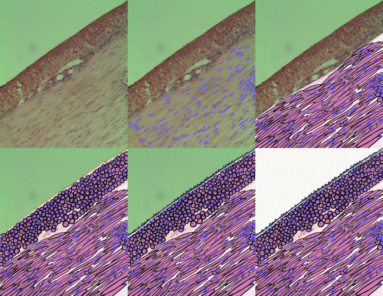
Smooth muscle is an involuntary non-striated muscle, so-called because it has no sarcomeres and therefore no striations (''bands'' or ''stripes''). It is divided into two subgroups, single-unit and multiunit smooth muscle. Within single-unit muscle, the whole bundle or sheet of smooth muscle cells contracts as a syncytium. Smooth muscle is found in the walls of hollow organs, including the stomach, intestines, bladder and uterus; in the walls of passageways, such as blood, and lymph vessels, and in the tracts of the respiratory, urinary, and reproductive systems. In the eyes, the ciliary muscles, a type of smooth muscle, dilate and contract the iris and alter the shape of the lens. In the skin, smooth muscle cells such as those of the arrector pili cause hair to stand erect in response to cold temperature or fear. Structure Gross anatomy Smooth muscle is grouped into two types: single-unit smooth muscle, also known as visceral smooth muscle, and multiunit smooth muscle. M ... [...More Info...] [...Related Items...] OR: [Wikipedia] [Google] [Baidu] |
Goblet Cell
Goblet cells are simple columnar epithelial cells that secrete gel-forming mucins, like mucin 5AC. The goblet cells mainly use the merocrine method of secretion, secreting vesicles into a duct, but may use apocrine methods, budding off their secretions, when under stress. The term ''goblet'' refers to the cell's goblet-like shape. The apical portion is shaped like a cup, as it is distended by abundant mucus laden granules; its basal portion lacks these granules and is shaped like a stem. The goblet cell is highly polarized with the nucleus and other organelles concentrated at the base of the cell and secretory granules containing mucin, at the apical surface. The apical plasma membrane projects short microvilli to give an increased surface area for secretion. Goblet cells are typically found in the respiratory, reproductive and gastrointestinal tracts and are surrounded by other columnar cells. Biased differentiation of airway basal cells in the respiratory epithelium, into gobl ... [...More Info...] [...Related Items...] OR: [Wikipedia] [Google] [Baidu] |
Vertebrate Trachea
The trachea, also known as the windpipe, is a cartilaginous tube that connects the larynx to the bronchi of the lungs, allowing the passage of air, and so is present in almost all air-breathing animals with lungs. The trachea extends from the larynx and branches into the two primary bronchi. At the top of the trachea the cricoid cartilage attaches it to the larynx. The trachea is formed by a number of horseshoe-shaped rings, joined together vertically by overlying ligaments, and by the trachealis muscle at their ends. The epiglottis closes the opening to the larynx during swallowing. The trachea begins to form in the second month of embryo development, becoming longer and more fixed in its position over time. It is epithelium lined with column-shaped cells that have hair-like extensions called cilia, with scattered goblet cells that produce protective mucins. The trachea can be affected by inflammation or infection, usually as a result of a viral illness affecting other parts ... [...More Info...] [...Related Items...] OR: [Wikipedia] [Google] [Baidu] |
Bronchus
A bronchus is a passage or airway in the lower respiratory tract that conducts air into the lungs. The first or primary bronchi pronounced (BRAN-KAI) to branch from the trachea at the carina are the right main bronchus and the left main bronchus. These are the widest bronchi, and enter the right lung, and the left lung at each hilum. The main bronchi branch into narrower secondary bronchi or lobar bronchi, and these branch into narrower tertiary bronchi or segmental bronchi. Further divisions of the segmental bronchi are known as 4th order, 5th order, and 6th order segmental bronchi, or grouped together as subsegmental bronchi. The bronchi, when too narrow to be supported by cartilage, are known as bronchioles. No gas exchange takes place in the bronchi. Structure The trachea (windpipe) divides at the carina into two main or primary bronchi, the left bronchus and the right bronchus. The carina of the trachea is located at the level of the sternal angle and the fifth thoracic vert ... [...More Info...] [...Related Items...] OR: [Wikipedia] [Google] [Baidu] |
Esophageal Glands
The esophageal glands are glands that are part of the digestive system of various animals, including humans. In humans Esophageal glands in humans are a part of a human digestive system. They are a small compound racemose exocrine glands of the mucous type. There are two types: * Esophageal glands proper- mucous glands located in the submucosa. They are compound tubulo-alveolar glands. Some serous cells are present. These glands are more numerous in the upper third of the esophagus. They secrete acid mucin for lubrication. * Esophageal cardiac glands- mucous glands located near the cardiac orifice (esophago-gastric junction) in the lamina propria mucosae. They secrete neutral mucin that protects the esophagus from acidic gastric juices. They are simple tubular or branched tubular glands. * There are also mucous glands present at the pharyngo-esophageal junction in the lamina propria mucosae. These are simple tubular or branched tubular glands. Each opens upon the surface by a l ... [...More Info...] [...Related Items...] OR: [Wikipedia] [Google] [Baidu] |
Brunner's Glands
Brunner's glands (or duodenal glands) are compound tubular submucosal glands found in that portion of the duodenum which is above the hepatopancreatic sphincter (i.e sphincter of Oddi). It also contains submucosa which creates special glands. The main function of these glands is to produce a mucus-rich alkaline secretion i.e. mucous (containing bicarbonate) in order to: * protect the duodenum from the acidic content of chyme (which is introduced into the duodenum from the stomach); * provide an alkaline condition for the intestinal enzymes to be active, thus enabling absorption to take place; * lubricate the intestinal walls. * They also secrete epidermal growth factor, which inhibits parietal and chief cells of the stomach from secreting acid and their digestive enzymes. This is another form of protection for the duodenum. They are the distinguishing feature of the duodenum, and are named for the Swiss physician who first described them, Johann Conrad Brunner. Structure Hi ... [...More Info...] [...Related Items...] OR: [Wikipedia] [Google] [Baidu] |
Glands
In animals, a gland is a group of cells in an animal's body that synthesizes substances (such as hormones) for release into the bloodstream (endocrine gland) or into cavities inside the body or its outer surface (exocrine gland). Structure Development Every gland is formed by an ingrowth from an epithelial surface. This ingrowth may in the beginning possess a tubular structure, but in other instances glands may start as a solid column of cells which subsequently becomes tubulated. As growth proceeds, the column of cells may split or give off offshoots, in which case a compound gland is formed. In many glands, the number of branches is limited, in others (salivary, pancreas) a very large structure is finally formed by repeated growth and sub-division. As a rule, the branches do not unite with one another, but in one instance, the liver, this does occur when a reticulated compound gland is produced. In compound glands the more typical or secretory epithelium is found forming t ... [...More Info...] [...Related Items...] OR: [Wikipedia] [Google] [Baidu] |


_and_foveolar_cells_in_incomplete_Barrett's_esophagus.jpg)


