|
Submasseteric Space
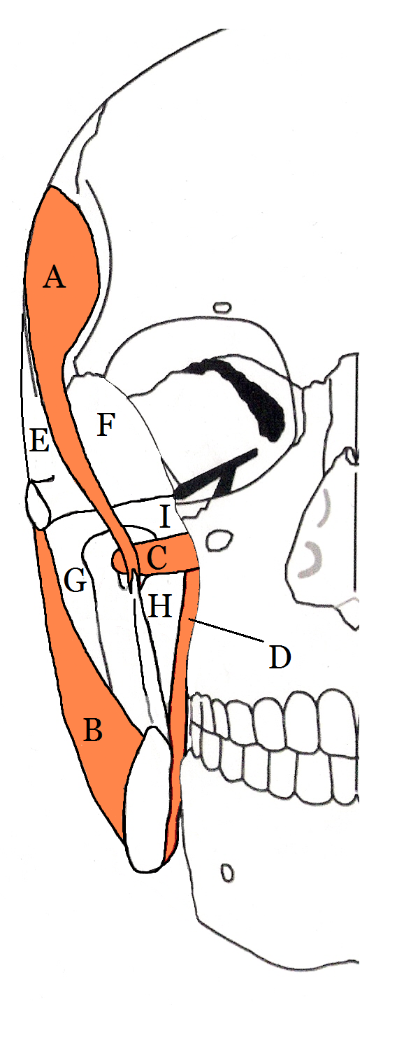
The submasseterric space (also termed the masseteric space) is a fascial space of the head and neck (sometimes also termed fascial spaces or tissue spaces). It is a potential space in the face over the angle of the jaw, and is paired on each side. It is located between the lateral aspect of the mandible and the medial aspect of the masseter muscle and its investing fascia. The term is derived from sub- meaning "under" in Latin and ''masseteric'' which refers to the masseter muscle. The submasseteric space is one of the four compartments of the masticator space. Sometimes the submasseteric space is described as a series of spaces, created because the masseter muscle has multiple insertions that cover most of the lateral surface of the ramus of the mandible. Structure Anatomic boundaries The boundaries of each submasseteric space are: * the anterior margin of the masseter muscle anteriorly, * the parotid gland posteriorly, * the zygomatic arch superiorly, * the inferior border o ... [...More Info...] [...Related Items...] OR: [Wikipedia] [Google] [Baidu] |
Fascial Spaces Of The Head And Neck
Fascial spaces (also termed fascial tissue spaces or tissue spaces) are potential spaces that exist between the fasciae and underlying organs and other tissues. In health, these spaces do not exist; they are only created by pathology, e.g. the spread of pus or cellulitis in an infection. The fascial spaces can also be opened during the dissection of a cadaver. The fascial spaces are different from the fasciae themselves, which are bands of connective tissue that surround structures, e.g. muscles. The opening of fascial spaces may be facilitated by pathogenic bacterial release of enzymes which cause tissue lysis (e.g. hyaluronidase and collagenase). The spaces filled with loose areolar connective tissue may also be termed clefts. Other contents such as salivary glands, blood vessels, nerves and lymph nodes are dependent upon the location of the space. Those containing neurovascular tissue (nerves and blood vessels) may also be termed compartments. Generally, the spread of infection ... [...More Info...] [...Related Items...] OR: [Wikipedia] [Google] [Baidu] |
Parotitis
Parotitis is an inflammation of one or both parotid glands, the major salivary glands located on either side of the face, in humans. The parotid gland is the salivary gland most commonly affected by inflammation. Etymology From Greek παρωτῖτις (νόσος), parōtĩtis (nósos) : (disease of the) parotid gland < παρωτίς (stem παρωτιδ-) : (gland) behind the ear < παρά - pará : behind, and οὖς - ous (stem ὠτ-, ōt-) : ear. Causes Dehydration ''Dehydration:'' This is a common, non-infectious cause of parotitis. It may occur in elderly or after surgery.Infectious parotitis ''Acute bacterial parotitis:'' is most often caused by a bacterial infection of but may be caused by any |
Otorhinolaryngology
Otorhinolaryngology ( , abbreviated ORL and also known as otolaryngology, otolaryngology–head and neck surgery (ORL–H&N or OHNS), or ear, nose, and throat (ENT)) is a surgical subspeciality within medicine that deals with the surgical and medical management of conditions of the head and neck. Doctors who specialize in this area are called otorhinolaryngologists, otolaryngologists, head and neck surgeons, or ENT surgeons or physicians. Patients seek treatment from an otorhinolaryngologist for diseases of the ear, nose, throat, base of the skull, head, and neck. These commonly include functional diseases that affect the senses and activities of eating, drinking, speaking, breathing, swallowing, and hearing. In addition, ENT surgery encompasses the surgical management of cancers and benign tumors and reconstruction of the head and neck as well as plastic surgery of the face and neck. Etymology The term is a combination of New Latin combining forms ('' oto-'' + ''rhino-'' + ... [...More Info...] [...Related Items...] OR: [Wikipedia] [Google] [Baidu] |
Fascial Spaces Of The Head And Neck
Fascial spaces (also termed fascial tissue spaces or tissue spaces) are potential spaces that exist between the fasciae and underlying organs and other tissues. In health, these spaces do not exist; they are only created by pathology, e.g. the spread of pus or cellulitis in an infection. The fascial spaces can also be opened during the dissection of a cadaver. The fascial spaces are different from the fasciae themselves, which are bands of connective tissue that surround structures, e.g. muscles. The opening of fascial spaces may be facilitated by pathogenic bacterial release of enzymes which cause tissue lysis (e.g. hyaluronidase and collagenase). The spaces filled with loose areolar connective tissue may also be termed clefts. Other contents such as salivary glands, blood vessels, nerves and lymph nodes are dependent upon the location of the space. Those containing neurovascular tissue (nerves and blood vessels) may also be termed compartments. Generally, the spread of infection ... [...More Info...] [...Related Items...] OR: [Wikipedia] [Google] [Baidu] |
Zygomaticus Minor Muscle
The zygomaticus minor muscle is a muscle of facial expression. It originates from the zygomatic bone, lateral to the rest of the levator labii superioris muscle, and inserts into the outer part of the upper lip. It draws the upper lip backward, upward, and outward and is used in smiling. It is innervated by the facial nerve (VII). Structure The zygomaticus minor muscle originates from the zygomatic bone. It inserts into the tissue around the upper lip, particularly blending its fibres with orbicularis oris muscle. It lies lateral to the rest of levator labii superioris muscle, and medial to its stronger synergist zygomaticus major muscle. It travels at an angle of approximately 30°. It has a mean width of around 0.5 cm. Nerve supply The zygomaticus minor muscle is supplied by the buccal branch of the facial nerve (VII). Variation The zygomaticus minor muscle may have either a straight or a curved course along its length. It may attach to both the upper lip and the latera ... [...More Info...] [...Related Items...] OR: [Wikipedia] [Google] [Baidu] |
Zygomaticus Major Muscle
The zygomaticus major muscle is a muscle of the human body. It extends from each zygomatic arch (cheekbone) to the corners of the mouth. It is a muscle of facial expression which draws the angle of the mouth superiorly and posteriorly to allow one to smile. Bifid zygomaticus major muscle is a notable variant, and may cause cheek dimples. Structure The zygomaticus major muscle originates from the upper margin of the temporal process, part of the lateral surface of the zygomatic bone. It inserts into tissue at the corner of the mouth. Nerve supply The zygomaticus major muscle is supplied by a buccal branch and a zygomatic branch of the facial nerve (VII). Variation The zygomaticus major muscle may occur in a bifid form, with two fascicles that are partially or completely separate from each other but adjacent. Usually a single unit, dimples are caused by variations in form. It is thought that cheek dimples are caused by bifid zygomaticus major muscle. Function The zygomat ... [...More Info...] [...Related Items...] OR: [Wikipedia] [Google] [Baidu] |
Platysma Muscle
The platysma muscle is a superficial muscle of the human neck that overlaps the sternocleidomastoid. It covers the anterior surface of the neck superficially. When it contracts, it produces a slight wrinkling of the neck, and a "bowstring" effect on either side of the neck. Structure The platysma muscle is a broad sheet of muscle arising from the fascia covering the upper parts of the pectoralis major muscle and deltoid muscle. Its fibers cross the clavicle, and proceed obliquely upward and medially along the side of the neck. This leaves the inferior part of the neck in the midline deficient of significant muscle cover. Fibres at the front of the muscle from the left and right sides intermingle together below and behind the mandibular symphysis, the junction where the two lateral halves of the mandible are fused at an early period of life (although not a true symphysis). Fibres at the back of the muscle cross the mandible, some being inserted into the bone below the oblique line ... [...More Info...] [...Related Items...] OR: [Wikipedia] [Google] [Baidu] |
Mandibular Third Molar
A third molar, commonly called wisdom tooth, is one of the three molars per quadrant of the human dentition. It is the most posterior of the three. The age at which wisdom teeth come through ( erupt) is variable, but this generally occurs between late teens and early twenties. Most adults have four wisdom teeth, one in each of the four quadrants, but it is possible to have none, fewer, or more, in which case the extras are called supernumerary teeth. Wisdom teeth may get stuck ( impacted) against other teeth if there is not enough space for them to come through normally. Impacted wisdom teeth are still sometimes removed for orthodontic treatment, believing that they move the other teeth and cause crowding, though this is not held anymore as true. Impacted wisdom teeth may suffer from tooth decay if oral hygiene becomes more difficult. Wisdom teeth which are partially erupted through the gum may also cause inflammation and infection in the surrounding gum tissues, termed pericoron ... [...More Info...] [...Related Items...] OR: [Wikipedia] [Google] [Baidu] |
Tooth Impaction
An impacted tooth is one that fails to erupt into the dental arch within the expected developmental window. Because impacted teeth do not erupt, they are retained throughout the individual's lifetime unless extracted or exposed surgically. Teeth may become impacted because of adjacent teeth, dense overlying bone, excessive soft tissue or a genetic abnormality. Most often, the cause of impaction is inadequate arch length and space in which to erupt. That is the total length of the alveolar arch is smaller than the tooth arch (the combined mesiodistal width of each tooth). The wisdom teeth (third molars) are frequently impacted because they are the last teeth to erupt in the oral cavity. Mandibular third molars are more commonly impacted than their maxillary counterparts. Some dentists believe that impacted teeth should be removed except, in certain cases, canine teeth: canines may just remain buried and give no further problems, thus not requiring surgical intervention. However ... [...More Info...] [...Related Items...] OR: [Wikipedia] [Google] [Baidu] |
Pericoronal Abscess
Pericoronitis is inflammation of the soft tissues surrounding the crown of a partially erupted tooth, including the gingiva (gums) and the dental follicle. The soft tissue covering a partially erupted tooth is known as an ''operculum'', an area which can be difficult to access with normal oral hygiene methods. The hyponym ''operculitis'' technically refers to inflammation of the operculum alone. Pericoronitis is caused by an accumulation of bacteria and debris beneath the operculum, or by mechanical trauma (e.g. biting the operculum with the opposing tooth). Pericoronitis is often associated with partially erupted and impacted mandibular third molars (lower wisdom teeth), often occurring at the age of wisdom tooth eruption (15-26). Other common causes of similar pain from the third molar region are food impaction causing periodontal pain, pulpitis from dental caries (tooth decay), and acute myofascial pain in temporomandibular joint disorder. Pericoronitis is classified into '' ... [...More Info...] [...Related Items...] OR: [Wikipedia] [Google] [Baidu] |
Odontogenic Infection
An odontogenic infection is an infection that originates within a tooth or in the closely surrounding tissues. The term is derived from '' odonto-'' (Ancient Greek: , – 'tooth') and '' -genic'' (Ancient Greek: , ; – 'birth'). The most common causes for odontogenic infection to be established are dental caries, deep fillings, failed root canal treatments, periodontal disease, and pericoronitis. Odontogenic infection starts as localised infection and may remain localised to the region where it started, or spread into adjacent or distant areas. It is estimated that 90-95% of all orofacial infections originate from the teeth or their supporting structures and are the most common infections in the oral and maxilofacial region. Odontogenic infections can be severe if not treated and are associated with mortality rate of 10 to 40%. Furthermore, about 70% of odontogenic infections occur as periapical inflammation, i.e. acute periapical periodontitis or a periapical abscess. The next m ... [...More Info...] [...Related Items...] OR: [Wikipedia] [Google] [Baidu] |
Incision And Drainage
Incision and drainage (I&D), also known as clinical lancing, are minor surgical procedures to release pus or pressure built up under the skin, such as from an abscess, boil, or infected paranasal sinus. It is performed by treating the area with an antiseptic, such as iodine-based solution, and then making a small incision to puncture the skin using a sterile instrument such as a sharp needle or a pointed scalpel. This allows the pus fluid to escape by draining out through the incision. Good medical practice for large abdominal abscesses requires insertion of a drainage tube, preceded by insertion of a PICC line to enable readiness of treatment for possible septic shock. Adjunct antibiotics Uncomplicated cutaneous abscesses do not need antibiotics after successful drainage. In incisional abscesses For incisional abscesses, it is recommended that incision and drainage is followed by covering the area with a thin layer of gauze followed by sterile dressing. The dressing should be ... [...More Info...] [...Related Items...] OR: [Wikipedia] [Google] [Baidu] |


