|
Subesophageal Ganglia
The suboesophageal ganglion (acronym: SOG; synonym: ''subesophageal ganglion'') of arthropods and in particular insects is part of the arthropod central nervous system (CNS). As indicated by its name, it is located ''below the'' ''oesophagus'', inside the head. As part of the ventral nerve cord, it is connected (via pairs of connections) to the brain (or supraoesophageal ganglion) and to the first thoracic ganglion (or protothoracic ganglion). Its nerves innervate the sensory organs and muscles of the mouthparts and the salivary glands. Neurons in the suboesophageal ganglion control movement of the head and neck as well. It is composed of three pairs of fused ganglia, each of which is associated with a pair of mouthparts. Therefore the fused parts are called the mandibular, maxillary and labial The term ''labial'' originates from '' Labium'' (Latin for "lip"), and is the adjective that describes anything of or related to lips, such as lip-like structures. Thus, it may refe ... [...More Info...] [...Related Items...] OR: [Wikipedia] [Google] [Baidu] |
Arthropod Central Nervous System
The central nervous system (CNS) is the part of the nervous system consisting primarily of the brain and spinal cord. The CNS is so named because the brain integrates the received information and coordinates and influences the activity of all parts of the bodies of bilaterally symmetric and triploblastic animals—that is, all multicellular animals except sponges and diploblasts. It is a structure composed of nervous tissue positioned along the rostral (nose end) to caudal (tail end) axis of the body and may have an enlarged section at the rostral end which is a brain. Only arthropods, cephalopods and vertebrates have a true brain (precursor structures exist in onychophorans, gastropods and lancelets). The rest of this article exclusively discusses the vertebrate central nervous system, which is radically distinct from all other animals. Overview In vertebrates, the brain and spinal cord are both enclosed in the meninges. The meninges provide a barrier to chemicals dissolved ... [...More Info...] [...Related Items...] OR: [Wikipedia] [Google] [Baidu] |
Oesophagus
The esophagus (American English) or oesophagus (British English; both ), non-technically known also as the food pipe or gullet, is an organ in vertebrates through which food passes, aided by peristaltic contractions, from the pharynx to the stomach. The esophagus is a fibromuscular tube, about long in adults, that travels behind the trachea and heart, passes through the diaphragm, and empties into the uppermost region of the stomach. During swallowing, the epiglottis tilts backwards to prevent food from going down the larynx and lungs. The word ''oesophagus'' is from Ancient Greek οἰσοφάγος (oisophágos), from οἴσω (oísō), future form of φέρω (phérō, “I carry”) + ἔφαγον (éphagon, “I ate”). The wall of the esophagus from the lumen outwards consists of mucosa, submucosa (connective tissue), layers of muscle fibers between layers of fibrous tissue, and an outer layer of connective tissue. The mucosa is a stratified squamous epithel ... [...More Info...] [...Related Items...] OR: [Wikipedia] [Google] [Baidu] |
Supraesophageal Ganglion
The supraesophageal ganglion (also "supraoesophageal ganglion", "arthropod brain" or "microbrain") is the first part of the arthropod, especially insect, central nervous system. It receives and processes information from the first, second, and third metameres. The supraesophageal ganglion lies dorsal to the esophagus and consists of three parts, each a pair of ganglia that may be more or less pronounced, reduced, or fused depending on the genus: * The ''protocerebrum'', associated with the eyes (compound eyes and ocelli). Directly associated with the eyes is the optic lobe, as the visual center of the brain. * The ''deutocerebrum'' processes sensory information from the antennae. It consists of two parts, the antennal lobe and the dorsal lobe. The dorsal lobe also contains motor neurons which control the antennal muscles. * The ''tritocerebrum'' integrates sensory inputs from the previous two pairs of ganglia. The lobes of the tritocerebrum split to circumvent the esophagus a ... [...More Info...] [...Related Items...] OR: [Wikipedia] [Google] [Baidu] |
Thoracic Ganglia
The thoracic ganglia are paravertebral ganglia. The thoracic portion of the sympathetic trunk typically has 12 thoracic ganglia. Emerging from the ganglia are thoracic splanchnic nerves (the cardiopulmonary, the greater, lesser, and least splanchnic nerves) that help provide sympathetic innervation to thoracic and abdominal structures. The thoracic part of sympathetic trunk lies posterior to the costovertebral pleura and is hence not a content of the posterior mediastinum Also, the ganglia of the thoracic sympathetic trunk have both white and gray rami communicantes. The white rami communicantes carry sympathetic fibers arising in the spinal cord into the sympathetic trunk, while the gray rami communicantes carry postganglionic nerve fibers of the sympathetic nervous system back to the spinal nerves A spinal nerve is a mixed nerve, which carries motor, sensory, and autonomic signals between the spinal cord and the body. In the human body there are 31 pairs of spinal nerves, ... [...More Info...] [...Related Items...] OR: [Wikipedia] [Google] [Baidu] |
Insect Mouthparts
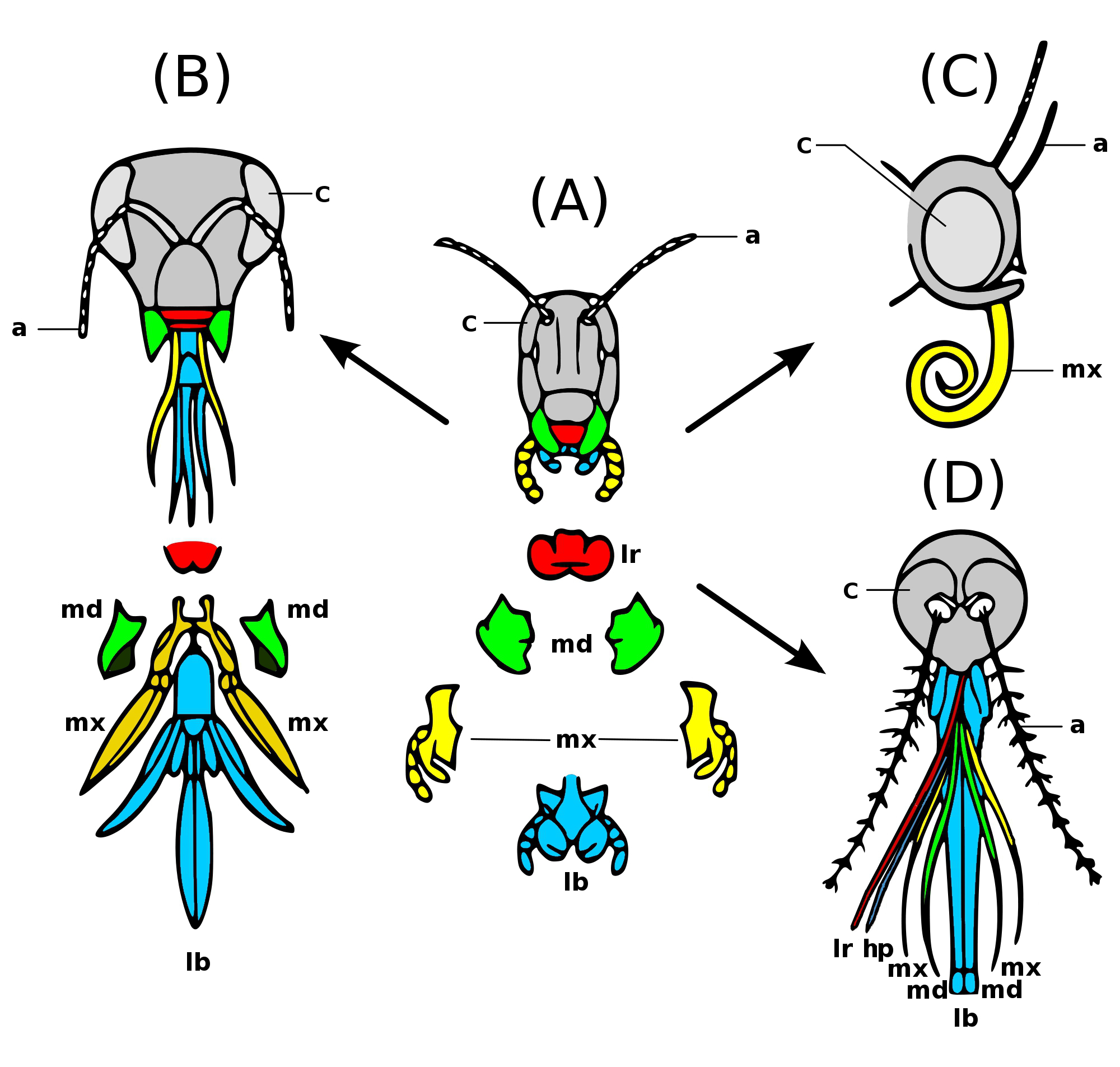
Insects have mouthparts that may vary greatly across insect species, as they are adapted to particular modes of feeding. The earliest insects had chewing mouthparts. Most specialisation of mouthparts are for piercing and sucking, and this mode of feeding has evolved a number of times idependently. For example, mosquitoes and aphids (which are true bugs) both pierce and suck, however female mosquitoes feed on animal blood whereas aphids feed on plant fluids. Evolution Like most external features of arthropods, the mouthparts of Hexapoda are highly derived. Insect mouthparts show a multitude of different functional mechanisms across the wide diversity of insect species. It is common for significant homology to be conserved, with matching structures forming from matching primordia, and having the same evolutionary origin. However, even if structures are almost physically and functionally identical, they may not be homologous; their analogous functions and appearance might be the pr ... [...More Info...] [...Related Items...] OR: [Wikipedia] [Google] [Baidu] |
Salivary Glands
The salivary glands in mammals are exocrine glands that produce saliva through a system of ducts. Humans have three paired major salivary glands (parotid, submandibular, and sublingual), as well as hundreds of minor salivary glands. Salivary glands can be classified as serous, mucous, or seromucous (mixed). In serous secretions, the main type of protein secreted is alpha-amylase, an enzyme that breaks down starch into maltose and glucose, whereas in mucous secretions, the main protein secreted is mucin, which acts as a lubricant. In humans, 1200 to 1500 ml of saliva are produced every day. The secretion of saliva (salivation) is mediated by parasympathetic stimulation; acetylcholine is the active neurotransmitter and binds to muscarinic receptors in the glands, leading to increased salivation. The fourth pair of salivary glands, the tubarial glands discovered in 2020, are named for their location, being positioned in front and over the torus tubarius. However, this finding ... [...More Info...] [...Related Items...] OR: [Wikipedia] [Google] [Baidu] |
Insect Anatomy Diagram
Insects (from Latin ') are pancrustacean hexapod invertebrates of the class Insecta. They are the largest group within the arthropod phylum. Insects have a chitinous exoskeleton, a three-part body ( head, thorax and abdomen), three pairs of jointed legs, compound eyes and one pair of antennae. Their blood is not totally contained in vessels; some circulates in an open cavity known as the haemocoel. Insects are the most diverse group of animals; they include more than a million described species and represent more than half of all known living organisms. The total number of extant species is estimated at between six and ten million; In: potentially over 90% of the animal life forms on Earth are insects. Insects may be found in nearly all environments, although only a small number of species reside in the oceans, which are dominated by another arthropod group, crustaceans, which recent research has indicated insects are nested within. Nearly all insects hatch from egg ... [...More Info...] [...Related Items...] OR: [Wikipedia] [Google] [Baidu] |
Ganglia
A ganglion is a group of neuron cell bodies in the peripheral nervous system. In the somatic nervous system this includes dorsal root ganglia and trigeminal ganglia among a few others. In the autonomic nervous system there are both sympathetic and parasympathetic ganglia which contain the cell bodies of postganglionic sympathetic and parasympathetic neurons respectively. A pseudoganglion looks like a ganglion, but only has nerve fibers and has no nerve cell bodies. Structure Ganglia are primarily made up of somata and dendritic structures which are bundled or connected. Ganglia often interconnect with other ganglia to form a complex system of ganglia known as a plexus. Ganglia provide relay points and intermediary connections between different neurological structures in the body, such as the peripheral and central nervous systems. Among vertebrates there are three major groups of ganglia: *Dorsal root ganglia (also known as the spinal ganglia) contain the cell bodies of se ... [...More Info...] [...Related Items...] OR: [Wikipedia] [Google] [Baidu] |
Mandible (insect Mouthpart)
Insect mandibles are a pair of appendages near the insect's mouth, and the most anterior of the three pairs of oral appendages (the labrum is more anterior, but is a single fused structure). Their function is typically to grasp, crush, or cut the insect's food, or to defend against predators or rivals. Insect mandibles, which appear to be evolutionarily derived from legs, move in the horizontal plane unlike those of vertebrates, which appear to be derived from gill arches and move vertically. Grasshoppers, crickets, and other simple insects The mouthparts of orthopteran insects are often used as a basic example of mandibulate (chewing) mouthparts, and the mandibles themselves are likewise generalized in structure. They are large and hardened, shaped like pinchers, with cutting surfaces on the distal portion and chewing or grinding surfaces basally. They are usually lined with teeth and move sideways. Large pieces of leaves can therefore be cut and then pulverized near the mouth ... [...More Info...] [...Related Items...] OR: [Wikipedia] [Google] [Baidu] |
Maxilla (arthropod Mouthpart)
In arthropods, the maxillae (singular maxilla) are paired structures present on the head as mouthparts in members of the clade Mandibulata, used for tasting and manipulating food. Embryologically, the maxillae are derived from the 4th and 5th segment of the head and the maxillary palps; segmented appendages extending from the base of the maxilla represent the former leg of those respective segments. In most cases, two pairs of maxillae are present and in different arthropod groups the two pairs of maxillae have been variously modified. In crustaceans, the first pair are called maxillulae (singular maxillula). Modified coxae at the base of the pedipalps in spiders are also called "maxillae", although they are not homologous with mandibulate maxillae. Myriapoda Millipedes In millipedes, the second maxillae have been lost, reducing the mouthparts to only the first maxillae which have fused together to form a gnathochilarium, acting as a lower lip to the buccal cavity and the man ... [...More Info...] [...Related Items...] OR: [Wikipedia] [Google] [Baidu] |
Labium (arthropod Mouthpart)
The mouthparts of arthropods have evolution, evolved into a number of forms, each adaptation, adapted to a different style or mode of feeding. Most mouthparts represent modified, paired appendages, which in ancestral forms would have appeared more like legs than mouthparts. In general, arthropods have mouthparts for cutting, chewing, piercing, sucking, shredding, siphoning, and filtering. This article outlines the basic elements of four arthropod groups: insects, myriapods, crustaceans and chelicerates. Insects are used as the model, with the novel mouthparts of the other groups introduced in turn. Insects are not, however, the Arthropod#Classification of arthropods, ancestral form of the other arthropods discussed here. Insects Insect mouthparts exhibit a range of forms. The earliest insects had chewing mouthparts. Specialisation includes mouthparts modified for siphoning, piercing, sucking and sponging. These modifications have evolved a number of times. For example, m ... [...More Info...] [...Related Items...] OR: [Wikipedia] [Google] [Baidu] |


