|
Stifle
The stifle joint (often simply stifle) is a complex joint in the hind limbs of quadruped mammals such as the sheep, horse or dog. It is the equivalent of the human knee and is often the largest synovial joint in the animal's body. The stifle joint joins three bones: the femur, patella, and tibia. The joint consists of three smaller ones: the femoropatellar joint, medial joint, and lateral femorotibial joint. The stifle joint consists of the femorotibial articulation ( femoral and tibial condyles), femoropatellar articulation (femoral trochlea and the patella), and the proximal articulation. The joint is stabilized by paired collateral ligaments which act to prevent abduction/adduction at the joint, as well as paired cruciate ligaments. The cranial cruciate ligament and the caudal cruciate ligament restrict cranial and caudal translation (respectively) of the tibia on the femur. The cranial cruciate also resists over-extension and inward rotation, and is the most commonly dama ... [...More Info...] [...Related Items...] OR: [Wikipedia] [Google] [Baidu] |
Dog Anatomy
Dog anatomy comprises the anatomical studies of the visible parts of the body of a domestic dog. Details of structures vary tremendously from breed to breed, more than in any other animal species, wild or domesticated, as dogs are highly variable in height and weight. The smallest known adult dog was a Yorkshire Terrier that stood only at the shoulder, in length along the head and body, and weighed only . The heaviest dog was an English Mastiff named Zorba which weighed . The tallest known adult dog is a Great Dane that stands at the shoulder. Anatomy Source: Muscles The following is a list of the muscles in the dog, along with their origin, insertion, action and innervation. ''Extrinsic muscles of the thoracic limb and related structures:'' Descending superficial pectoral: originates on the first sternebrae and inserts on the greater tubercle of the humerus. It both adducts the limb and also prevents the limb from being abducted during weight bearing. It is innervated b ... [...More Info...] [...Related Items...] OR: [Wikipedia] [Google] [Baidu] |
Cruciate Ligaments
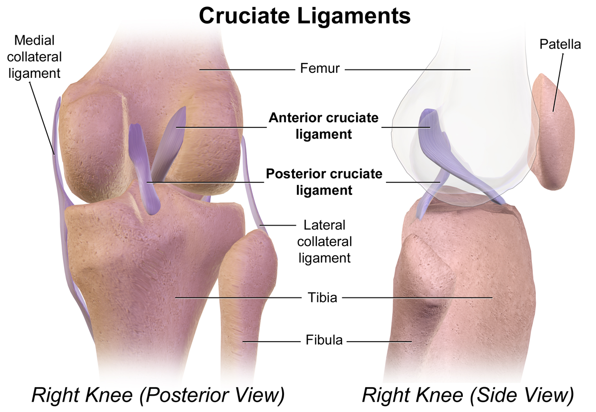
Cruciate ligaments (also cruciform ligaments) are pairs of ligaments arranged like a letter X. They occur in several joints of the body, such as the knee joint and the atlantoaxial joint, atlanto-axial joint. In a fashion similar to the cords in a toy Jacob's ladder (toy), Jacob's ladder, the crossed ligaments stabilize the joint while allowing a very large range of motion. Knee Structure Cruciate ligaments occur in the knee of humans and other bipedal animals and the corresponding Stifle joint, stifle of quadrupedal animals, and in the neck, fingers, and foot. * The cruciate ligaments of the knee are the anterior cruciate ligament (ACL) and the posterior cruciate ligament (PCL). These ligaments are two strong, rounded bands that extend from the head of the tibia to the intercondyloid notch of the femur. The ACL is lateral and the PCL is medial. They cross each other like the limbs of an X. They are named for their insertion into the tibia: the ACL attaches to the anterior ... [...More Info...] [...Related Items...] OR: [Wikipedia] [Google] [Baidu] |
Horse Anatomy
Equine anatomy encompasses the gross and microscopic anatomy of horses, ponies and other equids, including donkeys, mules and zebras. While all anatomical features of equids are described in the same terms as for other animals by the International Committee on Veterinary Gross Anatomical Nomenclature in the book ''Nomina Anatomica Veterinaria'', there are many horse-specific colloquial terms used by equestrians. External anatomy * Back: the area where the saddle sits, beginning at the end of the withers, extending to the last thoracic vertebrae (colloquially includes the loin or "coupling," though technically incorrect usage) * Barrel: the body of the horse, enclosing the rib cage and the major internal organs * Buttock: the part of the hindquarters behind the thighs and below the root of the tail * Cannon or cannon bone: the area between the knee or hock and the fetlock joint, sometimes called the "shin" of the horse, though technically it is the third metacarpal * Chestnut: ... [...More Info...] [...Related Items...] OR: [Wikipedia] [Google] [Baidu] |
Knee
In humans and other primates, the knee joins the thigh with the leg and consists of two joints: one between the femur and tibia (tibiofemoral joint), and one between the femur and patella (patellofemoral joint). It is the largest joint in the human body. The knee is a modified hinge joint, which permits flexion and extension as well as slight internal and external rotation. The knee is vulnerable to injury and to the development of osteoarthritis. It is often termed a ''compound joint'' having tibiofemoral and patellofemoral components. (The fibular collateral ligament is often considered with tibiofemoral components.) Structure The knee is a modified hinge joint, a type of synovial joint, which is composed of three functional compartments: the patellofemoral articulation, consisting of the patella, or "kneecap", and the patellar groove on the front of the femur through which it slides; and the medial and lateral tibiofemoral articulations linking the femur, or thigh bone ... [...More Info...] [...Related Items...] OR: [Wikipedia] [Google] [Baidu] |
Caudal Cruciate Ligament
The posterior cruciate ligament (PCL) is a ligament in each knee of humans and various other animals. It works as a counterpart to the anterior cruciate ligament (ACL). It connects the posterior intercondylar area of the tibia to the medial condyle of the femur. This configuration allows the PCL to resist forces pushing the tibia posteriorly relative to the femur. The PCL and ACL are intracapsular ligaments because they lie deep within the knee joint. They are both isolated from the fluid-filled synovial cavity, with the synovial membrane wrapped around them. The PCL gets its name by attaching to the posterior portion of the tibia. The PCL, ACL, MCL, and LCL are the four main ligaments of the knee in primates. Structure The PCL is located within the knee joint where it stabilizes the articulating bones, particularly the femur and the tibia, during movement. It originates from the lateral edge of the medial femoral condyle and the roof of the intercondyle notch then stretch ... [...More Info...] [...Related Items...] OR: [Wikipedia] [Google] [Baidu] |
Knee
In humans and other primates, the knee joins the thigh with the leg and consists of two joints: one between the femur and tibia (tibiofemoral joint), and one between the femur and patella (patellofemoral joint). It is the largest joint in the human body. The knee is a modified hinge joint, which permits flexion and extension as well as slight internal and external rotation. The knee is vulnerable to injury and to the development of osteoarthritis. It is often termed a ''compound joint'' having tibiofemoral and patellofemoral components. (The fibular collateral ligament is often considered with tibiofemoral components.) Structure The knee is a modified hinge joint, a type of synovial joint, which is composed of three functional compartments: the patellofemoral articulation, consisting of the patella, or "kneecap", and the patellar groove on the front of the femur through which it slides; and the medial and lateral tibiofemoral articulations linking the femur, or thigh bone ... [...More Info...] [...Related Items...] OR: [Wikipedia] [Google] [Baidu] |
Cranial Cruciate Ligament
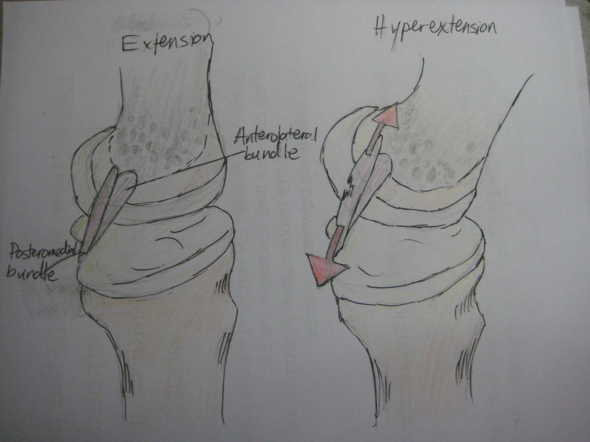
The anterior cruciate ligament (ACL) is one of a pair of cruciate ligaments (the other being the posterior cruciate ligament) in the human knee. The two ligaments are also called "cruciform" ligaments, as they are arranged in a crossed formation. In the quadruped stifle joint (analogous to the knee), based on its anatomical position, it is also referred to as the cranial cruciate ligament. The term cruciate translates to cross. This name is fitting because the ACL crosses the posterior cruciate ligament to form an “X”. It is composed of strong, fibrous material and assists in controlling excessive motion. This is done by limiting mobility of the joint. The anterior cruciate ligament is one of the four main ligaments of the knee, providing 85% of the restraining force to anterior tibial displacement at 30 and 90° of knee flexion. The ACL is the most injured ligament of the four located in the knee. Structure The ACL originates from deep within the notch of the distal femur. ... [...More Info...] [...Related Items...] OR: [Wikipedia] [Google] [Baidu] |
Meniscus (anatomy)
A meniscus is a crescent-shaped fibrocartilaginous anatomical structure that, in contrast to an articular disc, only partly divides a joint cavity.Platzer (2004), p 208 In humans they are present in the knee, wrist, acromioclavicular, sternoclavicular, and temporomandibular joints; in other animals they may be present in other joints. Generally, the term "meniscus" is used to refer to the cartilage of the knee, either to the lateral or medial meniscus. Both are cartilaginous tissues that provide structural integrity to the knee when it undergoes tension and torsion. The menisci are also known as "semi-lunar" cartilages, referring to their half-moon, crescent shape. The term "meniscus" is from the Ancient Greek word (), meaning "crescent". Structure The menisci of the knee are two pads of fibrocartilaginous tissue which serve to disperse friction in the knee joint between the lower leg (tibia) and the thigh (femur). They are concave on the top and flat on the bottom, articula ... [...More Info...] [...Related Items...] OR: [Wikipedia] [Google] [Baidu] |
Temporomandibular
In anatomy, the temporomandibular joints (TMJ) are the two joints connecting the jawbone to the skull. It is a bilateral Synovial joint, synovial articulation between the temporal bone of the skull above and the Human mandible, mandible below; it is from these bones that its name is derived. This joint is unique in that it is a bilateral joint that functions as one unit. Since the TMJ is connected to the mandible, the right and left joints must function together and therefore are not independent of each other. Structure The main components are the joint capsule, articular disc, mandibular condyles, articular surface of the temporal bone, temporomandibular ligament, stylomandibular ligament, sphenomandibular ligament, and lateral pterygoid muscle. Capsule The articular capsule (capsular ligament) is a thin, loose envelope, attached above to the circumference of the mandibular fossa and the articular tubercle immediately in front; below, to the neck of the condyle of the mandi ... [...More Info...] [...Related Items...] OR: [Wikipedia] [Google] [Baidu] |
Proprioception
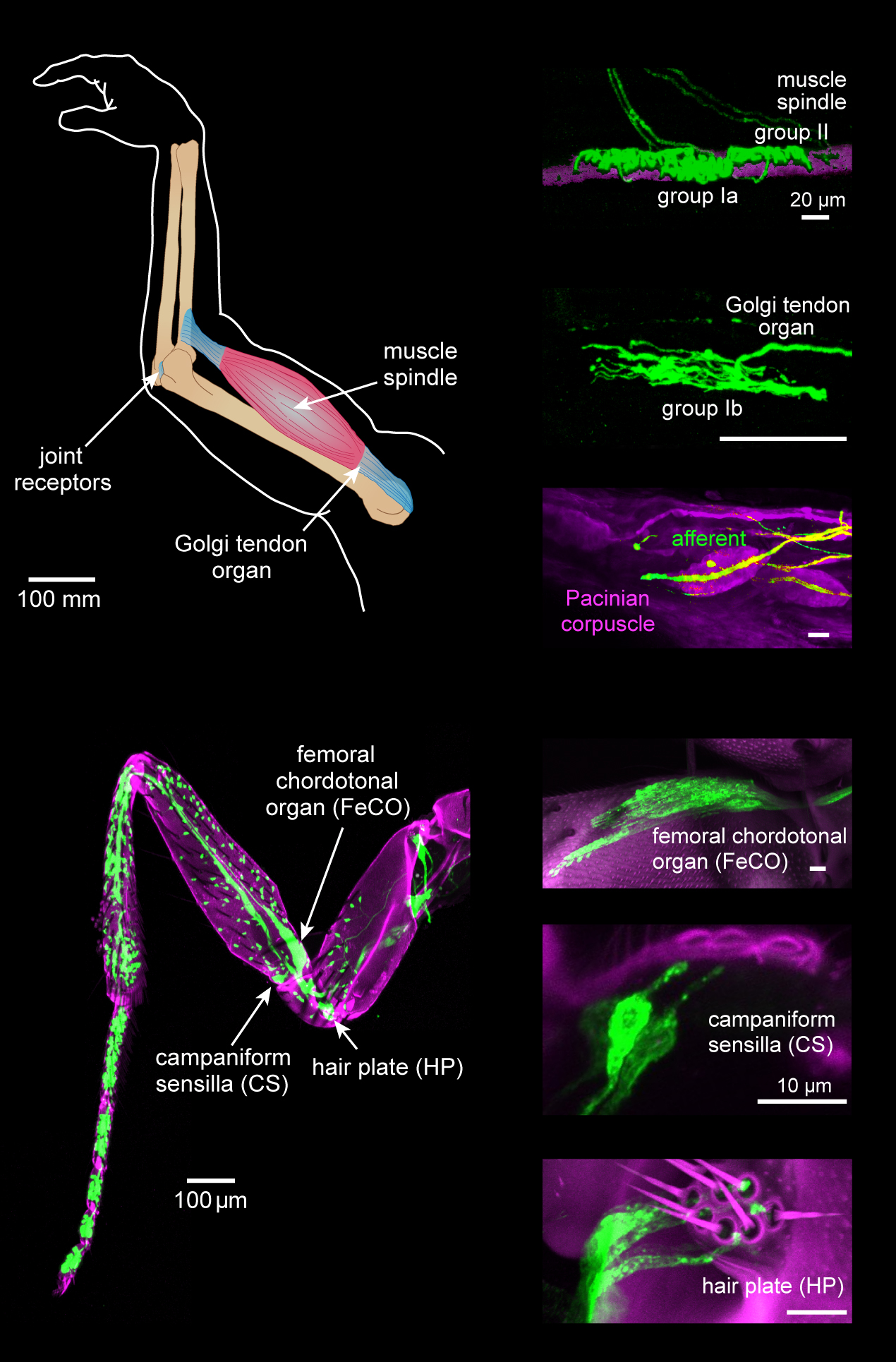
Proprioception ( ), also referred to as kinaesthesia (or kinesthesia), is the sense of self-movement, force, and body position. It is sometimes described as the "sixth sense". Proprioception is mediated by proprioceptors, mechanosensory neurons located within muscles, tendons, and joints. Most animals possess multiple subtypes of proprioceptors, which detect distinct kinematic parameters, such as joint position, movement, and load. Although all mobile animals possess proprioceptors, the structure of the sensory organs can vary across species. Proprioceptive signals are transmitted to the central nervous system, where they are integrated with information from other sensory systems, such as the visual system and the vestibular system, to create an overall representation of body position, movement, and acceleration. In many animals, sensory feedback from proprioceptors is essential for stabilizing body posture and coordinating body movement. System overview In vertebrates, limb ve ... [...More Info...] [...Related Items...] OR: [Wikipedia] [Google] [Baidu] |
Anatomy Dog
Anatomy () is the branch of biology concerned with the study of the structure of organisms and their parts. Anatomy is a branch of natural science that deals with the structural organization of living things. It is an old science, having its beginnings in prehistoric times. Anatomy is inherently tied to developmental biology, embryology, comparative anatomy, evolutionary biology, and phylogeny, as these are the processes by which anatomy is generated, both over immediate and long-term timescales. Anatomy and physiology, which study the structure and function of organisms and their parts respectively, make a natural pair of related disciplines, and are often studied together. Human anatomy is one of the essential basic sciences that are applied in medicine. The discipline of anatomy is divided into macroscopic and microscopic. Macroscopic anatomy, or gross anatomy, is the examination of an animal's body parts using unaided eyesight. Gross anatomy also includes the branch of s ... [...More Info...] [...Related Items...] OR: [Wikipedia] [Google] [Baidu] |
Sesamoid
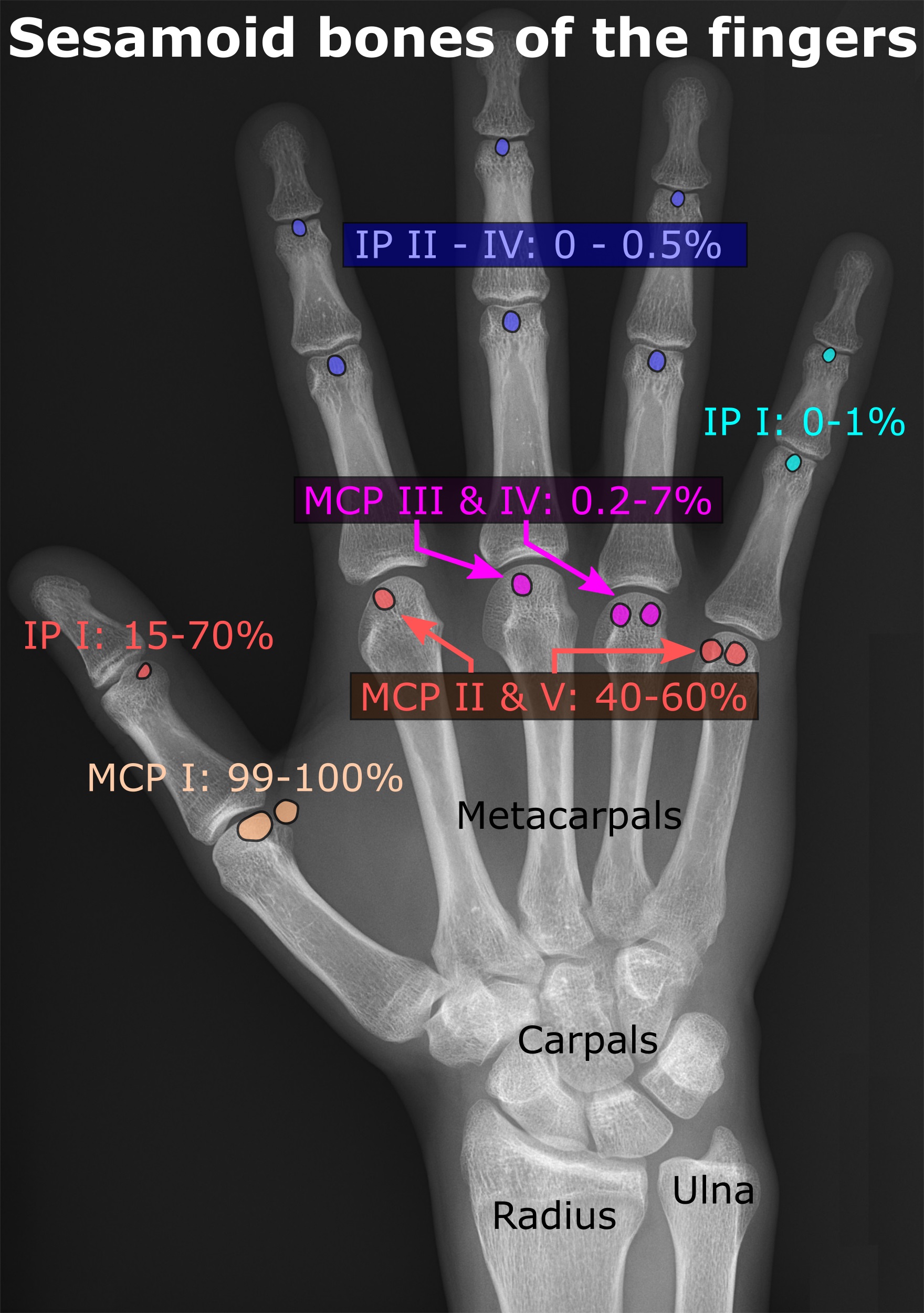
In anatomy, a sesamoid bone () is a bone embedded within a tendon or a muscle. Its name is derived from the Arabic word for 'sesame seed', indicating the small size of most sesamoids. Often, these bones form in response to strain, or can be present as a normal variant. The patella is the largest sesamoid bone in the body. Sesamoids act like pulleys, providing a smooth surface for tendons to slide over, increasing the tendon's ability to transmit muscular forces. Structure Sesamoid bones can be found on joints throughout the body, including: * In the knee—the patella (within the quadriceps tendon). This is the largest sesamoid bone. * In the hand—two sesamoid bones are commonly found in the distal portions of the first metacarpal bone (within the tendons of adductor pollicis and flexor pollicis brevis). There is also commonly a sesamoid bone in distal portions of the second metacarpal bone. * In the wrist—The pisiform of the wrist is a sesamoid bone (within the tendon o ... [...More Info...] [...Related Items...] OR: [Wikipedia] [Google] [Baidu] |





.jpg)
