|
Sternal Glands
The parasternal lymph nodes (or sternal glands) are placed at the anterior ends of the intercostal spaces, by the side of the internal thoracic artery. They derive afferents from the mamma; from the deeper structures of the anterior abdominal wall above the level of the umbilicus; from the upper surface of the liver through a small group of glands which lie behind the xiphoid process; and from the deeper parts of the anterior portion of the thoracic wall. Their efferents usually unite to form a single trunk on either side; this may open directly into the junction of the internal jugular and subclavian veins, or that of the right side may join the right subclavian trunk, and that of the left the thoracic duct In human anatomy, the thoracic duct is the larger of the two lymph ducts of the lymphatic system. It is also known as the ''left lymphatic duct'', ''alimentary duct'', ''chyliferous duct'', and ''Van Hoorne's canal''. The other duct is the right .... The parasternal l ... [...More Info...] [...Related Items...] OR: [Wikipedia] [Google] [Baidu] |
Bronchomediastinal Trunk
The efferent vessels of the tracheobronchial lymph nodes ascend upon the trachea and unite with efferents of the internal mammary and anterior mediastinal glands to form the right and left bronchomediastinal trunks. The right bronchomediastinal trunk may join the right lymphatic duct, and the left thoracic duct. More frequently, they open independently of these ducts into the junction of the internal jugular and subclavian vein The subclavian vein is a paired large vein, one on either side of the body, that is responsible for draining blood from the upper extremities, allowing this blood to return to the heart. The left subclavian vein plays a key role in the absorption ...s of their own side. References External links * https://web.archive.org/web/20071113205446/http://www.instantanatomy.net/thorax/vessels/lrightlymphatictrunk.html Lymphatics of the torso {{lymphatic-stub ... [...More Info...] [...Related Items...] OR: [Wikipedia] [Google] [Baidu] |
Intercostal Space
The intercostal space (ICS) is the anatomic space between two ribs (Lat. costa). Since there are 12 ribs on each side, there are 11 intercostal spaces, each numbered for the rib superior to it. Structures in intercostal space * several kinds of intercostal muscle * intercostal arteries and intercostal veins * intercostal lymph nodes * intercostal nerves Order of components Muscles There are 3 muscular layers in each intercostal space, consisting of the external intercostal muscle, the internal intercostal muscle, and the thinner innermost intercostal muscle. These muscles help to move the ribs during breathing. Neurovascular bundles Neurovascular bundles are located between the internal intercostal muscle and the innermost intercostal muscle. The neurovascular bundle has a strict order of vein-artery- nerve (VAN), from top to bottom. This neurovascular bundle runs high in the intercostal space, and the smaller collateral neurovascular bundle runs just superior ... [...More Info...] [...Related Items...] OR: [Wikipedia] [Google] [Baidu] |
Internal Thoracic Artery
In human anatomy, the internal thoracic artery (ITA), previously commonly known as the internal mammary artery (a name still common among surgeons), is an artery that supplies the anterior chest wall and the breasts. It is a paired artery, with one running along each side of the sternum, to continue after its bifurcation as the superior epigastric and musculophrenic arteries. Structure The internal thoracic artery arises from the anterior surface of the subclavian artery near its origin. It has a width of between 1-2 mm. It travels downward on the inside of the rib cage, approximately 1 cm from the sides of the sternum, and thus medial to the nipple. It is accompanied by the internal thoracic vein. It runs deep to the abdominal external oblique muscle, but superficial to the vagus nerve. In adults, internal thoracic artery lies closest to the sternum at the first intercoastal space. The gap between the artery and lateral border of the sternum increases when going downwards ... [...More Info...] [...Related Items...] OR: [Wikipedia] [Google] [Baidu] |
Mamma (anatomy)
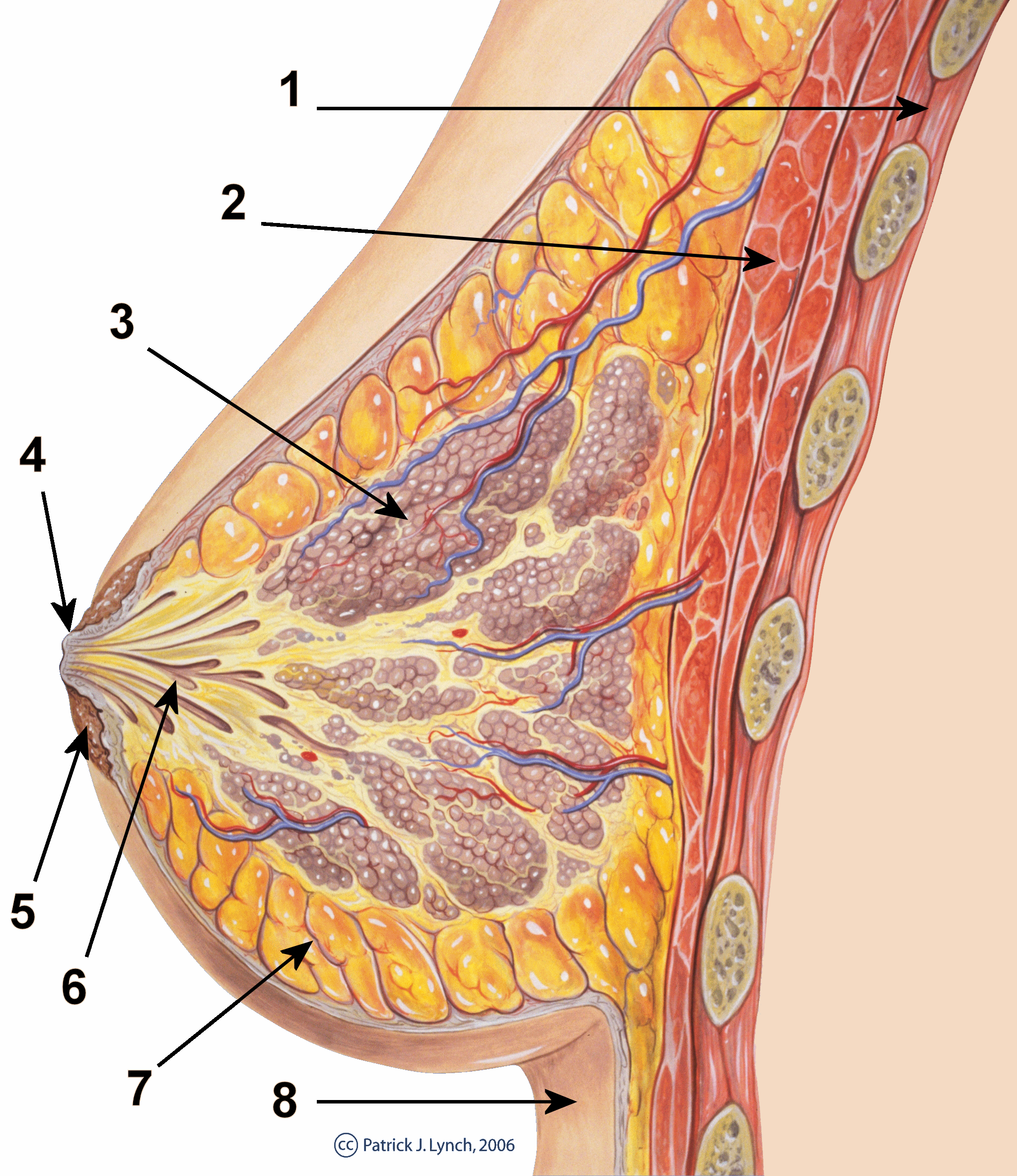
The breast is one of two prominences located on the upper ventral region of a primate's torso. Both females and males develop breasts from the same embryological tissues. In females, it serves as the mammary gland, which produces and secretes milk to feed infants. Subcutaneous fat covers and envelops a network of ducts that converge on the nipple, and these tissues give the breast its size and shape. At the ends of the ducts are lobules, or clusters of alveoli, where milk is produced and stored in response to hormonal signals. During pregnancy, the breast responds to a complex interaction of hormones, including estrogens, progesterone, and prolactin, that mediate the completion of its development, namely lobuloalveolar maturation, in preparation of lactation and breastfeeding. Humans are the only animals with permanent breasts. At puberty, estrogens, in conjunction with growth hormone, cause permanent breast growth in female humans. This happens only to a much lesser e ... [...More Info...] [...Related Items...] OR: [Wikipedia] [Google] [Baidu] |
Abdominal Wall
In anatomy, the abdominal wall represents the boundaries of the abdominal cavity. The abdominal wall is split into the anterolateral and posterior walls. There is a common set of layers covering and forming all the walls: the deepest being the visceral peritoneum, which covers many of the abdominal organs (most of the large and small intestines, for example), and the parietal peritoneum- which covers the visceral peritoneum below it, the extraperitoneal fat, the transversalis fascia, the internal and external oblique and transversus abdominis aponeurosis, and a layer of fascia, which has different names according to what it covers (e.g., transversalis, psoas fascia). In medical vernacular, the term 'abdominal wall' most commonly refers to the layers composing the anterior abdominal wall which, in addition to the layers mentioned above, includes the three layers of muscle: the transversus abdominis (transverse abdominal muscle), the internal (obliquus internus) and the external o ... [...More Info...] [...Related Items...] OR: [Wikipedia] [Google] [Baidu] |
Navel
The navel (clinically known as the umbilicus, commonly known as the belly button or tummy button) is a protruding, flat, or hollowed area on the abdomen at the attachment site of the umbilical cord. All placental mammals have a navel, although it is generally more conspicuous in humans. Structure The umbilicus is used to visually separate the abdomen into quadrants. The umbilicus is a prominent scar on the abdomen, with its position being relatively consistent among humans. The skin around the waist at the level of the umbilicus is supplied by the tenth thoracic spinal nerve (T10 dermatome). The umbilicus itself typically lies at a vertical level corresponding to the junction between the L3 and L4 vertebrae, with a normal variation among people between the L3 and L5 vertebrae. Parts of the adult navel include the "umbilical cord remnant" or "umbilical tip", which is the often protruding scar left by the detachment of the umbilical cord. This is located in the center of the ... [...More Info...] [...Related Items...] OR: [Wikipedia] [Google] [Baidu] |
Liver
The liver is a major Organ (anatomy), organ only found in vertebrates which performs many essential biological functions such as detoxification of the organism, and the Protein biosynthesis, synthesis of proteins and biochemicals necessary for digestion and growth. In humans, it is located in the quadrant (anatomy), right upper quadrant of the abdomen, below the thoracic diaphragm, diaphragm. Its other roles in metabolism include the regulation of Glycogen, glycogen storage, decomposition of red blood cells, and the production of hormones. The liver is an accessory digestive organ that produces bile, an alkaline fluid containing cholesterol and bile acids, which helps the fatty acid degradation, breakdown of fat. The gallbladder, a small pouch that sits just under the liver, stores bile produced by the liver which is later moved to the small intestine to complete digestion. The liver's highly specialized biological tissue, tissue, consisting mostly of hepatocytes, regulates a w ... [...More Info...] [...Related Items...] OR: [Wikipedia] [Google] [Baidu] |
Xiphoid Process
The xiphoid process , or xiphisternum or metasternum, is a small cartilaginous process (extension) of the inferior (lower) part of the sternum, which is usually ossified in the adult human. It may also be referred to as the ensiform process. Both the Greek-derived ''xiphoid'' and its Latin equivalent ''ensiform'' mean 'swordlike' or 'sword-shaped' Structure The xiphoid process is considered to be at the level of the 9th thoracic vertebra and the T7 dermatome. Development In newborns and young (especially small) infants, the tip of the xiphoid process may be both seen and felt as a lump just below the sternal notch. At 15 to 29 years old, the xiphoid usually fuses to the body of the sternum with a fibrous joint. Unlike the synovial articulation of major joints, this is non-movable. Ossification of the xiphoid process occurs around age 40. Variation The xiphoid process can be naturally bifurcated or sometimes perforated (xiphoidal foramen). These variances in morphology are inher ... [...More Info...] [...Related Items...] OR: [Wikipedia] [Google] [Baidu] |
Thoracic Wall
The thoracic wall or chest wall is the boundary of the thoracic cavity. Structure The bony skeletal part of the thoracic wall is the rib cage, and the rest is made up of muscle, skin, and fasciae. The chest wall has 10 layers, namely (from superficial to deep) skin (epidermis and dermis), superficial fascia, deep fascia and the invested extrinsic muscles (from the upper limbs), intrinsic muscles associated with the ribs (three layers of intercostal muscles), endothoracic fascia and parietal pleura. However, the extrinsic muscular layers vary according to the region of the chest wall. For example, the front and back sides may include attachments of large upper limb muscles like pectoralis major or latissimus dorsi, while the sides only have serratus anterior. Function Diving reflex When not breathing for long and dangerous periods of time in cold water, a person's body undergoes great temporary changes to try to prevent death. It achieves this through the activation of ... [...More Info...] [...Related Items...] OR: [Wikipedia] [Google] [Baidu] |
Internal Jugular Vein
The internal jugular vein is a paired jugular vein that collects blood from the brain and the superficial parts of the face and neck. This vein runs in the carotid sheath with the common carotid artery and vagus nerve. It begins in the posterior compartment of the jugular foramen, at the base of the skull. It is somewhat dilated at its origin, which is called the ''superior bulb''. This vein also has a common trunk into which drains the anterior branch of the retromandibular vein, the facial vein, and the lingual vein. It runs down the side of the neck in a vertical direction, being at one end lateral to the internal carotid artery, and then lateral to the common carotid artery, and at the root of the neck, it unites with the subclavian vein to form the brachiocephalic vein (innominate vein); a little above its termination is a second dilation, the ''inferior bulb''. Above, it lies upon the rectus capitis lateralis, behind the internal carotid artery and the nerves passing ... [...More Info...] [...Related Items...] OR: [Wikipedia] [Google] [Baidu] |
Subclavian Vein
The subclavian vein is a paired large vein, one on either side of the body, that is responsible for draining blood from the upper extremities, allowing this blood to return to the heart. The left subclavian vein plays a key role in the absorption of lipids, by allowing products that have been carried by lymph in the thoracic duct to enter the bloodstream. The diameter of the subclavian veins is approximately 1–2 cm, depending on the individual. Structure Each subclavian vein is a continuation of the axillary vein and runs from the outer border of the first rib to the medial border of anterior scalene muscle. From here it joins with the internal jugular vein to form the brachiocephalic vein (also known as "innominate vein"). The angle of union is termed the venous angle. The subclavian vein follows the subclavian artery and is separated from the subclavian artery by the insertion of anterior scalene. Thus, the subclavian vein lies anterior to the anterior scalene while the su ... [...More Info...] [...Related Items...] OR: [Wikipedia] [Google] [Baidu] |
Subclavian Trunk
The efferent vessels of the subclavicular group unite to form the subclavian trunk, which opens either directly into the junction of the internal jugular and subclavian veins or into the jugular lymphatic trunk; on the left side it may end in the thoracic duct In human anatomy, the thoracic duct is the larger of the two lymph ducts of the lymphatic system. It is also known as the ''left lymphatic duct'', ''alimentary duct'', ''chyliferous duct'', and ''Van Hoorne's canal''. The other duct is the right .... References External links * http://anatomy.uams.edu/anatomyhtml/lymph_thorax.html Lymphatics of the upper limb {{lymphatic-stub ... [...More Info...] [...Related Items...] OR: [Wikipedia] [Google] [Baidu] |


