|
Soleus
In humans and some other mammals, the soleus is a powerful muscle in the back part of the lower leg (the calf). It runs from just below the knee to the heel, and is involved in standing and walking. It is closely connected to the gastrocnemius muscle and some anatomists consider them to be a single muscle, the triceps surae. Its name is derived from the Latin word "solea", meaning "sandal". Structure The soleus is located in the superficial posterior compartment of the leg. The soleus exhibits significant morphological differences across species. It is unipennate in many species. In some animals, such as the rabbit, it is fused for much of its length with the gastrocnemius muscle. In humans, the soleus is a complex, multi-pennate muscle, usually having a separate (posterior) aponeurosis from the gastrocnemius muscle. A majority of soleus muscle fibers originate from each side of the anterior aponeurosis, attached to the tibia and fibula. Other fibers originate from the pos ... [...More Info...] [...Related Items...] OR: [Wikipedia] [Google] [Baidu] |
Triceps Surae Muscle
The triceps surae consists of two muscles located at the calf – the two-headed gastrocnemius and the soleus. These muscles both insert into the calcaneus, the bone of the heel of the human foot, and form the major part of the muscle of the posterior leg, commonly known as the calf muscle. Structure The triceps surae is connected to the foot through the Achilles tendon, and has 3 heads deriving from the 2 major masses of muscle. * The superficial portion (the gastrocnemius) gives off 2 heads attaching to the base of the femur directly above the knee. * The deep (profundus) mass of muscle (the soleus) forms the remaining head which attaches to the superior posterior area of the tibia. The triceps surae is innervated by the tibial nerve, specifically, nerve roots L5–S2. Function Contraction of the triceps surae induce plantar flexion (sagittal plane) and stabilization of the ankle complex in the transverse plane. Functional activities include primarily movement in the ... [...More Info...] [...Related Items...] OR: [Wikipedia] [Google] [Baidu] |
Achilles Tendon
The Achilles tendon or heel cord, also known as the calcaneal tendon, is a tendon at the back of the lower leg, and is the thickest in the human body. It serves to attach the plantaris, gastrocnemius (calf) and soleus muscles to the calcaneus (heel) bone. These muscles, acting via the tendon, cause plantar flexion of the foot at the ankle joint, and (except the soleus) flexion at the knee. Abnormalities of the Achilles tendon include inflammation ( Achilles tendinitis), degeneration, rupture, and becoming embedded with cholesterol deposits ( xanthomas). The Achilles tendon was named in 1693 after the Greek hero Achilles. History The oldest-known written record of the tendon being named for Achilles is in 1693 by the Flemish/Dutch anatomist Philip Verheyen. In his widely used text he described the tendon's location and said that it was commonly called "the cord of Achilles." The tendon has been described as early as the time of Hippocrates, who described it as the ... [...More Info...] [...Related Items...] OR: [Wikipedia] [Google] [Baidu] |
Gastrocnemius Muscle
The gastrocnemius muscle (plural ''gastrocnemii'') is a superficial two-headed muscle that is in the back part of the lower leg of humans. It runs from its two heads just above the knee to the heel, a three joint muscle (knee, ankle and subtalar joints). The muscle is named via Latin, from Greek γαστήρ (''gaster'') 'belly' or 'stomach' and κνήμη (''knḗmē'') 'leg', meaning 'stomach of the leg' (referring to the bulging shape of the calf). Structure The gastrocnemius is located with the soleus in the posterior (back) compartment of the leg. The lateral head originates from the lateral condyle of the femur, while the medial head originates from the medial condyle of the femur. Its other end forms a common tendon with the soleus muscle; this tendon is known as the calcaneal tendon or Achilles tendon and inserts onto the posterior surface of the calcaneus, or heel bone. It is considered a superficial muscle as it is located directly under skin, and its shape may o ... [...More Info...] [...Related Items...] OR: [Wikipedia] [Google] [Baidu] |
Plantaris Muscle
The plantaris is one of the superficial muscles of the superficial posterior compartment of the leg, one of the fascial compartments of the leg. It is composed of a thin muscle belly and a long thin tendon. While not as thick as the achilles tendon, the plantaris tendon (which tends to be between in length) is the longest tendon in the human body. Not including the tendon, the plantaris muscle is approximately long and is absent in 8-12% of the population. It is one of the plantar flexors in the posterior compartment of the leg, along with the gastrocnemius and soleus muscles. The plantaris is considered to have become an unimportant muscle when human ancestors switched from climbing trees to bipedalism and in anatomically modern humans it mainly acts with the gastrocnemius. Structure The plantaris muscle arises from the inferior part of the lateral supracondylar ridge of the femur at a position slightly superior to the origin of the lateral head of gastrocnemius. It passe ... [...More Info...] [...Related Items...] OR: [Wikipedia] [Google] [Baidu] |
Tibial Nerve
The tibial nerve is a branch of the sciatic nerve. The tibial nerve passes through the popliteal fossa to pass below the arch of soleus. Structure Popliteal fossa The tibial nerve is the larger terminal branch of the sciatic nerve with root values of L4, L5, S1, S2, and S3. It lies superficial (or posterior) to the popliteal vessels, extending from the superior angle to the inferior angle of the popliteal fossa, crossing the popliteal vessels from lateral to medial side. It gives off branches as shown below: * Muscular branches - Muscular branches arise from the distal part of the popliteal fossa. It supplies the medial and lateral heads of gastrocnemius, soleus, plantaris and popliteus muscles. Nerve to popliteus crosses the popliteus muscle, runs downwards and laterally, winds around the lower border of the popliteus to supply the deep (or anterior) surface of the popliteus. This nerve also supplies the tibialis posterior muscle, superior tibiofibular joint, tibia bone, in ... [...More Info...] [...Related Items...] OR: [Wikipedia] [Google] [Baidu] |
Fibula
The fibula or calf bone is a human leg, leg bone on the Lateral (anatomy), lateral side of the tibia, to which it is connected above and below. It is the smaller of the two bones and, in proportion to its length, the most slender of all the long bones. Its upper extremity is small, placed toward the back of the Upper extremity of tibia, head of the tibia, below the knee, knee joint and excluded from the formation of this joint. Its lower extremity inclines a little forward, so as to be on a plane anterior to that of the upper end; it projects below the tibia and forms the lateral part of the ankle, ankle joint. Structure The bone has the following components: * Lateral malleolus * Interosseous membrane connecting the fibula to the tibia, forming a syndesmosis joint * The superior tibiofibular articulation is an arthrodial joint between the lateral condyle of tibia, lateral condyle of the tibia and the head of the fibula. * The inferior tibiofibular articulation (tibiofibular synd ... [...More Info...] [...Related Items...] OR: [Wikipedia] [Google] [Baidu] |
Heel
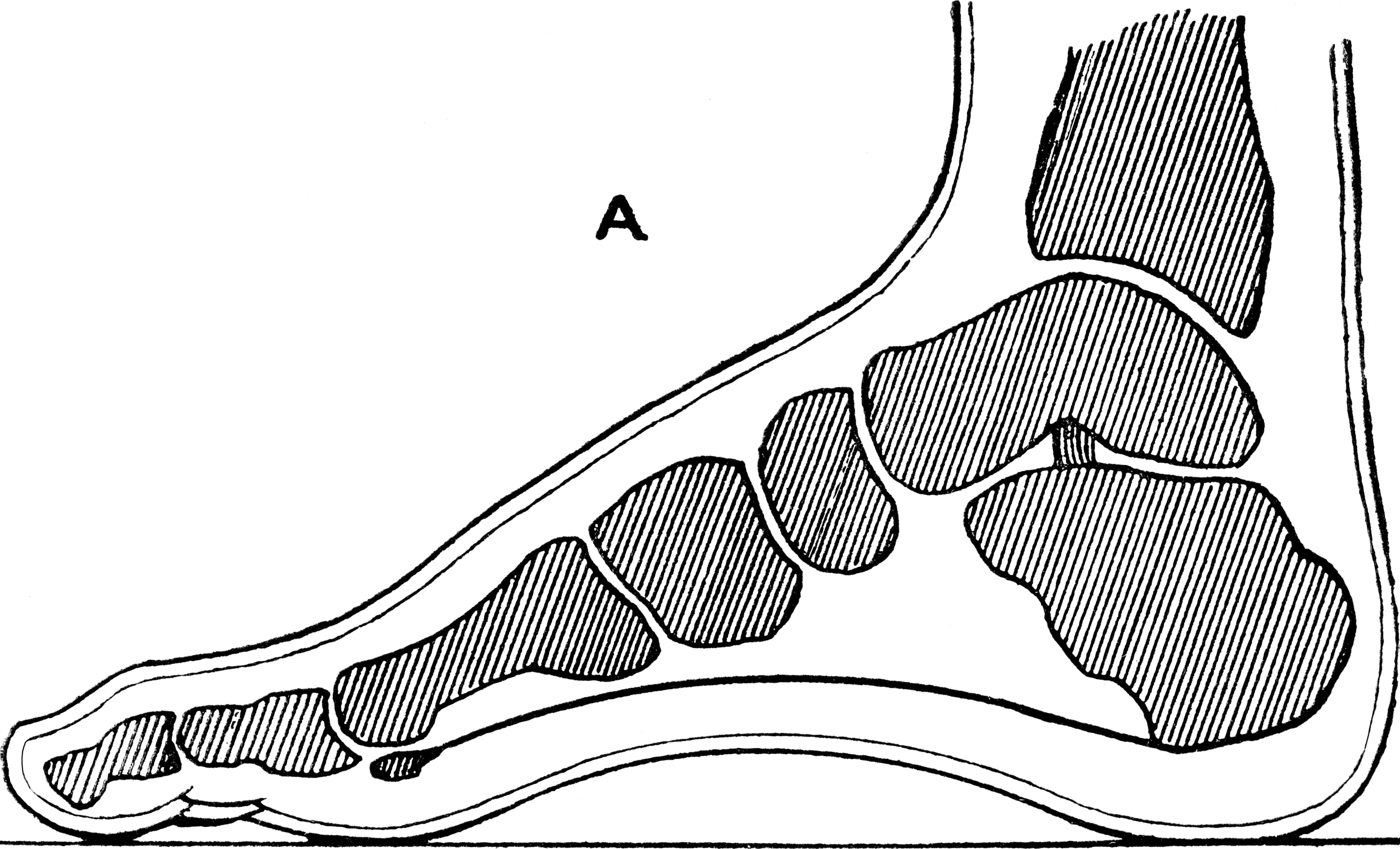
The heel is the prominence at the posterior end of the foot. It is based on the projection of one bone, the calcaneus or heel bone, behind the articulation of the bones of the lower leg. Structure To distribute the compressive forces exerted on the heel during gait, and especially the stance phase when the heel contacts the ground, the sole of the foot is covered by a layer of subcutaneous connective tissue up to 2 cm thick (under the heel). This tissue has a system of pressure chambers that both acts as a shock absorber and stabilises the sole. Each of these chambers contains fibrofatty tissue covered by a layer of tough connective tissue made of collagen fibers. These septa ("walls") are firmly attached both to the plantar aponeurosis above and the sole's skin below. The sole of the foot is one of the most highly vascularized regions of the body surface, and the dense system of blood vessels further stabilize the septa. The Achilles tendon is the muscle tendon of t ... [...More Info...] [...Related Items...] OR: [Wikipedia] [Google] [Baidu] |
Tibia
The tibia (; ), also known as the shinbone or shankbone, is the larger, stronger, and anterior (frontal) of the two bones in the leg below the knee in vertebrates (the other being the fibula, behind and to the outside of the tibia); it connects the knee with the ankle. The tibia is found on the medial side of the leg next to the fibula and closer to the median plane. The tibia is connected to the fibula by the interosseous membrane of leg, forming a type of fibrous joint called a syndesmosis with very little movement. The tibia is named for the flute '' tibia''. It is the second largest bone in the human body, after the femur. The leg bones are the strongest long bones as they support the rest of the body. Structure In human anatomy, the tibia is the second largest bone next to the femur. As in other vertebrates the tibia is one of two bones in the lower leg, the other being the fibula, and is a component of the knee and ankle joints. The ossification or formation of the bo ... [...More Info...] [...Related Items...] OR: [Wikipedia] [Google] [Baidu] |
Soleal Line
The soleal line, also known as the popliteal line (in older texts), is a prominent ridge on the posterior surface of the tibia. It is the site of many muscle origins and insertions, such as those of popliteus muscle, soleus muscle, flexor digitorum longus muscle, and tibialis posterior muscle. Structure The soleal line is a prominent ridge on the posterior surface of the tibia. It extends obliquely downward from the back part of the articular facet for the fibula to the medial border, at the junction of its upper and middle thirds. Development The soleal line becomes more prominent between childhood and adulthood. It is rarely seen in children between the ages of 6 and 8. Function The soleal line marks the lower limit of the insertion of the popliteus muscle. It is the attachment of the fascia covering this muscle. It is the origin of part of soleus muscle (along with a triangular area above it), flexor digitorum longus muscle, and tibialis posterior muscle The tibialis ... [...More Info...] [...Related Items...] OR: [Wikipedia] [Google] [Baidu] |
Popliteal Artery
The popliteal artery is a deeply placed continuation of the femoral artery opening in the distal portion of the adductor magnus muscle. It courses through the popliteal fossa and ends at the lower border of the popliteus muscle, where it branches into the anterior and posterior tibial arteries. The deepest (most anterior) structure in the fossa, the popliteal artery runs close to the joint capsule of the knee as it spans the intercondylar fossa. Five genicular branches of the popliteal artery supply the capsule and ligaments of the knee joint. The genicular arteries are the superior lateral, superior medial, middle, inferior lateral, and inferior medial genicular arteries. They participate in the formation of the periarticular genicular anastomosis, a network of vessels surrounding the knee that provides collateral circulation capable of maintaining blood supply to the leg during full knee flexion, which may kink the popliteal artery. Structure The popliteal artery is the contin ... [...More Info...] [...Related Items...] OR: [Wikipedia] [Google] [Baidu] |
Calf (anatomy)
The calf ( : calves; Latin: ''sura'') is the back portion of the lower leg in human anatomy. The muscles within the calf correspond to the posterior compartment of the leg. The two largest muscles within this compartment are known together as the calf muscle and attach to the heel via the Achilles tendon. Several other, smaller muscles attach to the knee, the ankle, and via long tendons to the toes. Etymology From Middle English ''calf'', ''kalf'', from Old Norse ''kalfi'', possibly derived from the same Germanic root as English ''calf'' ("young cow"). Cognate with Icelandic ''kálfi'' ("calf of the leg"). ''Calf'' and ''calf of the leg'' are documented in use in Middle English circa AD 1350 and AD 1425 respectively. Historically, the ''absence of calf'', meaning a lower leg without a prominent calf muscle, was regarded by some authors as a sign of inferiority: ''it is well known that monkeys have no calves, and still less do they exist among the lower orders of mammals''. ... [...More Info...] [...Related Items...] OR: [Wikipedia] [Google] [Baidu] |


