|
Shoulder Dystocia
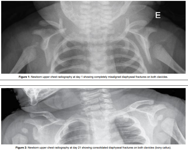
Shoulder dystocia is when, after vaginal delivery of the head, the baby's anterior shoulder gets caught above the mother's pubic bone. Signs include retraction of the baby's head back into the vagina, known as "turtle sign". Complications for the baby may include brachial plexus injury, or clavicle fracture. Complications for the mother may include vaginal or perineal tears, postpartum bleeding, or uterine rupture. Risk factors include gestational diabetes, previous history of the condition, operative vaginal delivery, obesity in the mother, an overly large baby, and epidural anesthesia. It is diagnosed when the body fails to deliver within one minute of delivery of the baby's head. It is a type of obstructed labour. Shoulder dystocia is an obstetric emergency. Initial efforts to release a shoulder typically include: with a woman on her back pushing the legs outward and upward, pushing on the abdomen above the pubic bone, and making a cut in the vagina. If these are not effec ... [...More Info...] [...Related Items...] OR: [Wikipedia] [Google] [Baidu] |
Suprapubic
The hypogastrium (also called the hypogastric region or suprapubic region) is a region of the abdomen located below the umbilical region. Etymology The roots of the word ''hypogastrium'' mean "below the stomach"; the roots of ''suprapubic'' mean "above the pubic bone". Boundaries The upper limit is the umbilicus while the pubis bone constitutes its lower limit. The lateral boundaries are formed are drawing straight lines through the midway between the anterior superior iliac spine and symphisis pubis The pubic symphysis is a secondary cartilaginous joint between the left and right superior rami of the pubis of the hip bones. It is in front of and below the urinary bladder. In males, the suspensory ligament of the penis attaches to the pubic .... References External links * Abdomen {{Anatomy-stub ... [...More Info...] [...Related Items...] OR: [Wikipedia] [Google] [Baidu] |
Cesarean Section
Caesarean section, also known as C-section or caesarean delivery, is the surgical procedure by which one or more babies are delivered through an incision in the mother's abdomen, often performed because vaginal delivery would put the baby or mother at risk. Reasons for the operation include obstructed labor, twin pregnancy, high blood pressure in the mother, breech birth, and problems with the placenta or umbilical cord. A caesarean delivery may be performed based upon the shape of the mother's pelvis or history of a previous C-section. A trial of vaginal birth after C-section may be possible. The World Health Organization recommends that caesarean section be performed only when medically necessary. Most C-sections are performed without a medical reason, upon request by someone, usually the mother. A C-section typically takes 45 minutes to an hour. It may be done with a spinal block, where the woman is awake, or under general anesthesia. A urinary catheter is used to drain t ... [...More Info...] [...Related Items...] OR: [Wikipedia] [Google] [Baidu] |
Intrauterine Hypoxia
Intrauterine hypoxia (also known as fetal hypoxia) occurs when the fetus is deprived of an adequate supply of oxygen. It may be due to a variety of reasons such as prolapse or occlusion of the umbilical cord, placental infarction, maternal diabetes (prepregnancy or gestational diabetes) and maternal smoking. Intrauterine growth restriction may cause or be the result of hypoxia. Intrauterine hypoxia can cause cellular damage that occurs within the central nervous system (the brain and spinal cord). This results in an increased mortality rate, including an increased risk of sudden infant death syndrome (SIDS). Oxygen deprivation in the fetus and neonate have been implicated as either a primary or as a contributing risk factor in numerous neurological and neuropsychiatric disorders such as epilepsy, attention deficit hyperactivity disorder, eating disorders and cerebral palsy. Presentation Maternal Consequences Complications arising from intrauterine hypoxia are some of the leadin ... [...More Info...] [...Related Items...] OR: [Wikipedia] [Google] [Baidu] |
Erb's Palsy
Erb's palsy is a paralysis of the arm caused by injury to the upper group of the arm's main nerves, specifically the severing of the upper trunk C5–C6 nerves. These form part of the brachial plexus, comprising the ventral rami of spinal nerves C5–C8 and thoracic nerve T1. pp.1037–1047 pp.370–374 pp.76–77 These injuries arise most commonly, but not exclusively, from shoulder dystocia during a difficult birth.A.D.A.M Healthcare center Depending on the nature of the damage, the paralysis can either resolve on its own over a period of months, necessitate rehabilitative therapy, or require surgery. Presentation The paralysis can be partial or complete; the damage to each nerve can range from bruising to tearing. The most commonly involved root is C5 (aka[...More Info...] [...Related Items...] OR: [Wikipedia] [Google] [Baidu] |
Klumpke Paralysis
Klumpke's paralysis is a variety of partial palsy of the lower roots of the brachial plexus. p.1046 The brachial plexus is a network of spinal nerves that originates in the back of the neck, extends through the axilla (armpit), and gives rise to nerves to the upper limb.Warwick, R., & Williams, P.L. (1973). pp.1037-1047 pp.370-374 pp.76-77Shenaq S.M., & Spiegel A.J. Hand, Brachial Plexus Surgery. eMedicine.com. URLhttp://www.emedicine.com/plastic/topic450.htm Accessed on: April 13, 2007. The paralytic condition is named after Augusta Déjerine-Klumpke. Signs and symptoms Symptoms include intrinsic minus hand deformity, paralysis of intrinsic hand muscles, and C8/T1 Dermatome distribution numbness. Involvement of T1 may result in Horner's syndrome, with ptosis, and miosis. Weakness or lack of ability to use specific muscles of the shoulder or arm. pp.576, 667 It can be contrasted to Erb-Duchenne's palsy, which affects C5 and C6. Cause Klumpke's paralysis is a form of paralysi ... [...More Info...] [...Related Items...] OR: [Wikipedia] [Google] [Baidu] |
Ventral Root Of Spinal Nerve
In anatomy and neurology, the ventral root of spinal nerve, anterior root, or motor root is the efferent motor root of a spinal nerve. At its distal end, the ventral root joins with the dorsal root The dorsal root of spinal nerve (or posterior root of spinal nerve or sensory root) is one of two "roots" which emerge from the spinal cord. It emerges directly from the spinal cord, and travels to the dorsal root ganglion. Nerve fibres with the v ... to form a mixed spinal nerve. Additional images Image:Cervical vertebra english.png, Cervical vertebra Image:Medulla spinalis - Section - English.svg, Medulla spinalis Image:Gray675.png, A spinal nerve with its anterior and posterior. Image:Gray764.png, The motor tract. Image:Gray770-en.svg, Diagrammatic transverse section of the medulla spinalis and its membranes. Image:Gray796.png, A portion of the spinal cord, showing its right lateral surface. The dura is opened and arranged to show the nerve roots. Image:Gray799.svg, Sch ... [...More Info...] [...Related Items...] OR: [Wikipedia] [Google] [Baidu] |
Pelvis
The pelvis (plural pelves or pelvises) is the lower part of the trunk, between the abdomen and the thighs (sometimes also called pelvic region), together with its embedded skeleton (sometimes also called bony pelvis, or pelvic skeleton). The pelvic region of the trunk includes the bony pelvis, the pelvic cavity (the space enclosed by the bony pelvis), the pelvic floor, below the pelvic cavity, and the perineum, below the pelvic floor. The pelvic skeleton is formed in the area of the back, by the sacrum and the coccyx and anteriorly and to the left and right sides, by a pair of hip bones. The two hip bones connect the spine with the lower limbs. They are attached to the sacrum posteriorly, connected to each other anteriorly, and joined with the two femurs at the hip joints. The gap enclosed by the bony pelvis, called the pelvic cavity, is the section of the body underneath the abdomen and mainly consists of the reproductive organs (sex organs) and the rectum, while the pelvic f ... [...More Info...] [...Related Items...] OR: [Wikipedia] [Google] [Baidu] |
Obstructed Labour
Obstructed labour, also known as labour dystocia, is the baby not exiting the pelvis because it is physically block during childbirth although the uterus contracts normally. Complications for the baby include not getting enough oxygen which may result in death. It increases the risk of the mother getting an infection, having uterine rupture, or having post-partum bleeding. Long-term complications for the mother include obstetrical fistula. Obstructed labour is said to result in prolonged labour, when the active phase of labour is longer than 12 hours. The main causes of obstructed labour include a large or abnormally positioned baby, a small pelvis, and problems with the birth canal. Abnormal positioning includes shoulder dystocia where the anterior shoulder does not pass easily below the pubic bone. Risk factors for a small pelvis include malnutrition and a lack of exposure to sunlight causing vitamin D deficiency. It is also more common in adolescence as the pelvis may not ha ... [...More Info...] [...Related Items...] OR: [Wikipedia] [Google] [Baidu] |
Gestational Diabetes
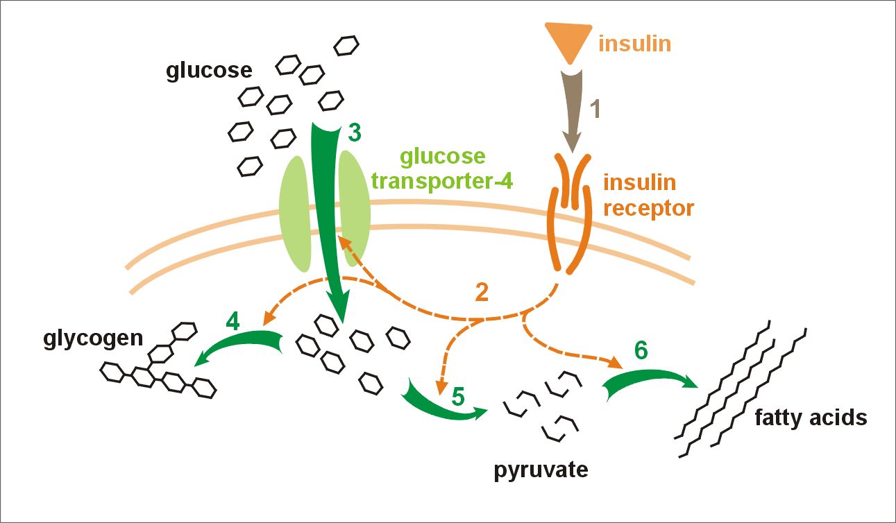
Gestational diabetes is a condition in which a woman without diabetes develops high blood sugar levels during pregnancy. Gestational diabetes generally results in few symptoms; however, it increases the risk of pre-eclampsia, depression, and of needing a Caesarean section. Babies born to mothers with poorly treated gestational diabetes are at increased risk of macrosomia, of having hypoglycemia after birth, and of jaundice. If untreated, diabetes can also result in stillbirth. Long term, children are at higher risk of being overweight and of developing type 2 diabetes. Gestational diabetes can occur during pregnancy because of insulin resistance or reduced production of insulin. Risk factors include being overweight, previously having gestational diabetes, a family history of type 2 diabetes, and having polycystic ovarian syndrome. Diagnosis is by blood tests. For those at normal risk, screening is recommended between 24 and 28 weeks' gestation. For those at high risk, testin ... [...More Info...] [...Related Items...] OR: [Wikipedia] [Google] [Baidu] |
Uterine Rupture
Uterine rupture is when the muscular wall of the uterus tears during pregnancy or childbirth. Symptoms, while classically including increased pain, vaginal bleeding, or a change in contractions, are not always present. Disability or death of the mother or baby may result. Risk factors include vaginal birth after cesarean section (VBAC), other uterine scars, obstructed labor, induction of labor, trauma, and cocaine use. While typically rupture occurs during labor it may occasionally happen earlier in pregnancy. Diagnosis may be suspected based on a rapid drop in the baby's heart rate during labor. Uterine dehiscence is a less severe condition in which there is only incomplete separation of the old scar. Treatment involves rapid surgery to control bleeding and delivery of the baby. A hysterectomy may be required to control the bleeding. Blood transfusions may be given to replace blood loss. Women who have had a prior rupture are generally recommended to have C-sections in subs ... [...More Info...] [...Related Items...] OR: [Wikipedia] [Google] [Baidu] |
Brachial Plexus Injury
A brachial plexus injury (BPI), also known as brachial plexus lesion, is an injury to the brachial plexus, the network of nerves that conducts signals from the spinal cord to the shoulder, arm and hand. These nerves originate in the fifth, sixth, seventh and eighth cervical (C5–C8), and first thoracic (T1) spinal nerves, and innervate the muscles and skin of the chest, shoulder, arm and hand. Brachial plexus injuries can occur as a result of shoulder trauma, tumours, or inflammation, or obstetric. Obstetric injuries may occur from mechanical injury involving shoulder dystocia during difficult childbirth, with a prevalence of 1 in 1000 births. "The brachial plexus may be injured by falls from a height on to the side of the head and shoulder, whereby the nerves of the plexus are violently stretched. The brachial plexus may also be injured by direct violence or gunshot wounds, by violent traction on the arm, or by efforts at reducing a dislocation of the shoulder joint". The rare ... [...More Info...] [...Related Items...] OR: [Wikipedia] [Google] [Baidu] |


