|
Scintimammography
Scintimammography is a type of breast imaging test that is used to detect cancer cells in the breasts of some women who have had abnormal mammograms, or for those who have dense breast tissue, post-operative scar tissue or breast implants. Scintimammography is not used for screening or in place of a mammogram. Rather, it is used when the detection of breast abnormalities is not possible or not reliable on the basis of mammography and ultrasound. Procedure In the scintimammography procedure, a woman receives an injection of a small amount of a radioactive substance which is taken up by cancer cells, and a gamma camera is used to take pictures of the breasts. Technetium-99m sestamibi (99mTc-MIBI) is the most commonly used radiopharmaceutical. When performed with MIBI the procedure may also be known as a Miraluma test. The typical dose is 740–1,110 megabecquerels (MBq) with the patient lying prone on the bed for imaging approximately 10 minutes after injection. 99mTc-DMSA and 9 ... [...More Info...] [...Related Items...] OR: [Wikipedia] [Google] [Baidu] |
Breast Imaging
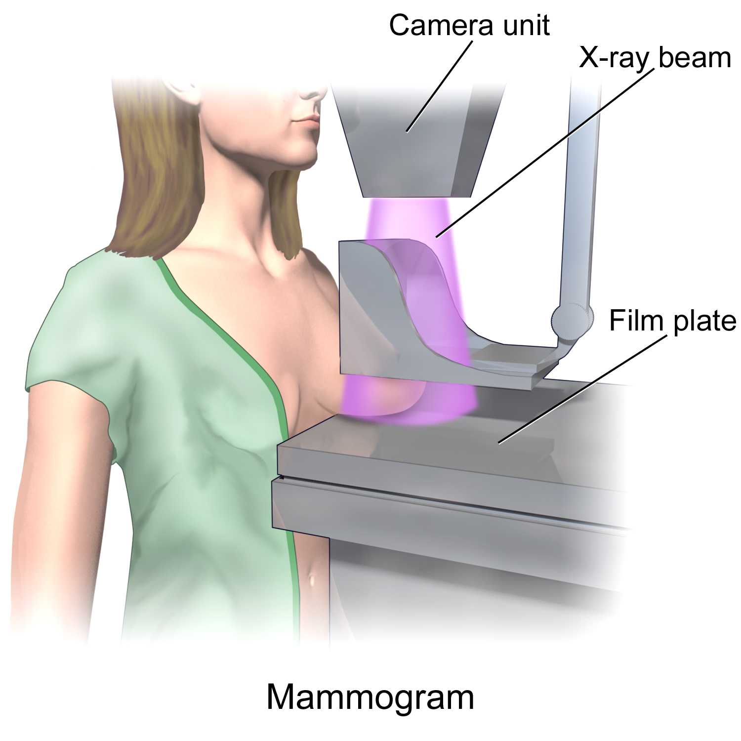
In medicine, breast imaging is a sub-speciality of diagnostic radiology that involves imaging of the breasts for screening or diagnostic purposes. There are various methods of breast imaging using a variety of technologies as described in detail below. Traditional screening and diagnostic mammography (“2D mammography”) uses x-ray technology and has been the mainstay of breast imaging for many decades. Breast tomosynthesis (“3D mammography”) is a relatively new digital x-ray mammography technique that produces multiple image slices of the breast similar to, but distinct from, CT scan, Computed Tomography (CT). Xeromammography and Galactography are somewhat outdated technologies that also use x-ray technology and are now used infrequently in the detection of breast cancer. Breast ultrasound is another technology employed in diagnosis and screening that can help differentiate between fluid filled and solid lesions, an important factor to determine if a lesion may be cancerous. ... [...More Info...] [...Related Items...] OR: [Wikipedia] [Google] [Baidu] |
Breast Cancer Screening
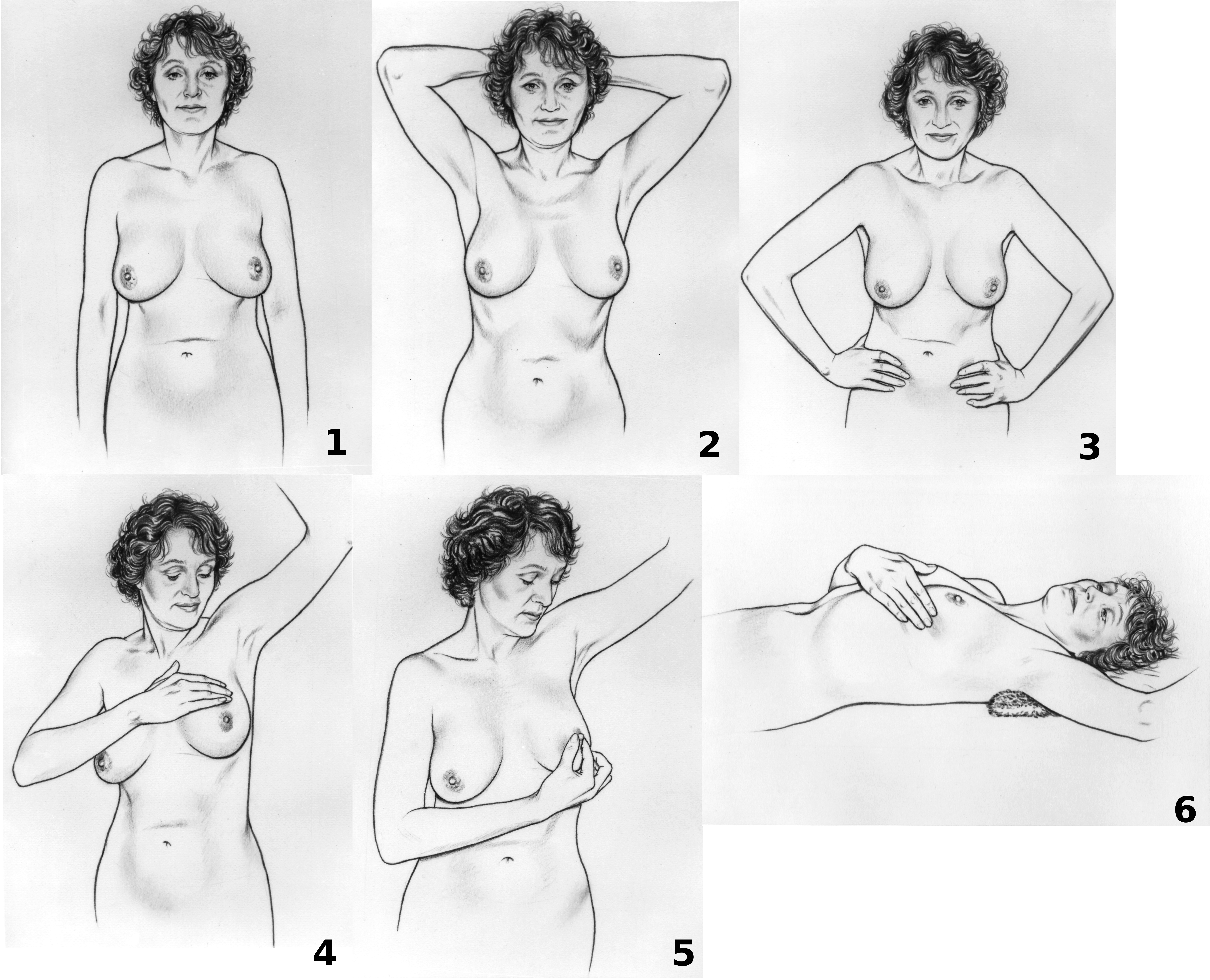
Breast cancer screening is the medical screening of asymptomatic, apparently healthy women for breast cancer in an attempt to achieve an earlier diagnosis. The assumption is that early detection will improve outcomes. A number of screening tests have been employed, including clinical and self breast exams, mammography, genetic screening, ultrasound, and magnetic resonance imaging. A clinical or self breast exam involves feeling the breast for breast lump, lumps or other abnormalities. Medical evidence, however, does not support its use in women with a typical risk for breast cancer. Universal screening with mammography is controversial as it may not reduce all-cause mortality and may cause harms through unnecessary treatments and medical procedures. Many national organizations recommend it for most older women. The United States Preventive Services Task Force recommends screening mammography in women at normal risk for breast cancer, every two years between the ages of 50 and 74 ... [...More Info...] [...Related Items...] OR: [Wikipedia] [Google] [Baidu] |
Megabecquerel
The becquerel (; symbol: Bq) is the unit of radioactivity in the International System of Units (SI). One becquerel is defined as the activity of a quantity of radioactive material in which one nucleus decays per second. For applications relating to human health this is a small quantity, and SI multiples of the unit are commonly used. The becquerel is named after Henri Becquerel, who shared a Nobel Prize in Physics with Pierre and Marie Skłodowska Curie in 1903 for their work in discovering radioactivity. Definition 1 Bq = 1 s−1 A special name was introduced for the reciprocal second (s−1) to represent radioactivity to avoid potentially dangerous mistakes with prefixes. For example, 1 µs−1 would mean 106 disintegrations per second: 1·(10−6 s)−1 = 106 s−1, whereas 1 µBq would mean 1 disintegration per 1 million seconds. Other names considered were hertz (Hz), a special name already in use for the reciprocal second, and Fourier ... [...More Info...] [...Related Items...] OR: [Wikipedia] [Google] [Baidu] |
2d Nuclear Medical Imaging
D, or d, is the fourth letter in the Latin alphabet, used in the modern English alphabet, the alphabets of other western European languages and others worldwide. Its name in English is ''dee'' (pronounced ), plural ''dees''. History The Semitic letter Dāleth may have developed from the logogram for a fish or a door. There are many different Egyptian hieroglyphs that might have inspired this. In Semitic, Ancient Greek and Latin, the letter represented ; in the Etruscan alphabet the letter was archaic, but still retained (see letter B). The equivalent Greek letter is Delta, Δ. Architecture The minuscule (lower-case) form of 'd' consists of a lower-story left bowl and a stem ascender. It most likely developed by gradual variations on the majuscule (capital) form 'D', and today now composed as a stem with a full lobe to the right. In handwriting, it was common to start the arc to the left of the vertical stroke, resulting in a serif at the top of the arc. This serif w ... [...More Info...] [...Related Items...] OR: [Wikipedia] [Google] [Baidu] |
Positron Emission Mammography
Positron emission mammography (PEM) is a nuclear medicine imaging modality used to detect or characterise breast cancer. Mammography typically refers to x-ray imaging of the breast, while PEM uses an injected positron emitting isotope and a dedicated scanner to locate breast tumors. Scintimammography is another nuclear medicine breast imaging technique, however it is performed using a gamma camera. Breasts can be imaged on standard whole-body PET scanners, however dedicated PEM scanners offer advantages including improved Image resolution, resolution. PEM is not recommended for routine use or for breast cancer screening, in part due to higher radiation dose compared to other modalities. Compared to breast MRI, PEM offers higher Sensitivity and specificity, specificity. Specific indications can include "high-risk patients with masses > 2 cm or aggressive malignancy and serum tumor marker elevation". Fludeoxyglucose (18F), 18F-FDG is the most common radiopharmaceutical used for PEM. ... [...More Info...] [...Related Items...] OR: [Wikipedia] [Google] [Baidu] |
Nuclear Medicine
Nuclear medicine or nucleology is a medical specialty involving the application of radioactive substances in the diagnosis and treatment of disease. Nuclear imaging, in a sense, is "radiology done inside out" because it records radiation emitting from within the body rather than radiation that is generated by external sources like X-rays. In addition, nuclear medicine scans differ from radiology, as the emphasis is not on imaging anatomy, but on the function. For such reason, it is called a physiological imaging modality. Single photon emission computed tomography (SPECT) and positron emission tomography (PET) scans are the two most common imaging modalities in nuclear medicine. Diagnostic medical imaging Diagnostic In nuclear medicine imaging, radiopharmaceuticals are taken internally, for example, through inhalation, intravenously or orally. Then, external detectors (gamma cameras) capture and form images from the radiation emitted by the radiopharmaceuticals. This process ... [...More Info...] [...Related Items...] OR: [Wikipedia] [Google] [Baidu] |
Technetium (99mTc) Sestamibi
Technetium (99mTc) sestamibi (INN) (commonly sestamibi; USP: technetium Tc 99m sestamibi; trade name Cardiolite) is a pharmaceutical agent used in nuclear medicine imaging. The drug is a coordination complex consisting of the radioisotope technetium-99m bound to six (sesta=6) methoxyisobutylisonitrile (MIBI) ligands. The anion is not defined. The generic drug became available late September 2008. A scan of a patient using MIBI is commonly known as a "MIBI scan". Sestamibi is taken up by tissues with large numbers of mitochondria and negative plasma membrane potentials. Sestamibi is mainly used to image the myocardium (heart muscle). It is also used in the work-up of primary hyperparathyroidism to identify parathyroid adenomas, for radioguided surgery of the parathyroid and in the work-up of possible breast cancer. Cardiac imaging (MIBI scan) A ''MIBI scan'' or ''sestamibi scan'' is now a common method of cardiac imaging. Technetium (99mTc) sestamibi is a lipophilic cation w ... [...More Info...] [...Related Items...] OR: [Wikipedia] [Google] [Baidu] |
X-ray
An X-ray, or, much less commonly, X-radiation, is a penetrating form of high-energy electromagnetic radiation. Most X-rays have a wavelength ranging from 10 picometers to 10 nanometers, corresponding to frequencies in the range 30 petahertz to 30 exahertz ( to ) and energies in the range 145 eV to 124 keV. X-ray wavelengths are shorter than those of UV rays and typically longer than those of gamma rays. In many languages, X-radiation is referred to as Röntgen radiation, after the German scientist Wilhelm Conrad Röntgen, who discovered it on November 8, 1895. He named it ''X-radiation'' to signify an unknown type of radiation.Novelline, Robert (1997). ''Squire's Fundamentals of Radiology''. Harvard University Press. 5th edition. . Spellings of ''X-ray(s)'' in English include the variants ''x-ray(s)'', ''xray(s)'', and ''X ray(s)''. The most familiar use of X-rays is checking for fractures (broken bones), but X-rays are also used in other ways. ... [...More Info...] [...Related Items...] OR: [Wikipedia] [Google] [Baidu] |
Image Resolution
Image resolution is the detail an image holds. The term applies to digital images, film images, and other types of images. "Higher resolution" means more image detail. Image resolution can be measured in various ways. Resolution quantifies how close lines can be to each other and still be visibly ''resolved''. Resolution units can be tied to physical sizes (e.g. lines per mm, lines per inch), to the overall size of a picture (lines per picture height, also known simply as lines, TV lines, or TVL), or to angular subtense. Instead of single lines, line pairs are often used, composed of a dark line and an adjacent light line; for example, a resolution of 10 lines per millimeter means 5 dark lines alternating with 5 light lines, or 5 line pairs per millimeter (5 LP/mm). Photographic lens and film resolution are most often quoted in line pairs per millimeter. Types The resolution of digital cameras can be described in many different ways. Pixel count The term ''resolution'' is o ... [...More Info...] [...Related Items...] OR: [Wikipedia] [Google] [Baidu] |
Field Of View
The field of view (FoV) is the extent of the observable world that is seen at any given moment. In the case of optical instruments or sensors it is a solid angle through which a detector is sensitive to electromagnetic radiation. Humans and animals In the context of human and primate vision, the term "field of view" is typically only used in the sense of a restriction to what is visible by external apparatus, like when wearing spectacles or virtual reality goggles. Note that eye movements are allowed in the definition but do not change the field of view when understood this way. If the analogy of the eye's retina working as a sensor is drawn upon, the corresponding concept in human (and much of animal vision) is the visual field. It is defined as "the number of degrees of visual angle during stable fixation of the eyes".Strasburger, Hans; Pöppel, Ernst (2002). Visual Field. In G. Adelman & B.H. Smith (Eds): ''Encyclopedia of Neuroscience''; 3rd edition, on CD-ROM. El ... [...More Info...] [...Related Items...] OR: [Wikipedia] [Google] [Baidu] |
Sensitivity And Specificity
''Sensitivity'' and ''specificity'' mathematically describe the accuracy of a test which reports the presence or absence of a condition. Individuals for which the condition is satisfied are considered "positive" and those for which it is not are considered "negative". *Sensitivity (true positive rate) refers to the probability of a positive test, conditioned on truly being positive. *Specificity (true negative rate) refers to the probability of a negative test, conditioned on truly being negative. If the true condition can not be known, a " gold standard test" is assumed to be correct. In a diagnostic test, sensitivity is a measure of how well a test can identify true positives and specificity is a measure of how well a test can identify true negatives. For all testing, both diagnostic and screening, there is usually a trade-off between sensitivity and specificity, such that higher sensitivities will mean lower specificities and vice versa. If the goal is to return the ratio at w ... [...More Info...] [...Related Items...] OR: [Wikipedia] [Google] [Baidu] |
Technetium (99mTc) Medronic Acid
Technetium (99mTc) medronic acid is a pharmaceutical product used in nuclear medicine to localize bone metastases as well as other diseases that can alter the natural turn-over in the bone by bone scintigraphy. Chemistry The drug is a complex of medronic acid (MDP, methylene diphosphonate), the simplest bisphosphonate, with technetium-99m (99mTc), a radionuclide that emits gamma rays. The exact structure of the complex is not known. Manufacture 99mTc-MDP must be prepared in a radiopharmacy. It is usually supplied as a "cold kit" to which radioactive 99mTc from a generator is added. Kit composition may vary between suppliers, but contents typically includes medronic acid, stannous chloride dihydrate and sometimes ascorbic acid. Pertechnetate, eluted from the generator is added to the kit vial, which is swirled and left to stand. The labelling efficiency, an indication of how much 99mTc remains in pertechnetate form rather than bound to the MDP, can be measured using chromatograp ... [...More Info...] [...Related Items...] OR: [Wikipedia] [Google] [Baidu] |


