|
Saccule
The saccule is a bed of sensory cells in the inner ear. It translates head movements into neural impulses for the brain to interpret. The saccule detects linear accelerations and head tilts in the vertical plane. When the head moves vertically, the sensory cells of the saccule are disturbed and the neurons connected to them begin transmitting impulses to the brain. These impulses travel along the vestibular portion of the eighth cranial nerve to the vestibular nuclei in the brainstem. The vestibular system is important in maintaining balance, or equilibrium. The vestibular system includes the saccule, utricle, and the three semicircular canals. The vestibule is the name of the fluid-filled, membranous duct that contains these organs of balance. The vestibule is encased in the temporal bone of the skull. Structure The saccule, or sacculus, is the smaller of the two vestibular sacs. It is globular in form and lies in the recessus sphæricus near the opening of the ve ... [...More Info...] [...Related Items...] OR: [Wikipedia] [Google] [Baidu] |
Otoconia
An otolith ( grc-gre, ὠτο-, ' ear + , ', a stone), also called statoconium or otoconium or statolith, is a calcium carbonate structure in the saccule or utricle of the inner ear, specifically in the vestibular system of vertebrates. The saccule and utricle, in turn, together make the ''otolith organs''. These organs are what allows an organism, including humans, to perceive linear acceleration, both horizontally and vertically (gravity). They have been identified in both extinct and extant vertebrates. Counting the annual growth rings on the otoliths is a common technique in estimating the age of fish. Description Endolymphatic infillings such as otoliths are structures in the saccule and utricle of the inner ear, specifically in the vestibular labyrinth of all vertebrates (fish, amphibians, reptiles, mammals and birds). In vertebrates, the saccule and utricle together make the ''otolith organs''. Both statoconia and otoliths are used as gravity, balance, movement, and ... [...More Info...] [...Related Items...] OR: [Wikipedia] [Google] [Baidu] |
Otoliths
An otolith ( grc-gre, ὠτο-, ' ear + , ', a stone), also called statoconium or otoconium or statolith, is a calcium carbonate structure in the saccule or utricle of the inner ear, specifically in the vestibular system of vertebrates. The saccule and utricle, in turn, together make the ''otolith organs''. These organs are what allows an organism, including humans, to perceive linear acceleration, both horizontally and vertically (gravity). They have been identified in both extinct and extant vertebrates. Counting the annual growth rings on the otoliths is a common technique in estimating the age of fish. Description Endolymphatic infillings such as otoliths are structures in the saccule and utricle of the inner ear, specifically in the vestibular labyrinth of all vertebrates (fish, amphibians, reptiles, mammals and birds). In vertebrates, the saccule and utricle together make the ''otolith organs''. Both statoconia and otoliths are used as gravity, balance, movement, and ... [...More Info...] [...Related Items...] OR: [Wikipedia] [Google] [Baidu] |
Inner Ear
The inner ear (internal ear, auris interna) is the innermost part of the vertebrate ear. In vertebrates, the inner ear is mainly responsible for sound detection and balance. In mammals, it consists of the bony labyrinth, a hollow cavity in the temporal bone of the skull with a system of passages comprising two main functional parts: * The cochlea, dedicated to hearing; converting sound pressure patterns from the outer ear into electrochemical impulses which are passed on to the brain via the auditory nerve. * The vestibular system, dedicated to balance The inner ear is found in all vertebrates, with substantial variations in form and function. The inner ear is innervated by the eighth cranial nerve in all vertebrates. Structure The labyrinth can be divided by layer or by region. Bony and membranous labyrinths The bony labyrinth, or osseous labyrinth, is the network of passages with bony walls lined with periosteum. The three major parts of the bony labyrinth are the ve ... [...More Info...] [...Related Items...] OR: [Wikipedia] [Google] [Baidu] |
Vestibulocochlear Nerve
The vestibulocochlear nerve or auditory vestibular nerve, also known as the eighth cranial nerve, cranial nerve VIII, or simply CN VIII, is a cranial nerve that transmits sound and equilibrium (balance) information from the inner ear to the brain. Through olivocochlear fibers, it also transmits motor and modulatory information from the superior olivary complex in the brainstem to the cochlea. Structure The vestibulocochlear nerve consists mostly of bipolar neurons and splits into two large divisions: the cochlear nerve and the vestibular nerve. Cranial nerve 8, the vestibulocochlear nerve, goes to the middle portion of the brainstem called the pons (which then is largely composed of fibers going to the cerebellum). The 8th cranial nerve runs between the base of the pons and medulla oblongata (the lower portion of the brainstem). This junction between the pons, medulla, and cerebellum that contains the 8th nerve is called the cerebellopontine angle. The vestibulocochlear ne ... [...More Info...] [...Related Items...] OR: [Wikipedia] [Google] [Baidu] |
Ductus Cochlearis
The cochlear duct (bounded by the scala media) is an endolymph filled cavity inside the cochlea, located between the tympanic duct and the vestibular duct, separated by the basilar membrane and the vestibular membrane (Reissner's membrane) respectively. The cochlear duct houses the organ of Corti. Structure The cochlear duct is part of the cochlea. It is separated from the tympanic duct (scala tympani) by the basilar membrane. It is separated from the vestibular duct (scala vestibuli) by the vestibular membrane (Reissner's membrane). The stria vascularis is located in the wall of the cochlear duct. Development The cochlear duct develops from the ventral otic vesicle (otocyst). It grows slightly flattened between the middle and outside of the body. This development may be regulated by the genes EYA1, SIX1, GATA3, and TBX1. The organ of Corti develops inside the cochlear duct. Function The cochlear duct contains the organ of Corti. This is attached to the basilar mem ... [...More Info...] [...Related Items...] OR: [Wikipedia] [Google] [Baidu] |
Canal Of Henson
The ductus reuniens also the canalis reuniens of Hensen is part of the human inner ear. It connects the lower part of the saccule to the cochlear duct near its vestibular extremity. See also * Victor Hensen Christian Andreas Victor Hensen (10 February 1835 – 5 April 1924) was a German zoologist and marine biologist (planktology). He coined the term ''plankton'' and laid the foundation for biological oceanography and quantitative studies. Family ... References Ear {{anatomy-stub ... [...More Info...] [...Related Items...] OR: [Wikipedia] [Google] [Baidu] |
Dura Mater
In neuroanatomy, dura mater is a thick membrane made of dense irregular connective tissue that surrounds the brain and spinal cord. It is the outermost of the three layers of membrane called the meninges that protect the central nervous system. The other two meningeal layers are the arachnoid mater and the pia mater. It envelops the arachnoid mater, which is responsible for keeping in the cerebrospinal fluid. It is derived primarily from the neural crest cell population, with postnatal contributions of the paraxial mesoderm. Structure The dura mater has several functions and layers. The dura mater is a membrane that envelops the arachnoid mater. It surrounds and supports the dural sinuses (also called dural venous sinuses, cerebral sinuses, or cranial sinuses) and carries blood from the brain toward the heart. Cranial dura mater has two layers called '' lamellae'', a superficial layer (also called the periosteal layer), which serves as the skull's inner periosteum, called t ... [...More Info...] [...Related Items...] OR: [Wikipedia] [Google] [Baidu] |
Petrous Portion Of The Temporal Bone
The petrous part of the temporal bone is pyramid-shaped and is wedged in at the base of the skull between the sphenoid and occipital bones. Directed medially, forward, and a little upward, it presents a base, an apex, three surfaces, and three angles, and houses in its interior, the components of the inner ear. The petrous portion is among the most basal elements of the skull and forms part of the endocranium. Petrous comes from the Latin word ''petrosus'', meaning "stone-like, hard". It is one of the densest bones in the body. The petrous bone is important for studies of ancient DNA from skeletal remains, as it tends to contain extremely well-preserved DNA. Base The base is fused with the internal surfaces of the squamous and mastoid parts. Apex The apex, which is rough and uneven, is received into the angular interval between the posterior border of the great wing of the sphenoid bone and the basilar part of the occipital bone; it presents the anterior or internal openi ... [...More Info...] [...Related Items...] OR: [Wikipedia] [Google] [Baidu] |
Endolymphatic Sac
From the posterior wall of the saccule a canal, the endolymphatic duct, is given off; this duct is joined by the utriculosaccular duct, and then passes along the vestibular aqueduct and ends in a blind pouch, the endolymphatic sac, on the posterior surface of the petrous portion of the temporal bone, where it is in contact with the dura mater. Studies suggest that the endolymphatic duct and endolymphatic sac perform both absorptive and secretory 440px Secretion is the movement of material from one point to another, such as a secreted chemical substance from a cell or gland. In contrast, excretion is the removal of certain substances or waste products from a cell or organism. The classica ..., as well as phagocytic and immunodefensive, functions.Wackym PA, Friberg U, Linthicum FH Jr, et al. Human endolymphatic sac: morphologic evidence of immunologic function. Ann Otol Rhinol Laryngol 1987;96:276–282 Neoplasms of the endolymphatic sac are very rare tumors. References E ... [...More Info...] [...Related Items...] OR: [Wikipedia] [Google] [Baidu] |
Endolymphatic Duct
From the posterior wall of the saccule a canal, the endolymphatic duct, is given off; this duct is joined by the ductus utriculosaccularis, and then passes along the aquaeductus vestibuli and ends in a blind pouch (endolymphatic sac) on the posterior surface of the Petrous portion of the temporal bone, petrous portion of the temporal bone, where it is in contact with the dura mater. Disorders of the endolymphatic duct include Meniere's Disease and Enlarged Vestibular Aqueduct. Additional images File:Gray902.png, Transverse section through head of fetal sheep, in the region of the labyrinth. X 30. File:Gray927.png, Transverse section of a human semicircular canal and duct References External links *The Endolymphatic Duct and Sac Vestibular system {{anatomy-stub ... [...More Info...] [...Related Items...] OR: [Wikipedia] [Google] [Baidu] |
Tip Link
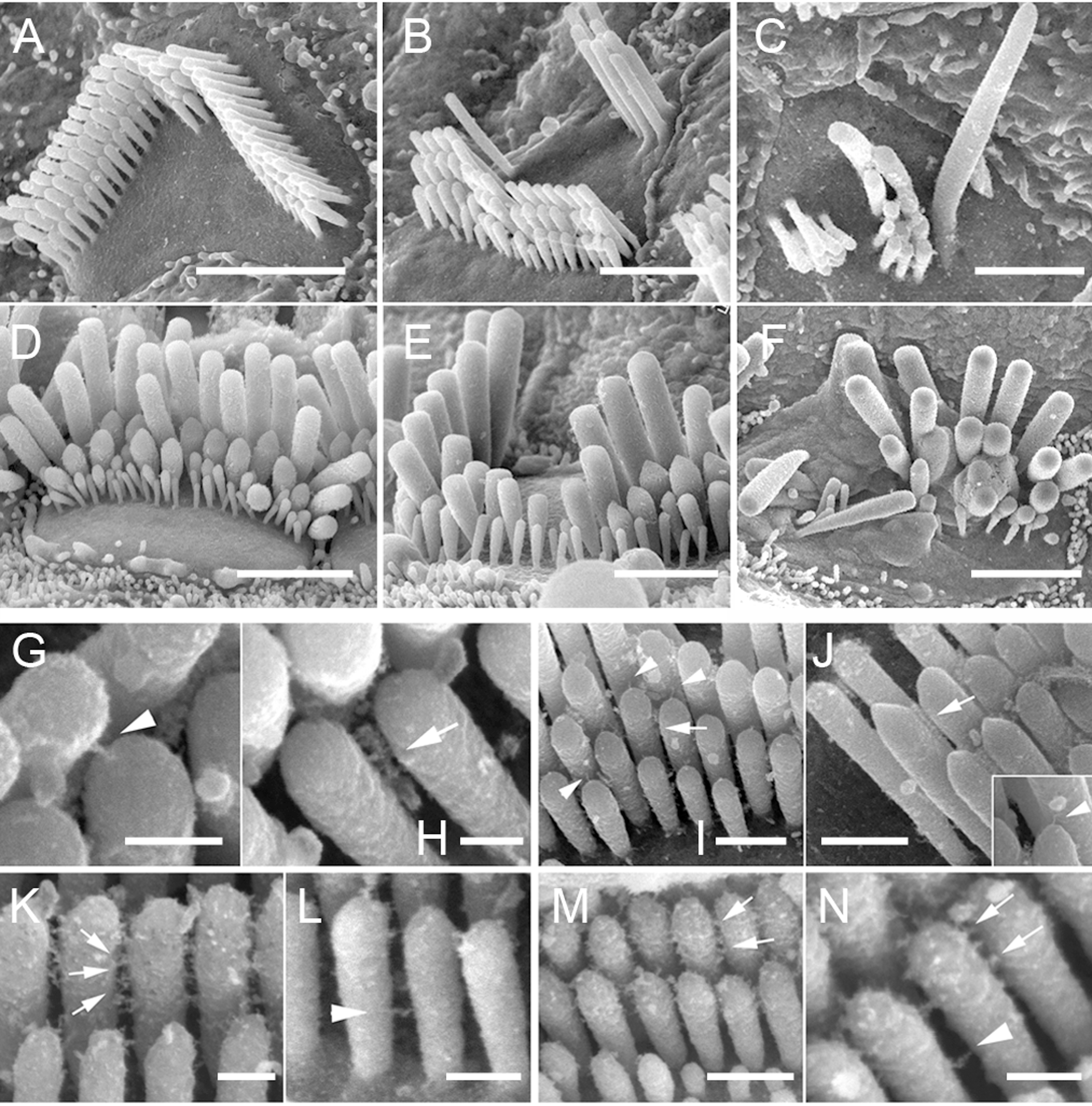
Tip links are extracellular filaments that connect stereocilia to each other or to the kinocilium in the hair cells of the inner ear.Pickles JO, Comis SD, Osborne MP. 1984.Cross-links between stereocilia in the guinea pig organ of Corti, and their possible relation to sensory transduction. Hearing Research 15:103-112. Mechanotransduction is thought to occur at the site of the tip links, which connect to spring-gated ion channels. These channels are cation-selective transduction channels that allow potassium and calcium ions to enter the hair cell from the endolymph that bathes its apical end. When the hair cells are deflected toward the kinocilium, depolarization occurs; when deflection is away from the kinocilium, hyperpolarization occurs. The tip link is made of two different cadherin molecules, protocadherin 15 and cadherin 23.Lewin GR, Moshourab R. 2004. Mechanosensation and pain. Journal of Neurobiology 61:30-44 It has been found that the tip links are relatively stiff, so ... [...More Info...] [...Related Items...] OR: [Wikipedia] [Google] [Baidu] |


