|
Right Main Bronchus
A bronchus is a passage or airway in the lower respiratory tract that conducts Atmosphere of Earth, air into the lungs. The first or primary bronchi pronounced (BRAN-KAI) to branch from the trachea at the Carina of trachea, carina are the right main bronchus and the left main bronchus. These are the widest bronchi, and enter the right lung, and the left lung at each Root of the lung, hilum. The main bronchi branch into narrower secondary bronchi or lobar bronchi, and these branch into narrower tertiary bronchi or segmental bronchi. Further divisions of the segmental bronchi are known as 4th order, 5th order, and 6th order segmental bronchi, or grouped together as subsegmental bronchi. The bronchi, when too narrow to be supported by cartilage, are known as bronchioles. No gas exchange takes place in the bronchi. Structure The trachea (windpipe) divides at the carina of trachea, carina into two main or primary bronchi, the left bronchus and the right bronchus. The carina of the trach ... [...More Info...] [...Related Items...] OR: [Wikipedia] [Google] [Baidu] |
Lower Respiratory Tract
The respiratory tract is the subdivision of the respiratory system involved with the process of respiration in mammals. The respiratory tract is lined with respiratory epithelium as respiratory mucosa. Air is breathed in through the nose to the nasal cavity, where a layer of nasal mucosa acts as a filter and traps pollutants and other harmful substances found in the air. Next, air moves into the pharynx, a passage that contains the intersection between the oesophagus and the larynx. The opening of the larynx has a special flap of cartilage, the epiglottis, that opens to allow air to pass through but closes to prevent food from moving into the airway. From the larynx, air moves into the trachea and down to the intersection known as the carina that branches to form the right and left primary (main) bronchi. Each of these bronchi branches into a secondary (lobar) bronchus that branches into tertiary (segmental) bronchi, that branch into smaller airways called bronchioles that ev ... [...More Info...] [...Related Items...] OR: [Wikipedia] [Google] [Baidu] |
Azygos Vein
The azygos vein is a vein running up the right side of the thoracic vertebral column draining itself towards the superior vena cava. It connects the systems of superior vena cava and inferior vena cava and can provide an alternative path for blood to the right atrium when either of the venae cavae is blocked. Structure The azygos vein transports deoxygenated blood from the posterior walls of the thorax and abdomen into the superior vena cava. It is formed by the union of the ascending lumbar veins with the right subcostal veins at the level of the 12th thoracic vertebra, ascending to the right of the descending aorta and thoracic duct, passing behind the right crus of diaphragm, anterior to the vertebral bodies of T12 to T5 and right posterior intercostal arteries. At the level of T4 vertebrae, it arches over the root of the right lung from behind to the front to join the superior vena cava. The trachea and oesophagus is located medially to the arch of the azygous vein. The ... [...More Info...] [...Related Items...] OR: [Wikipedia] [Google] [Baidu] |
Respiratory Bronchiole
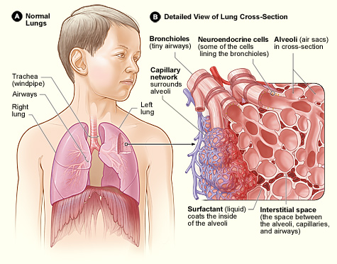
The bronchioles or bronchioli (pronounced ''bron-kee-oh-lee'') are the smaller branches of the bronchial airways in the lower respiratory tract. They include the terminal bronchioles, and finally the respiratory bronchioles that mark the start of the respiratory zone delivering air to the gas exchanging units of the alveoli. The bronchioles no longer contain the cartilage that is found in the bronchi, or glands in their submucosa. Structure The pulmonary lobule is the portion of the lung ventilated by one bronchiole. Bronchioles are approximately 1 mm or less in diameter and their walls consist of ciliated cuboidal epithelium and a layer of smooth muscle. Bronchioles divide into even smaller bronchioles, called ''terminal'', which are 0.5 mm or less in diameter. Terminal bronchioles in turn divide into smaller respiratory bronchioles which divide into alveolar ducts. Terminal bronchioles mark the end of the conducting division of air flow in the respiratory syste ... [...More Info...] [...Related Items...] OR: [Wikipedia] [Google] [Baidu] |
Terminal Bronchiole
The bronchioles or bronchioli (pronounced ''bron-kee-oh-lee'') are the smaller branches of the bronchial airways in the lower respiratory tract. They include the terminal bronchioles, and finally the respiratory bronchioles that mark the start of the respiratory zone delivering air to the gas exchanging units of the alveoli. The bronchioles no longer contain the cartilage that is found in the bronchi, or glands in their submucosa. Structure The pulmonary lobule is the portion of the lung ventilated by one bronchiole. Bronchioles are approximately 1 mm or less in diameter and their walls consist of ciliated cuboidal epithelium and a layer of smooth muscle. Bronchioles divide into even smaller bronchioles, called ''terminal'', which are 0.5 mm or less in diameter. Terminal bronchioles in turn divide into smaller respiratory bronchioles which divide into alveolar ducts. Terminal bronchioles mark the end of the conducting division of air flow in the respiratory syste ... [...More Info...] [...Related Items...] OR: [Wikipedia] [Google] [Baidu] |
Connective Tissue
Connective tissue is one of the four primary types of animal tissue, along with epithelial tissue, muscle tissue, and nervous tissue. It develops from the mesenchyme derived from the mesoderm the middle embryonic germ layer. Connective tissue is found in between other tissues everywhere in the body, including the nervous system. The three meninges, membranes that envelop the brain and spinal cord are composed of connective tissue. Most types of connective tissue consists of three main components: elastic and collagen fibers, ground substance, and cells. Blood, and lymph are classed as specialized fluid connective tissues that do not contain fiber. All are immersed in the body water. The cells of connective tissue include fibroblasts, adipocytes, macrophages, mast cells and leucocytes. The term "connective tissue" (in German, ''Bindegewebe'') was introduced in 1830 by Johannes Peter Müller. The tissue was already recognized as a distinct class in the 18th century. ... [...More Info...] [...Related Items...] OR: [Wikipedia] [Google] [Baidu] |
Septum
In biology, a septum (Latin for ''something that encloses''; plural septa) is a wall, dividing a cavity or structure into smaller ones. A cavity or structure divided in this way may be referred to as septate. Examples Human anatomy * Interatrial septum, the wall of tissue that is a sectional part of the left and right atria of the heart * Interventricular septum, the wall separating the left and right ventricles of the heart * Lingual septum, a vertical layer of fibrous tissue that separates the halves of the tongue. *Nasal septum: the cartilage wall separating the nostrils of the nose * Alveolar septum: the thin wall which separates the alveoli from each other in the lungs * Orbital septum, a palpebral ligament in the upper and lower eyelids * Septum pellucidum or septum lucidum, a thin structure separating two fluid pockets in the brain * Uterine septum, a malformation of the uterus * Vaginal septum, a lateral or transverse partition inside the vagina * Intermuscular sep ... [...More Info...] [...Related Items...] OR: [Wikipedia] [Google] [Baidu] |
Bronchopulmonary Segment
A bronchopulmonary segment is a portion of lung supplied by a specific segmental bronchus and its vessels. These arteries branch from the pulmonary and bronchial arteries, and run together through the center of the segment. Veins and lymphatic vessels drain along the edges of the segment. The segments are separated from each other by layers of connective tissue that forms them into discrete anatomical and functional units. This separation means that a bronchopulmonary segment can be surgically removed without affecting the function of the others. There are ten bronchopulmonary segments in the right lung: three in the superior lobe, two in the middle lobe, and five in the inferior lobe. Some of the segments may fuse in the left lung to form usually eight to nine segments (four to five in the upper lobe and four to five in the lower lobe. The delineation of the bronchopulmonary segments was made by Chevalier Jackson and John Franklin Huber at Temple University Hospital. Right lung ... [...More Info...] [...Related Items...] OR: [Wikipedia] [Google] [Baidu] |
Right Lung
The lungs are the primary Organ (anatomy), organs of the respiratory system in humans and most other animals, including some snails and a small number of fish. In mammals and most other vertebrates, two lungs are located near the vertebral column, backbone on either side of the heart. Their function in the respiratory system is to extract oxygen from the air and transfer it into the bloodstream, and to release carbon dioxide from the bloodstream into the Atmosphere of Earth, atmosphere, in a process of gas exchange. Respiration (physiology), Respiration is driven by different muscle, muscular systems in different species. Mammals, reptiles and birds use their different muscles to support and foster breathing. In earlier tetrapods, air was driven into the lungs by the pharyngeal muscles via buccal pumping, a mechanism still seen in amphibians. In humans, the main muscles of respiration, muscle of respiration that drives breathing is the thoracic diaphragm, diaphragm. The lungs a ... [...More Info...] [...Related Items...] OR: [Wikipedia] [Google] [Baidu] |
Eparterial Branch
The eparterial bronchus (right superior lobar bronchus) is a branch of the right main bronchus given off about 2.5 cm from the bifurcation of the trachea. This branch supplies the superior lobe of the right lung and is the most superior of all secondary bronchi. It arises above the level of the right pulmonary artery, and for this reason is named the eparterial bronchus. All other distributions falling below the pulmonary artery are termed ''hyparterial''. The eparterial bronchus is the only secondary bronchus with a specific name apart from the name of its corresponding lobe. Name The classification of ''eparterial'' and ''hyparterial'' is attributed to Swiss anatomist and anthropologist Christoph Theodor Aeby Christoph Theodor Aeby (25 February 1835 – 7 July 1885) was a Swiss anatomist, anthropologist, and academic. His main scientific interest comparative anatomy and his studies were said to be facilitated by a large collection of bones, which he ..., and is ce ... [...More Info...] [...Related Items...] OR: [Wikipedia] [Google] [Baidu] |
Descending Aorta
In human anatomy, the descending aorta is part of the aorta, the largest artery in the body. The descending aorta begins at the aortic arch and runs down through the chest and abdomen. The descending aorta anatomically consists of two portions or segments, the thoracic and the abdominal aorta, in correspondence with the two great cavities of the trunk in which it is situated. Within the abdomen, the descending aorta branches into the two common iliac arteries which serve the pelvis and eventually legs. The ductus arteriosus connects to the junction between the pulmonary artery and the descending aorta in foetal life. This artery later regresses as the ligamentum arteriosum. See also *Abbott artery Abbott's artery describes an anomalous artery that arises from the posteromedial aspect of the proximal part of the descending aorta. Normally a minor congenital abnormality, its presence is important during surgical repair of coarctation of the ... References External links ... [...More Info...] [...Related Items...] OR: [Wikipedia] [Google] [Baidu] |
Thoracic Duct
In human anatomy, the thoracic duct is the larger of the two lymph ducts of the lymphatic system. It is also known as the ''left lymphatic duct'', ''alimentary duct'', ''chyliferous duct'', and ''Van Hoorne's canal''. The other duct is the right lymphatic duct. The thoracic duct carries chyle, a liquid containing both lymph and emulsified fats, rather than pure lymph. It also collects most of the lymph in the body other than from the right thorax, arm, head, and neck (which are drained by the right lymphatic duct). The thoracic duct usually starts from the level of the twelfth thoracic vertebra (T12) and extends to the root of the neck. It drains into the systemic (blood) circulation at the junction of the left subclavian and internal jugular veins, at the commencement of the brachiocephalic vein. When the duct ruptures, the resulting flood of liquid into the pleural cavity is known as chylothorax. Structure In adults, the thoracic duct is typically 38–45 cm in length an ... [...More Info...] [...Related Items...] OR: [Wikipedia] [Google] [Baidu] |
Esophagus
The esophagus (American English) or oesophagus (British English; both ), non-technically known also as the food pipe or gullet, is an organ in vertebrates through which food passes, aided by peristaltic contractions, from the pharynx to the stomach. The esophagus is a fibromuscular tube, about long in adults, that travels behind the trachea and heart, passes through the diaphragm, and empties into the uppermost region of the stomach. During swallowing, the epiglottis tilts backwards to prevent food from going down the larynx and lungs. The word ''oesophagus'' is from Ancient Greek οἰσοφάγος (oisophágos), from οἴσω (oísō), future form of φέρω (phérō, “I carry”) + ἔφαγον (éphagon, “I ate”). The wall of the esophagus from the lumen outwards consists of mucosa, submucosa (connective tissue), layers of muscle fibers between layers of fibrous tissue, and an outer layer of connective tissue. The mucosa is a stratified squamous epithel ... [...More Info...] [...Related Items...] OR: [Wikipedia] [Google] [Baidu] |


