|
Reverse Sneezing
Reverse sneezing, also known as inspiratory paroxysmal respiration, is a clinical event that occurs in dogs. It is possibly caused by a muscle spasm at the back of the dog's mouth, more specifically where the muscle and throat meet. Other hypotheses state that it occurs when the dog's soft palate gets irritated. The irritation causes spasms in the soft palate muscle thus narrowing the trachea. Because the trachea is narrowed, the dog isn't able to inhale a full breath of air, resulting in forceful attempts to inhale through their nose. This causes the dog to experience reverse sneezing. The clinical symptoms seem to occur more in brachycephalic dog breeds such as Boxer, English- and French bulldogs. The specific cause of reverse sneezing is unknown but there could be a link between nasal, pharyngeal or sinus irritation which increases the production of mucus. In attempt to remove this excess mucus, reverse sneezing can be observed. Another hypothesis is based on the overexciteme ... [...More Info...] [...Related Items...] OR: [Wikipedia] [Google] [Baidu] |
Pug Reverse Sneezing After A Bath
The Pug is a breed of dog originally from China, with physically distinctive features of a wrinkly, short-muzzled face and curled tail. The breed has a fine, glossy coat that comes in a variety of colors, most often light brown (fawn) or black, and a compact, square body with well developed and thick muscles all over the body. Pugs were brought from China to Europe in the sixteenth century and were popularized in Western Europe by the House of Orange of the Netherlands, and the House of Stuart. In the United Kingdom, in the nineteenth century, Queen Victoria developed a passion for pugs which she passed on to other members of the Royal Family. Pugs are known for being sociable and gentle companion dogs. The American Kennel Club describes the breed's personality as "even-tempered and charming". Pugs remain popular into the twenty-first century, with some famous celebrity owners. Description Physical characteristics While the pugs that are depicted in eighteenth century pr ... [...More Info...] [...Related Items...] OR: [Wikipedia] [Google] [Baidu] |
Soft Palate
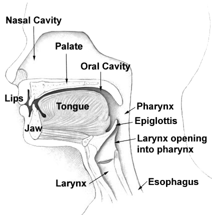
The soft palate (also known as the velum, palatal velum, or muscular palate) is, in mammals, the soft tissue constituting the back of the roof of the mouth. The soft palate is part of the palate of the mouth; the other part is the hard palate. The soft palate is distinguished from the hard palate at the front of the mouth in that it does not contain bone. Structure Muscles The five muscles of the soft palate play important roles in swallowing and breathing. The muscles are: # Tensor veli palatini, which is involved in swallowing # Palatoglossus, involved in swallowing # Palatopharyngeus, involved in breathing # Levator veli palatini, involved in swallowing # Musculus uvulae, which moves the uvula These muscles are innervated by the pharyngeal plexus via the vagus nerve, with the exception of the tensor veli palatini. The tensor veli palatini is innervated by the mandibular division of the trigeminal nerve (V3). Function The soft palate is moveable, consisting of muscle f ... [...More Info...] [...Related Items...] OR: [Wikipedia] [Google] [Baidu] |
Brachycephaly
Brachycephaly (derived from the Ancient Greek '' βραχύς'', 'short' and '' κεφαλή'', 'head') is the shape of a skull shorter than typical for its species. It is perceived as a desirable trait in some domesticated dog and cat breeds, notably the pug and Persian, and can be normal or abnormal in other animal species. In humans, the cephalic disorder is known as flat head syndrome, and results from premature fusion of the coronal sutures, or from external deformation. The coronal suture is the fibrous joint that unites the frontal bone with the two parietal bones of the skull. The parietal bones form the top and sides of the skull. This feature can be seen in Down syndrome. In anthropology, human populations have been characterized as either dolichocephalic (long-headed), mesaticephalic (moderate-headed), or brachycephalic (short-headed). The usefulness of the cephalic index was questioned by Giuseppe Sergi, who argued that cranial morphology provided a better mean ... [...More Info...] [...Related Items...] OR: [Wikipedia] [Google] [Baidu] |
Boxer (dog)
The Boxer is a medium to large, short-haired dog breed of mastiff-type, developed in Germany. The coat is smooth and tight-fitting; colors are fawn, brindled, or white, with or without white markings. Boxers are brachycephalic (they have broad, short skulls), have a square muzzle, mandibular prognathism (an underbite), very strong jaws, and a powerful bite ideal for hanging on to large prey. The Boxer was bred from the Old English Bulldog and the now extinct Bullenbeisser, which became extinct by crossbreeding rather than by a decadence of the breed. The Boxer is a member of both The Kennel Club and American Kennel Club (AKC) Working Group.http://www.akc.org/dog-breeds/boxer/#standard "Get to Know the Boxer", 'The American Kennel Club', Retrieved 14 May 2014 The first Boxer club was founded in 1895, with Boxers being first exhibited in a dog show for St. Bernards in Munich the next year. Based on 2013 AKC statistics, Boxers held steady as the seventh-most popular breed of dog ... [...More Info...] [...Related Items...] OR: [Wikipedia] [Google] [Baidu] |
Bulldog
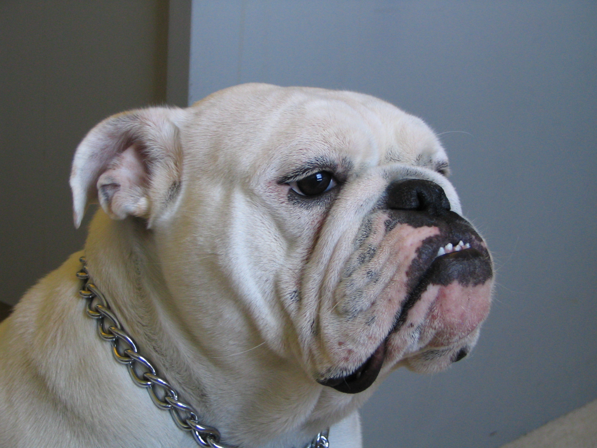
The Bulldog is a British breed of dog of mastiff type. It may also be known as the English Bulldog or British Bulldog. It is of medium size, a muscular, hefty dog with a wrinkled face and a distinctive pushed-in nose."Get to Know the Bulldog" , 'The American Kennel Club'. Retrieved 29 May 2014 It is commonly kept as a ; in 2013 it was in twelfth place on a list of the breeds most frequently registered worldwide. The Bulldog has a longstanding association with ; the |
Mucus
Mucus ( ) is a slippery aqueous secretion produced by, and covering, mucous membranes. It is typically produced from cells found in mucous glands, although it may also originate from mixed glands, which contain both serous and mucous cells. It is a viscous colloid containing inorganic salts, antimicrobial enzymes (such as lysozymes), immunoglobulins (especially IgA), and glycoproteins such as lactoferrin and mucins, which are produced by goblet cells in the mucous membranes and submucosal glands. Mucus serves to protect epithelial cells in the linings of the respiratory, digestive, and urogenital systems, and structures in the visual and auditory systems from pathogenic fungi, bacteria and viruses. Most of the mucus in the body is produced in the gastrointestinal tract. Amphibians, fish, snails, slugs, and some other invertebrates also produce external mucus from their epidermis as protection against pathogens, and to help in movement and is also produced in fish to line the ... [...More Info...] [...Related Items...] OR: [Wikipedia] [Google] [Baidu] |
Tracheal Collapse
Tracheal collapse in dogs is a condition characterized by incomplete formation or weakening of the cartilaginous rings of the trachea resulting in flattening of the trachea. It can be congenital or acquired, and extrathoracic or intrathoracic (inside or outside the thoracic cavity). Tracheal collapse is a dynamic condition. Collapse of the cervical trachea or extrathoracic (in the neck) occurs during inspiration; collapse of the thoracic trachea or intrathoracic (in the chest) occurs during expiration. Tracheal collapse is most commonly found in small dog breeds, including the Chihuahua, Pomeranian, Toy Poodle, Shih Tzu, Lhasa Apso, Maltese, Pug, and Yorkshire Terrier. Congenital tracheal collapse appears to be caused by a deficiency of normal components of tracheal ring cartilage like glycosaminoglycans, glycoproteins, calcium, and chondroitin. Acquired tracheal collapse can be caused by Cushing's syndrome, heart disease, and chronic respiratory disease and infection. ... [...More Info...] [...Related Items...] OR: [Wikipedia] [Google] [Baidu] |
Nasal Cavity
The nasal cavity is a large, air-filled space above and behind the nose in the middle of the face. The nasal septum divides the cavity into two cavities, also known as fossae. Each cavity is the continuation of one of the two nostrils. The nasal cavity is the uppermost part of the respiratory system and provides the nasal passage for inhaled air from the nostrils to the nasopharynx and rest of the respiratory tract. The paranasal sinuses surround and drain into the nasal cavity. Structure The term "nasal cavity" can refer to each of the two cavities of the nose, or to the two sides combined. The lateral wall of each nasal cavity mainly consists of the maxilla. However, there is a deficiency that is compensated for by the perpendicular plate of the palatine bone, the medial pterygoid plate, the labyrinth of ethmoid and the inferior concha. The paranasal sinuses are connected to the nasal cavity through small orifices called ostia. Most of these ostia communicate with the n ... [...More Info...] [...Related Items...] OR: [Wikipedia] [Google] [Baidu] |
Pharynx
The pharynx (plural: pharynges) is the part of the throat behind the mouth and nasal cavity, and above the oesophagus and trachea (the tubes going down to the stomach and the lungs). It is found in vertebrates and invertebrates, though its structure varies across species. The pharynx carries food and air to the esophagus and larynx respectively. The flap of cartilage called the epiglottis stops food from entering the larynx. In humans, the pharynx is part of the digestive system and the conducting zone of the respiratory system. (The conducting zone—which also includes the nostrils of the nose, the larynx, trachea, bronchi, and bronchioles—filters, warms and moistens air and conducts it into the lungs). The human pharynx is conventionally divided into three sections: the nasopharynx, oropharynx, and laryngopharynx. It is also important in vocalization. In humans, two sets of pharyngeal muscles form the pharynx and determine the shape of its lumen. They are arranged as an ... [...More Info...] [...Related Items...] OR: [Wikipedia] [Google] [Baidu] |
Elongated Soft Palate
An elongated soft palate is a congenital hereditary disorder that negatively affect dogs and cats' breathing and eating. A soft palate is considered elongated when it extends past the top of the epiglottis and/or past the middle of the tonsillar crypts. The soft palate is made up of muscle and connective tissue located in the posterior portion on the roof of the mouth. The soft palate creates a barrier between the mouth (oral cavity) and nose (nasal cavity). This continuation between the cavities makes it possible to chew and breathe at the same time. The soft palate only blocks the nasal cavity while swallowing. At rest the soft palate should only stretch caudally from the hard palate to the tip of the epiglottis leaving an opening between the nasal and oral cavities. When the soft palate is elongated, it partially blocks the throat thereby creating breathing and feeding-related issues. The elongation and other accompanying symptoms occur in breeds characterized with “smooshed face ... [...More Info...] [...Related Items...] OR: [Wikipedia] [Google] [Baidu] |
Spasm
A spasm is a sudden involuntary contraction of a muscle, a group of muscles, or a hollow organ such as the bladder. A spasmodic muscle contraction may be caused by many medical conditions, including dystonia. Most commonly, it is a muscle cramp which is accompanied by a sudden burst of pain. A muscle cramp is usually harmless and ceases after a few minutes. It is typically caused by ion imbalance or muscle overload. There are other causes of involuntary muscle contractions, and some of these may cause a health problem. Description and causes Various kinds of involuntary muscle activity may be referred to as a "spasm". A spasm may be a muscle contraction caused by abnormal nerve stimulation or by abnormal activity of the muscle itself. A spasm may lead to muscle strains or tears in tendons and ligaments if the force of the spasm exceeds the tensile strength of the underlying connective tissue. This can occur with a particularly strong spasm or with weakened connective ti ... [...More Info...] [...Related Items...] OR: [Wikipedia] [Google] [Baidu] |


