|
Recti
The rectus abdominis muscle, ( la, straight abdominal) also known as the "abdominal muscle" or simply the "abs", is a paired straight muscle. It is a paired muscle, separated by a midline band of connective tissue called the linea alba. It extends from the pubic symphysis, pubic crest and pubic tubercle inferiorly, to the xiphoid process and costal cartilages of ribs V to VII superiorly. The proximal attachments are the pubic crest and the pubic symphysis. It attaches distally at the costal cartilages of ribs 5-7 and the xiphoid process of the sternum. The rectus abdominis muscle is contained in the rectus sheath, which consists of the aponeuroses of the lateral abdominal muscles. The outer, most lateral line, defining the rectus is the linea semilunaris. Bands of connective tissue traverse the rectus abdominis, separating it into distinct muscle bellies. In the abdomens of people with low body fat, these muscle bellies can be viewed externally. They can appear in sets of a ... [...More Info...] [...Related Items...] OR: [Wikipedia] [Google] [Baidu] |
Connective Tissue
Connective tissue is one of the four primary types of animal tissue, along with epithelial tissue, muscle tissue, and nervous tissue. It develops from the mesenchyme derived from the mesoderm the middle embryonic germ layer. Connective tissue is found in between other tissues everywhere in the body, including the nervous system. The three meninges, membranes that envelop the brain and spinal cord are composed of connective tissue. Most types of connective tissue consists of three main components: elastic and collagen fibers, ground substance, and cells. Blood, and lymph are classed as specialized fluid connective tissues that do not contain fiber. All are immersed in the body water. The cells of connective tissue include fibroblasts, adipocytes, macrophages, mast cells and leucocytes. The term "connective tissue" (in German, ''Bindegewebe'') was introduced in 1830 by Johannes Peter Müller. The tissue was already recognized as a distinct class in the 18th century. ... [...More Info...] [...Related Items...] OR: [Wikipedia] [Google] [Baidu] |
Human Body
The human body is the structure of a Human, human being. It is composed of many different types of Cell (biology), cells that together create Tissue (biology), tissues and subsequently organ systems. They ensure homeostasis and the life, viability of the human body. It comprises a human head, head, hair, neck, Trunk (anatomy), trunk (which includes the thorax and abdomen), arms and hands, human leg, legs and feet. The study of the human body involves anatomy, physiology, histology and embryology. The body anatomical variability, varies anatomically in known ways. Physiology focuses on the systems and organs of the human body and their functions. Many systems and mechanisms interact in order to maintain homeostasis, with safe levels of substances such as sugar and oxygen in the blood. The body is studied by health professionals, physiologists, anatomists, and by artists to assist them in their work. Composition The composition of the human body, human body is composed of ... [...More Info...] [...Related Items...] OR: [Wikipedia] [Google] [Baidu] |
Muscle
Skeletal muscles (commonly referred to as muscles) are organs of the vertebrate muscular system and typically are attached by tendons to bones of a skeleton. The muscle cells of skeletal muscles are much longer than in the other types of muscle tissue, and are often known as muscle fibers. The muscle tissue of a skeletal muscle is striated – having a striped appearance due to the arrangement of the sarcomeres. Skeletal muscles are voluntary muscles under the control of the somatic nervous system. The other types of muscle are cardiac muscle which is also striated and smooth muscle which is non-striated; both of these types of muscle tissue are classified as involuntary, or, under the control of the autonomic nervous system. A skeletal muscle contains multiple fascicles – bundles of muscle fibers. Each individual fiber, and each muscle is surrounded by a type of connective tissue layer of fascia. Muscle fibers are formed from the fusion of developmental myoblasts in ... [...More Info...] [...Related Items...] OR: [Wikipedia] [Google] [Baidu] |
Costal Cartilages
The costal cartilages are bars of hyaline cartilage that serve to prolong the ribs forward and contribute to the elasticity of the walls of the thorax. Costal cartilage is only found at the anterior ends of the ribs, providing medial extension. Differences from Ribs 1-12 The first seven pairs are connected with the sternum; the next three are each articulated with the lower border of the cartilage of the preceding rib; the last two have pointed extremities, which end in the wall of the abdomen. Like the ribs, the costal cartilages vary in their length, breadth, and direction. They increase in length from the first to the seventh, then gradually decrease to the twelfth. Their breadth, as well as that of the intervals between them, diminishes from the first to the last. They are broad at their attachments to the ribs, and taper toward their sternal extremities, excepting the first two, which are of the same breadth throughout, and the sixth, seventh, and eighth, which are enlarg ... [...More Info...] [...Related Items...] OR: [Wikipedia] [Google] [Baidu] |
Pelvis
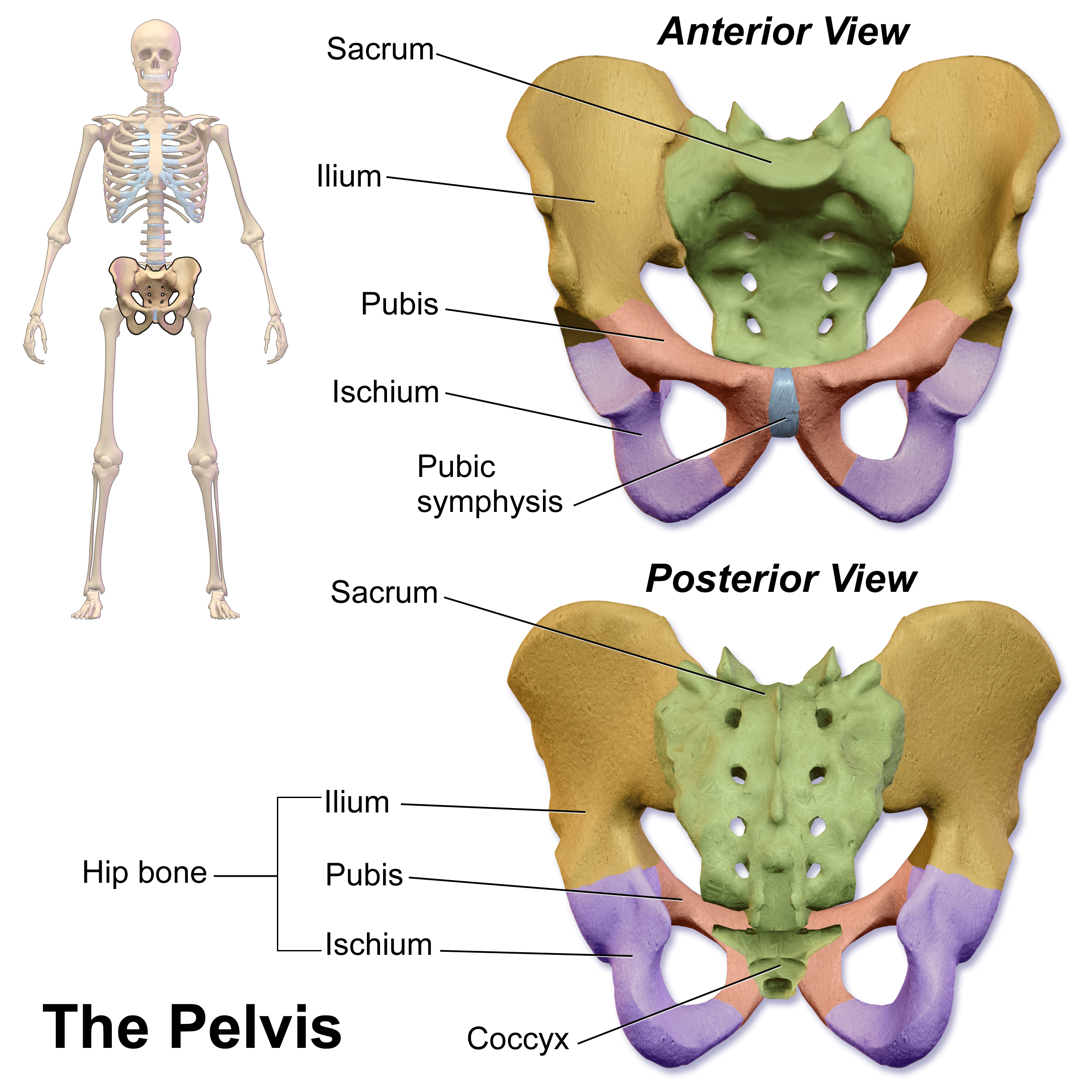
The pelvis (plural pelves or pelvises) is the lower part of the trunk, between the abdomen and the thighs (sometimes also called pelvic region), together with its embedded skeleton (sometimes also called bony pelvis, or pelvic skeleton). The pelvic region of the trunk includes the bony pelvis, the pelvic cavity (the space enclosed by the bony pelvis), the pelvic floor, below the pelvic cavity, and the perineum, below the pelvic floor. The pelvic skeleton is formed in the area of the back, by the sacrum and the coccyx and anteriorly and to the left and right sides, by a pair of hip bones. The two hip bones connect the spine with the lower limbs. They are attached to the sacrum posteriorly, connected to each other anteriorly, and joined with the two femurs at the hip joints. The gap enclosed by the bony pelvis, called the pelvic cavity, is the section of the body underneath the abdomen and mainly consists of the reproductive organs (sex organs) and the rectum, while the pelvic f ... [...More Info...] [...Related Items...] OR: [Wikipedia] [Google] [Baidu] |
Xiphoid Process
The xiphoid process , or xiphisternum or metasternum, is a small cartilaginous process (extension) of the inferior (lower) part of the sternum, which is usually ossified in the adult human. It may also be referred to as the ensiform process. Both the Greek-derived ''xiphoid'' and its Latin equivalent ''ensiform'' mean 'swordlike' or 'sword-shaped' Structure The xiphoid process is considered to be at the level of the 9th thoracic vertebra and the T7 dermatome. Development In newborns and young (especially small) infants, the tip of the xiphoid process may be both seen and felt as a lump just below the sternal notch. At 15 to 29 years old, the xiphoid usually fuses to the body of the sternum with a fibrous joint. Unlike the synovial articulation of major joints, this is non-movable. Ossification of the xiphoid process occurs around age 40. Variation The xiphoid process can be naturally bifurcated or sometimes perforated (xiphoidal foramen). These variances in morphology are inher ... [...More Info...] [...Related Items...] OR: [Wikipedia] [Google] [Baidu] |
Tendinous Intersections
The rectus abdominis muscle is crossed by three fibrous bands called the tendinous intersections or tendinous inscriptions. One is usually situated at the level of the umbilicus, one at the extremity of the xiphoid process, and the third about midway between the two. These intersections pass transversely or obliquely across the muscle; they rarely extend completely through its substance and may pass only halfway across it; they are intimately adherent in front to the sheath of the muscle. Sometimes one or two additional intersections, generally incomplete, are present below the umbilicus. Colloquial reference If well-defined, the rectus abdominis is colloquially called a "six-pack". This is due to tendinous intersections within the muscle, usually at the level of the umbilicus (belly-button), the xiphisternum, and about halfway between. An extremely well defined abdominal section can appear to be an "eight pack", as all eight sections of the abdominal muscle become defined. Th ... [...More Info...] [...Related Items...] OR: [Wikipedia] [Google] [Baidu] |
Rectus Sheath
The rectus sheath, also called the rectus fascia,. is formed by the aponeuroses of the transverse abdominal and the internal and external oblique muscles. It contains the rectus abdominis and pyramidalis muscles. Structure The rectus sheath can be divided into anterior and posterior laminae. The arrangement of the layers has important variations at different locations in the body. Below the costal margin For context, above the sheath are the following two layers: # Camper's fascia (anterior part of Superficial fascia) # Scarpa's fascia (posterior part of the Superficial fascia) Within the sheath, the layers vary: Below the sheath are the following three layers: # transversalis fascia # extraperitoneal fat # parietal peritoneum The rectus, in the situation where its sheath is deficient below, is separated from the peritoneum only by the transversalis fascia, in contrast to the upper layers, where part of the internal oblique also runs beneath the rectus. Because of the ... [...More Info...] [...Related Items...] OR: [Wikipedia] [Google] [Baidu] |
Abdomen
The abdomen (colloquially called the belly, tummy, midriff, tucky or stomach) is the part of the body between the thorax (chest) and pelvis, in humans and in other vertebrates. The abdomen is the front part of the abdominal segment of the torso. The area occupied by the abdomen is called the abdominal cavity. In arthropods it is the posterior (anatomy), posterior tagma (biology), tagma of the body; it follows the thorax or cephalothorax. In humans, the abdomen stretches from the thorax at the thoracic diaphragm to the pelvis at the pelvic brim. The pelvic brim stretches from the lumbosacral joint (the intervertebral disc between Lumbar vertebrae, L5 and Vertebra#Sacrum, S1) to the pubic symphysis and is the edge of the pelvic inlet. The space above this inlet and under the thoracic diaphragm is termed the abdominal cavity. The boundary of the abdominal cavity is the abdominal wall in the front and the peritoneal surface at the rear. In vertebrates, the abdomen is a large body c ... [...More Info...] [...Related Items...] OR: [Wikipedia] [Google] [Baidu] |
Pubic Symphysis
The pubic symphysis is a secondary cartilaginous joint between the left and right superior rami of the pubis of the hip bones. It is in front of and below the urinary bladder. In males, the suspensory ligament of the penis attaches to the pubic symphysis. In females, the pubic symphysis is close to the clitoris. In most adults it can be moved roughly 2 mm and with 1 degree rotation. This increases for women at the time of childbirth. The name comes from the Greek word ''symphysis'', meaning 'growing together'. Structure The pubic symphysis is a nonsynovial amphiarthrodial joint. The width of the pubic symphysis at the front is 3–5 mm greater than its width at the back. This joint is connected by fibrocartilage and may contain a fluid-filled cavity; the center is avascular, possibly due to the nature of the compressive forces passing through this joint, which may lead to harmful vascular disease. The ends of both pubic bones are covered by a thin layer of hyaline ... [...More Info...] [...Related Items...] OR: [Wikipedia] [Google] [Baidu] |
Lumbar Spine
The lumbar vertebrae are, in human anatomy, the five vertebrae between the rib cage and the pelvis. They are the largest segments of the vertebral column and are characterized by the absence of the foramen transversarium within the transverse process (since it is only found in the cervical region) and by the absence of facets on the sides of the body (as found only in the thoracic region). They are designated L1 to L5, starting at the top. The lumbar vertebrae help support the weight of the body, and permit movement. Human anatomy General characteristics The adjacent figure depicts the general characteristics of the first through fourth lumbar vertebrae. The fifth vertebra contains certain peculiarities, which are detailed below. As with other vertebrae, each lumbar vertebra consists of a ''vertebral body'' and a ''vertebral arch''. The vertebral arch, consisting of a pair of ''pedicles'' and a pair of ''laminae'', encloses the ''vertebral foramen'' (opening) and sup ... [...More Info...] [...Related Items...] OR: [Wikipedia] [Google] [Baidu] |
Erector Spinae
The erector spinae ( ) or spinal erectors is a set of muscles that straighten and rotate the back. The spinal erectors work together with the glutes (gluteus maximus, gluteus medius and gluteus minimus) to maintain stable posture standing or sitting. Structure The erector spinae is not just one muscle, but a group of muscles and tendons which run more or less the length of the spine on the left and the right, from the sacrum, or sacral region, and hips to the base of the skull. They are also known as the sacrospinalis group of muscles. These muscles lie on either side of the spinous processes of the vertebrae and extend throughout the lumbar, thoracic, and cervical regions. The erector spinae is covered in the lumbar and thoracic regions by the thoracolumbar fascia, and in the cervical region by the nuchal ligament. This large muscular and tendinous mass varies in size and structure at different parts of the vertebral column. In the sacral region, it is narrow and pointed, and at ... [...More Info...] [...Related Items...] OR: [Wikipedia] [Google] [Baidu] |


