|
Ranine
The lingual veins begin on the dorsum, sides, and under surface of the tongue, and, passing backward along the course of the lingual artery, end in the internal jugular vein. The vena comitans of the hypoglossal nerve (ranine vein), a branch of considerable size, begins below the tip of the tongue, and may join the lingual; generally, however, it passes backward on the hyoglossus, and joins the common facial. The lingual veins are important clinically as they are capable of rapid absorption of drugs; for this reason, nitroglycerin is given under the tongue to patients suspected of having angina pectoris. Tributaries # Sublingual vein # Deep lingual vein # Dorsal lingual veins # Suprahyoid vein The suprahyoid muscles are four muscles located above the hyoid bone in the neck. They are the digastric, stylohyoid, geniohyoid, and mylohyoid muscles. They are all pharyngeal muscles, with the exception of the geniohyoid muscle. The digastric is ... External links Photo of model (frog ... [...More Info...] [...Related Items...] OR: [Wikipedia] [Google] [Baidu] |
Lingual Artery
The lingual artery arises from the external carotid artery between the superior thyroid artery and facial artery. It can be located easily in the tongue. Structure The lingual artery first branches off from the external carotid artery. It runs obliquely upward and medially to the greater horns of the hyoid bone. It then curves downward and forward, forming a loop which is crossed by the hypoglossal nerve. It then passes beneath the digastric muscle and stylohyoid muscle running horizontally forward, beneath the hyoglossus. This takes it through the sublingual space. Finally, ascending almost perpendicularly to the tongue, it turns forward on its lower surface as far as the tip of the tongue, now called the deep lingual artery (profunda linguae). Branches The lingual artery gives 4 main branches: the deep lingual artery, the sublingual artery, the suprahyoid branch, and the dorsal lingual branch. Deep lingual artery The deep lingual artery (or ranine artery) is the terminal p ... [...More Info...] [...Related Items...] OR: [Wikipedia] [Google] [Baidu] |
Tongue
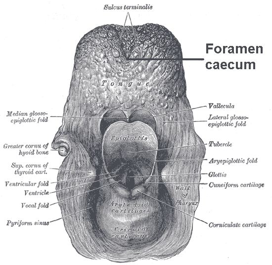
The tongue is a muscular organ (anatomy), organ in the mouth of a typical tetrapod. It manipulates food for mastication and swallowing as part of the digestive system, digestive process, and is the primary organ of taste. The tongue's upper surface (dorsum) is covered by taste buds housed in numerous lingual papillae. It is sensitive and kept moist by saliva and is richly supplied with nerves and blood vessels. The tongue also serves as a natural means of oral hygiene, cleaning the teeth. A major function of the tongue is the enabling of speech in humans and animal communication, vocalization in other animals. The human tongue is divided into two parts, an oral cavity, oral part at the front and a pharynx, pharyngeal part at the back. The left and right sides are also separated along most of its length by a vertical section of connective tissue, fibrous tissue (the lingual septum) that results in a groove, the median sulcus, on the tongue's surface. There are two groups of muscle ... [...More Info...] [...Related Items...] OR: [Wikipedia] [Google] [Baidu] |
Internal Jugular Vein
The internal jugular vein is a paired jugular vein that collects blood from the brain and the superficial parts of the face and neck. This vein runs in the carotid sheath with the common carotid artery and vagus nerve. It begins in the posterior compartment of the jugular foramen, at the base of the skull. It is somewhat dilated at its origin, which is called the ''superior bulb''. This vein also has a common trunk into which drains the anterior branch of the retromandibular vein, the facial vein, and the lingual vein. It runs down the side of the neck in a vertical direction, being at one end lateral to the internal carotid artery, and then lateral to the common carotid artery, and at the root of the neck, it unites with the subclavian vein to form the brachiocephalic vein (innominate vein); a little above its termination is a second dilation, the ''inferior bulb''. Above, it lies upon the rectus capitis lateralis, behind the internal carotid artery and the nerves passing ... [...More Info...] [...Related Items...] OR: [Wikipedia] [Google] [Baidu] |
Vena Comitans
Vena comitans is Latin for accompanying vein. It refers to a vein that is usually paired, with both veins lying on the sides of an artery. They are found in close proximity to arteries so that the pulsations of the artery aid venous return. Because they are generally found in pairs, they are often referred to by their plural form: venae comitantes. Venae comitantes are usually found with certain smaller arteries, especially those in the extremities. Larger arteries, on the other hand, generally do not have venae comitantes. They usually have a single, similarly sized vein which is not as intimately associated with the artery. Examples of arteries and their venae comitantes: * Radial artery and radial veins * Ulnar artery and ulnar veins * Brachial artery and brachial veins * Anterior tibial artery and anterior tibial veins * Posterior tibial artery and Posterior tibial veins * Fibular artery and Fibular veins Examples of arteries that do not have venae comitantes (i.e. thos ... [...More Info...] [...Related Items...] OR: [Wikipedia] [Google] [Baidu] |
Hypoglossal Nerve
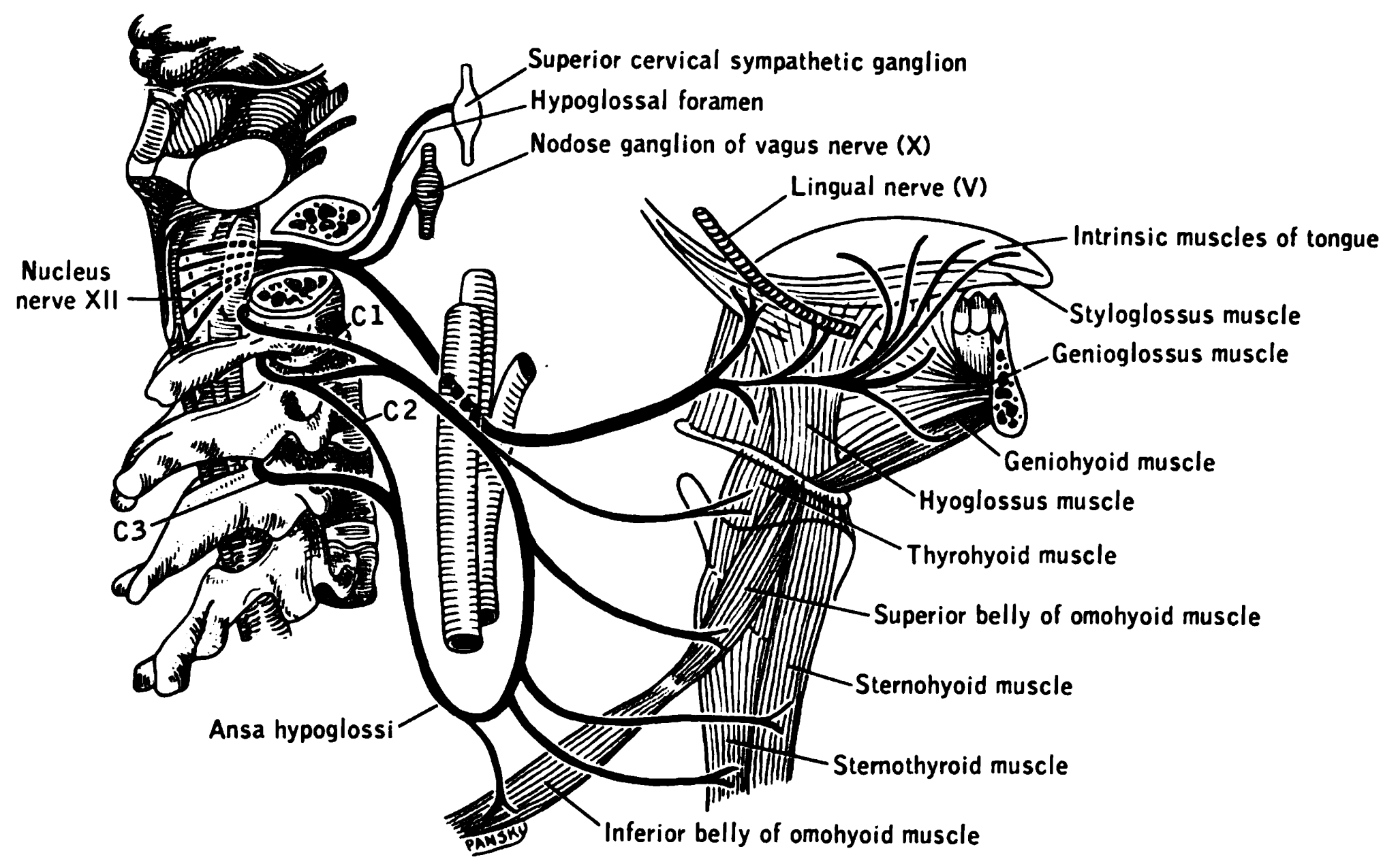
The hypoglossal nerve, also known as the twelfth cranial nerve, cranial nerve XII, or simply CN XII, is a cranial nerve that innervates all the extrinsic and intrinsic muscles of the tongue except for the palatoglossus, which is innervated by the vagus nerve. CN XII is a nerve with a solely motor function. The nerve arises from the hypoglossal nucleus in the medulla as a number of small rootlets, passes through the hypoglossal canal and down through the neck, and eventually passes up again over the tongue muscles it supplies into the tongue. The nerve is involved in controlling tongue movements required for speech and swallowing, including sticking out the tongue and moving it from side to side. Damage to the nerve or the neural pathways which control it can affect the ability of the tongue to move and its appearance, with the most common sources of damage being injury from trauma or surgery, and motor neuron disease. The first recorded description of the nerve is by Herophil ... [...More Info...] [...Related Items...] OR: [Wikipedia] [Google] [Baidu] |
Hyoglossus
The hyoglossus, thin and quadrilateral, arises from the side of the body and from the whole length of the greater cornu of the hyoid bone, and passes almost vertically upward to enter the side of the tongue, between the styloglossus and the inferior longitudinal muscle of the tongue. It forms a part of the floor of submandibular triangle. Structure The fibers arising from the body of the hyoid bone overlap those from the greater cornu. Structures that are medial/deep to the hyoglossus are the glossopharyngeal nerve (cranial nerve 9), the stylohyoid ligament and the lingual artery and lingual vein. The lingual vein passes medial to the hyoglossus, and the lingual artery passes deep to the hyoglossus. Laterally, in between the hyoglossus muscle and the mylohyoid muscle lay several important structures (from upper to lower): sublingual gland, submandibular duct, lingual nerve, vena comitans of hypoglossal nerve, and the hypoglossal nerve. Note, posteriorly, the lingual nerve is ... [...More Info...] [...Related Items...] OR: [Wikipedia] [Google] [Baidu] |
Common Facial
The facial vein usually unites with the anterior branch of the retromandibular vein to form the common facial vein, which crosses the external carotid artery and enters the internal jugular vein at a variable point below the hyoid bone. From near its termination a communicating branch often runs down the anterior border of the sternocleidomastoideus to join the lower part of the anterior jugular vein The anterior jugular vein is a vein in the neck. Structure The anterior jugular vein lies lateral to the cricothyroid ligament. It begins near the hyoid bone by the confluence of several superficial veins from the submandibular region. Its trib .... The common facial vein is not present in all individuals. References External links * () Veins of the head and neck Common vein {{Portal bar, Anatomy ... [...More Info...] [...Related Items...] OR: [Wikipedia] [Google] [Baidu] |
Glyceryl Trinitrate (pharmacology)
Nitroglycerin, also known as glyceryl trinitrate (GTN), is a medication used for heart failure, high blood pressure, anal fissures, painful periods, and to treat and prevent chest pain caused by decreased blood flow to the heart (angina) or due to the recreational use of cocaine. This includes chest pain from a heart attack. It is taken by mouth, under the tongue, applied to the skin, or by injection into a vein. Common side effects include headache and low blood pressure. The low blood pressure can be severe. It is unclear if use in pregnancy is safe for the baby. It should not be used together with medications within the PDE5 inhibitor family such as sildenafil due to the risk of low blood pressure. Nitroglycerin is in the nitrate family of medications. While it is not entirely clear how it works, it is believed to function by dilating blood vessels. Nitroglycerin was written about as early as 1846 and came into medical use in 1878. It is on the World Health Organizat ... [...More Info...] [...Related Items...] OR: [Wikipedia] [Google] [Baidu] |
Angina Pectoris
Angina, also known as angina pectoris, is chest pain or pressure, usually caused by insufficient blood flow to the heart muscle (myocardium). It is most commonly a symptom of coronary artery disease. Angina is typically the result of obstruction or spasm of the arteries that supply blood to the heart muscle. The main mechanism of coronary artery obstruction is atherosclerosis as part of coronary artery disease. Other causes of angina include abnormal heart rhythms, heart failure and, less commonly, anemia. The term derives from the Latin ''angere'' ("to strangle") and ''pectus'' ("chest"), and can therefore be translated as "a strangling feeling in the chest". There is a weak relationship between severity of angina and degree of oxygen deprivation in the heart muscle, however, the severity of angina does not always match the degree of oxygen deprivation to the heart or the risk of a myocardial infarction (heart attack). Some people may experience severe pain even though the ... [...More Info...] [...Related Items...] OR: [Wikipedia] [Google] [Baidu] |


