|
Rostral Ventrolateral Medulla
The rostral ventrolateral medulla (RVLM), also known as the pressor area of the medulla, is a brain region that is responsible for basal and reflex control of sympathetic activity associated with cardiovascular function. Abnormally elevated sympathetic activity in the RVLM is associated with various cardiovascular diseases, such as heart failure and hypertension. The RVLM is notably involved in the baroreflex. It receives inhibitory GABAergic input from the caudal ventrolateral medulla (CVLM). The RVLM is a primary regulator of the sympathetic nervous system; it sends catecholaminergic projections to the sympathetic preganglionic neurons in the intermediolateral nucleus of the spinal cord via reticulospinal tract. Physostigmine, a choline-esterase inhibitor, elevates endogenous levels of acetylcholine and causes a rise in blood pressure by stimulation of the RVLM. Orexinergic neurons from the lateral hypothalamus The lateral hypothalamus (LH), also called the lateral hy ... [...More Info...] [...Related Items...] OR: [Wikipedia] [Google] [Baidu] |
Pressor
An antihypotensive agent, also known as a vasopressor agent or simply vasopressor, or pressor, is any substance, whether endogenous or a medication, that tends to raise low blood pressure. Some antihypotensive drugs act as vasoconstrictors to increase total peripheral resistance, others sensitize adrenoreceptors to catecholamines - glucocorticoids, and the third class increase cardiac output - dopamine, dobutamine. If low blood pressure is due to blood loss, then preparations increasing volume of blood circulation—plasma-substituting solutions such as colloid and crystalloid solutions (salt solutions)—will raise the blood pressure without any direct vasopressor activity. Packed red blood cells, plasma or whole blood should not be used solely for volume expansion or to increase oncotic pressure of circulating blood. Blood products should only be used if reduced oxygen carrying capacity or coagulopathy is present. Other causes of either absolute (dehydration, loss of plasma ... [...More Info...] [...Related Items...] OR: [Wikipedia] [Google] [Baidu] |
Reticulospinal Tract
The reticular formation is a set of interconnected nuclei that are located throughout the brainstem. It is not anatomically well defined, because it includes neurons located in different parts of the brain. The neurons of the reticular formation make up a complex set of networks in the core of the brainstem that extend from the upper part of the midbrain to the lower part of the medulla oblongata. The reticular formation includes ascending pathways to the cortex in the ascending reticular activating system (ARAS) and descending pathways to the spinal cord via the reticulospinal tracts. Neurons of the reticular formation, particularly those of the ascending reticular activating system, play a crucial role in maintaining behavioral arousal and consciousness. The overall functions of the reticular formation are modulatory and premotor, involving somatic motor control, cardiovascular control, pain modulation, sleep and consciousness, and habituation. The modulatory functions are pri ... [...More Info...] [...Related Items...] OR: [Wikipedia] [Google] [Baidu] |
Reflexes
In biology, a reflex, or reflex action, is an involuntary, unplanned sequence or action and nearly instantaneous response to a stimulus. Reflexes are found with varying levels of complexity in organisms with a nervous system. A reflex occurs via neural pathways in the nervous system called reflex arcs. A stimulus initiates a neural signal, which is carried to a synapse. The signal is then transferred across the synapse to a motor neuron which evokes a target response. These neural signals do not always travel to the brain, so many reflexes are an automatic response to a stimulus that does not receive or need conscious thought. Many reflexes are fine-tuned to increase organism survival and self-defense. This is observed in reflexes such as the startle reflex, which provides an automatic response to an unexpected stimuli, and the feline righting reflex, which reorients a cat's body when falling to ensure safe landing. The simplest type of reflex, a short-latency reflex, has a s ... [...More Info...] [...Related Items...] OR: [Wikipedia] [Google] [Baidu] |
Sympathetic Nervous System
The sympathetic nervous system (SNS) is one of the three divisions of the autonomic nervous system, the others being the parasympathetic nervous system and the enteric nervous system. The enteric nervous system is sometimes considered part of the autonomic nervous system, and sometimes considered an independent system. The autonomic nervous system functions to regulate the body's unconscious actions. The sympathetic nervous system's primary process is to stimulate the body's fight or flight response. It is, however, constantly active at a basic level to maintain homeostasis. The sympathetic nervous system is described as being antagonistic to the parasympathetic nervous system which stimulates the body to "feed and breed" and to (then) "rest-and-digest". Structure There are two kinds of neurons involved in the transmission of any signal through the sympathetic system: pre-ganglionic and post-ganglionic. The shorter preganglionic neurons originate in the thoracolumbar divisio ... [...More Info...] [...Related Items...] OR: [Wikipedia] [Google] [Baidu] |
Vasomotor Center
The vasomotor center (VMC) is a portion of the medulla oblongata. Together with the cardiovascular center and respiratory center, it regulates blood pressure. It also has a more minor role in other homeostatic processes. Upon increase in carbon dioxide level at central chemoreceptors, it stimulates the sympathetic system to constrict vessels. This is opposite to carbon dioxide in tissues causing vasodilatation, especially in the brain. Cranial nerves IX (glossopharyngeal nerve) and X (vagus nerve) both feed into the vasomotor centre and are themselves involved in the regulation of blood pressure.vasomotor center has other actions also Structure The vasomotor center is a collection of integrating neurons in the medulla oblongata of the middle brain stem. The term "vasomotor center" is not truly accurate, since this function relies not on a single brain structure ("center") but rather represents a network of interacting neurons. Afferent fibres The vasomotor center integrate ... [...More Info...] [...Related Items...] OR: [Wikipedia] [Google] [Baidu] |
Lateral Hypothalamus
The lateral hypothalamus (LH), also called the lateral hypothalamic area (LHA), contains the primary orexinergic nucleus within the hypothalamus that widely projects throughout the nervous system; this system of neurons mediates an array of cognitive and physical processes, such as promoting feeding behavior and arousal, reducing pain perception, and regulating body temperature, digestive functions, and blood pressure, among many others. Clinically significant disorders that involve dysfunctions of the orexinergic projection system include narcolepsy, motility disorders or functional gastrointestinal disorders involving visceral hypersensitivity (e.g., irritable bowel syndrome), and eating disorders. The neurotransmitter glutamate and the endocannabinoids (e.g., anandamide) and the orexin neuropeptides orexin-A and orexin-B are the primary signaling neurochemicals in orexin neurons; pathway-specific neurochemicals include GABA, melanin-concentrating hormone, nocice ... [...More Info...] [...Related Items...] OR: [Wikipedia] [Google] [Baidu] |
Orexinergic
Orexin (), also known as hypocretin, is a neuropeptide that regulates arousal, wakefulness, and appetite. The most common form of narcolepsy, type 1, in which the individual experiences brief losses of muscle tone ("drop attacks" or cataplexy), is caused by a lack of orexin in the brain due to destruction of the cells that produce it.Stanford Center for NarcolepsFAQ(retrieved 27-Mar-2012) It exists in the forms of Orexin-A and Orexin-B. There are 50,000–80,000 orexin-producing neurons in the human brain, located predominantly in the perifornical area and lateral hypothalamus. They project widely throughout the central nervous system, regulating wakefulness, feeding, and other behaviours. There are two types of orexin peptide and two types of orexin receptor. Orexin was discovered in 1998 almost simultaneously by two independent groups of researchers working on the rat brain. One group named it orexin, from ''orexis,'' meaning "appetite" in Greek; the other group named it h ... [...More Info...] [...Related Items...] OR: [Wikipedia] [Google] [Baidu] |
Acetylcholine
Acetylcholine (ACh) is an organic chemical that functions in the brain and body of many types of animals (including humans) as a neurotransmitter. Its name is derived from its chemical structure: it is an ester of acetic acid and choline. Parts in the body that use or are affected by acetylcholine are referred to as cholinergic. Substances that increase or decrease the overall activity of the cholinergic system are called cholinergics and anticholinergics, respectively. Acetylcholine is the neurotransmitter used at the neuromuscular junction—in other words, it is the chemical that motor neurons of the nervous system release in order to activate muscles. This property means that drugs that affect cholinergic systems can have very dangerous effects ranging from paralysis to convulsions. Acetylcholine is also a neurotransmitter in the autonomic nervous system, both as an internal transmitter for the sympathetic nervous system and as the final product released by the para ... [...More Info...] [...Related Items...] OR: [Wikipedia] [Google] [Baidu] |
Endogenous
Endogenous substances and processes are those that originate from within a living system such as an organism, tissue, or cell. In contrast, exogenous substances and processes are those that originate from outside of an organism. For example, estradiol is an endogenous estrogen hormone produced within the body, whereas ethinylestradiol is an exogenous synthetic estrogen, commonly used in birth control pills Oral contraceptives, abbreviated OCPs, also known as birth control pills, are medications taken by mouth for the purpose of birth control. Female Two types of female oral contraceptive pill, taken once per day, are widely available: * The combin .... References External links *{{Wiktionary-inline, endogeny Biology ... [...More Info...] [...Related Items...] OR: [Wikipedia] [Google] [Baidu] |
Physostigmine
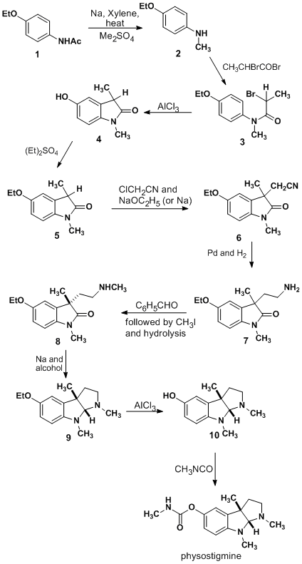
Physostigmine (also known as eserine from ''éséré'', the West African name for the Calabar bean) is a highly toxic parasympathomimetic alkaloid, specifically, a reversible cholinesterase inhibitor. It occurs naturally in the Calabar bean and the fruit of the Manchineel tree. The chemical was synthesized for the first time in 1935 by Percy Lavon Julian and Josef Pikl. It is available in the U.S. under the trade names Antilirium and Isopto Eserine, and as eserine salicylate and eserine sulfate. Today, physostigmine is most commonly used for its medicinal value. However, before its discovery by Sir Robert Christison in 1846, it was much more prevalent as an ordeal poison. The positive medical applications of the drug were first suggested in the gold medal-winning final thesis of Thomas Richard Fraser at the University of Edinburgh in 1862. Medical uses Physostigmine is used to treat glaucoma and delayed gastric emptying. Because it enhances the transmission of acetylch ... [...More Info...] [...Related Items...] OR: [Wikipedia] [Google] [Baidu] |
Intermediolateral Nucleus
The intermediolateral nucleus (IML) is a region of grey matter found in one of the three grey columns of the spinal cord, the lateral grey column. This is Rexed lamina VII. The intermediolateral cell column exists at vertebral levels T1 – L3. It mediates the entire sympathetic innervation of the body, but the nucleus resides in the grey matter of the spinal cord. Rexed Lamina VII contains several well defined nuclei including the nucleus dorsalis (Clarke's column), the intermediolateral nucleus, and the sacral autonomic nucleus. It extends from T1 to L3, and contains the autonomic motor neurons that give rise to the preganglionic fibers of the sympathetic nervous system, (preganglionic sympathetic general visceral efferent General visceral efferent fibers (GVE) or visceral efferents or autonomic efferents, are the efferent nerve fibers of the autonomic nervous system (also known as the ''visceral efferent nervous system'' that provide motor innervation to smooth mu ...s) ... [...More Info...] [...Related Items...] OR: [Wikipedia] [Google] [Baidu] |
Medulla Oblongata
The medulla oblongata or simply medulla is a long stem-like structure which makes up the lower part of the brainstem. It is anterior and partially inferior to the cerebellum. It is a cone-shaped neuronal mass responsible for autonomic (involuntary) functions, ranging from vomiting to sneezing. The medulla contains the cardiac, respiratory, vomiting and vasomotor centers, and therefore deals with the autonomic functions of breathing, heart rate and blood pressure as well as the sleep–wake cycle. During embryonic development, the medulla oblongata develops from the myelencephalon. The myelencephalon is a secondary vesicle which forms during the maturation of the rhombencephalon, also referred to as the hindbrain. The bulb is an archaic term for the medulla oblongata. In modern clinical usage, the word bulbar (as in bulbar palsy) is retained for terms that relate to the medulla oblongata, particularly in reference to medical conditions. The word bulbar can refer to the ... [...More Info...] [...Related Items...] OR: [Wikipedia] [Google] [Baidu] |


