|
Retromandibular Vein
The retromandibular vein (temporomaxillary vein, posterior facial vein) is a major vein of the face. Anatomy Origin The retromandibular vein is formed by the union of the superficial temporal and maxillary veins. Course It descends in the substance of the parotid gland, superficial to the external carotid artery (but beneath the facial nerve), between the ramus of the mandible and the sternocleidomastoideus muscle. It terminates by dividing into two branches: * an ''anterior'', which passes forward and joins anterior facial vein, to form the common facial vein, which then drains into the internal jugular vein. * a ''posterior'', which is joined by the posterior auricular vein and becomes the external jugular vein. Function The retromandibular vein provides venous drainage to the superior cranium, and significant drainage to the ear. Clinical significance Parrot's sign is a sensation of pain when pressure is applied to the retromandibular region. Additional images ... [...More Info...] [...Related Items...] OR: [Wikipedia] [Google] [Baidu] |
Head And Neck Anatomy
This article describes the anatomy of the head and neck of the human body, including the brain, bones, muscles, blood vessels, nerves, glands, nose, mouth, teeth, tongue, and throat. Structure Bones The head rests on the top part of the vertebral column, with the skull joining at C1 (the first cervical vertebra known as the atlas). The skeletal section of the head and neck forms the top part of the axial skeleton and is made up of the skull, hyoid bone, auditory ossicles, and cervical spine. The skull can be further subdivided into: # the cranium (8 bones: frontal, 2-parietal, occipital, 2-temporal, sphenoid, ethmoid), and # the facial bones (14 bones: 2-zygomatic, 2-maxillary, 2-palatine, 2-nasal, 2-lacrimal, vomer, 2-inferior conchae, mandible). The occipital bone joins with the atlas near the foramen magnum, a large hole () at the base of the skull. The atlas joins with the occipital condyle above and the axis below. The spinal cord passes through the foramen magnum. Musc ... [...More Info...] [...Related Items...] OR: [Wikipedia] [Google] [Baidu] |
Ramus Of The Mandible
In anatomy, the mandible, lower jaw or jawbone is the largest, strongest and lowest bone in the human facial skeleton. It forms the lower jaw and holds the lower teeth in place. The mandible sits beneath the maxilla. It is the only movable bone of the skull (discounting the ossicles of the middle ear). It is connected to the temporal bones by the temporomandibular joints. The bone is formed in the fetus from a fusion of the left and right mandibular prominences, and the point where these sides join, the mandibular symphysis, is still visible as a faint ridge in the midline. Like other symphyses in the body, this is a midline articulation where the bones are joined by fibrocartilage, but this articulation fuses together in early childhood.Illustrated Anatomy of the Head and Neck, Fehrenbach and Herring, Elsevier, 2012, p. 59 The word "mandible" derives from the Latin word ''mandibula'', "jawbone" (literally "one used for chewing"), from '' mandere'' "to chew" and ''-bula'' (i ... [...More Info...] [...Related Items...] OR: [Wikipedia] [Google] [Baidu] |
Parrot's Sign
Parrot's sign refers to at least two medical signs; one relating to the skull and another to a pupil reaction. Background Marie Jules Parrot was a French physician in Paris, whose early work concentrated on the brain, followed by tuberculosis and later syphilis. Skull Parrot's sign, also known as 'Parrot's nodes' and 'Parrot's bosses', refers to the bony growth noted at autopsy by Marie Jules Parrot and Jonathan Hutchinson on the skulls of children with congenital syphilis (CS) in the 19th century. Later publications also describe it as the frontal bossing that presents in the late type CS. Initially thought to be indicative of congenital syphilis, it was noted to be present in other conditions, particularly rickets. A description of bone findings in CS by Parrot was published in ''The Lancet'' in 1879 following his presentation at a meeting hosted by Jonathan Hutchinson and Thomas Barlow in London. In 1883 Barlow referred to the overgrowth of skull bone seen in CS as 'Parrot's ... [...More Info...] [...Related Items...] OR: [Wikipedia] [Google] [Baidu] |
Skull
The skull is a bone protective cavity for the brain. The skull is composed of four types of bone i.e., cranial bones, facial bones, ear ossicles and hyoid bone. However two parts are more prominent: the cranium and the mandible. In humans, these two parts are the neurocranium and the viscerocranium ( facial skeleton) that includes the mandible as its largest bone. The skull forms the anterior-most portion of the skeleton and is a product of cephalisation—housing the brain, and several sensory structures such as the eyes, ears, nose, and mouth. In humans these sensory structures are part of the facial skeleton. Functions of the skull include protection of the brain, fixing the distance between the eyes to allow stereoscopic vision, and fixing the position of the ears to enable sound localisation of the direction and distance of sounds. In some animals, such as horned ungulates (mammals with hooves), the skull also has a defensive function by providing the mount (on the front ... [...More Info...] [...Related Items...] OR: [Wikipedia] [Google] [Baidu] |
External Jugular Vein
The external jugular vein receives the greater part of the blood from the exterior of the cranium and the deep parts of the face, being formed by the junction of the posterior division of the retromandibular vein with the posterior auricular vein. Structure It commences in the substance of the parotid gland, on a level with the angle of the mandible, and runs perpendicularly down the neck, in the direction of a line drawn from the angle of the mandible to the middle of the clavicle superficial to the sternocleidomastoideus. In its course it crosses the sternocleidomastoideus obliquely, and in the subclavian triangle perforates the deep fascia, and ends in the subclavian vein lateral to or in front of the scalenus anterior, piercing the roof of the posterior triangle. It is separated from the sternocleidomastoideus by the investing layer of the deep cervical fascia, and is covered by the platysma, the superficial fascia, and the integument; it crosses the cutaneous cervical nerv ... [...More Info...] [...Related Items...] OR: [Wikipedia] [Google] [Baidu] |
Posterior Auricular Vein
The posterior auricular vein is a vein of the head. It begins from a plexus with the occipital vein and the superficial temporal vein, descends behind the auricle, and drains into the external jugular vein. Structure The posterior auricular vein begins upon the side of the head, in a plexus which communicates with the tributaries of the occipital vein and the superficial temporal vein. It descends behind the auricle. It joins the posterior division of the retromandibular vein. It drains into the external jugular vein. It receive the stylomastoid vein, and some tributaries from the cranial surface of the auricle. Variation The posterior auricular vein may drain into the internal jugular vein or a posterior jugular vein if there are variations in the external jugular vein The external jugular vein receives the greater part of the blood from the exterior of the cranium and the deep parts of the face, being formed by the junction of the posterior division of the retromandib ... [...More Info...] [...Related Items...] OR: [Wikipedia] [Google] [Baidu] |
Internal Jugular Vein
The internal jugular vein is a paired jugular vein that collects blood from the brain and the superficial parts of the face and neck. This vein runs in the carotid sheath with the common carotid artery and vagus nerve. It begins in the posterior compartment of the jugular foramen, at the base of the skull. It is somewhat dilated at its origin, which is called the ''superior bulb''. This vein also has a common trunk into which drains the anterior branch of the retromandibular vein, the facial vein, and the lingual vein. It runs down the side of the neck in a vertical direction, being at one end lateral to the internal carotid artery, and then lateral to the common carotid artery, and at the root of the neck, it unites with the subclavian vein to form the brachiocephalic vein (innominate vein); a little above its termination is a second dilation, the ''inferior bulb''. Above, it lies upon the rectus capitis lateralis, behind the internal carotid artery and the nerves passing ... [...More Info...] [...Related Items...] OR: [Wikipedia] [Google] [Baidu] |
Common Facial Vein
The facial vein usually unites with the anterior branch of the retromandibular vein to form the common facial vein, which crosses the external carotid artery and enters the internal jugular vein at a variable point below the hyoid bone. From near its termination a communicating branch often runs down the anterior border of the sternocleidomastoideus to join the lower part of the anterior jugular vein The anterior jugular vein is a vein in the neck. Structure The anterior jugular vein lies lateral to the cricothyroid ligament. It begins near the hyoid bone by the confluence of several superficial veins from the submandibular region. Its tr .... The common facial vein is not present in all individuals. References External links * () Veins of the head and neck Common vein {{Portal bar, Anatomy ... [...More Info...] [...Related Items...] OR: [Wikipedia] [Google] [Baidu] |
Anterior Facial Vein
Standard anatomical terms of location are used to unambiguously describe the anatomy of animals, including humans. The terms, typically derived from Latin or Greek roots, describe something in its standard anatomical position. This position provides a definition of what is at the front ("anterior"), behind ("posterior") and so on. As part of defining and describing terms, the body is described through the use of anatomical planes and anatomical axes. The meaning of terms that are used can change depending on whether an organism is bipedal or quadrupedal. Additionally, for some animals such as invertebrates, some terms may not have any meaning at all; for example, an animal that is radially symmetrical will have no anterior surface, but can still have a description that a part is close to the middle ("proximal") or further from the middle ("distal"). International organisations have determined vocabularies that are often used as standard vocabularies for subdisciplines of anatom ... [...More Info...] [...Related Items...] OR: [Wikipedia] [Google] [Baidu] |
Sternocleidomastoideus
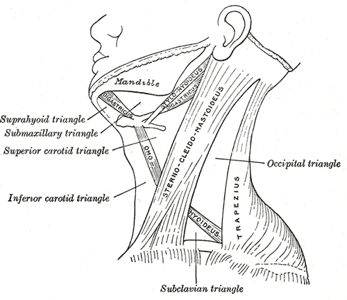
The sternocleidomastoid muscle is one of the largest and most superficial cervical muscles. The primary actions of the muscle are rotation of the head to the opposite side and flexion of the neck. The sternocleidomastoid is innervated by the accessory nerve. Etymology and location It is given the name ''sternocleidomastoid'' because it originates at the manubrium of the sternum (''sterno-'') and the clavicle (''cleido-'') and has an insertion at the mastoid process of the temporal bone of the skull. Structure The sternocleidomastoid muscle originates from two locations: the manubrium of the sternum and the clavicle. It travels obliquely across the side of the neck and inserts at the mastoid process of the temporal bone of the skull by a thin aponeurosis. The sternocleidomastoid is thick and narrow at its centre, and broader and thinner at either end. The sternal head is a round fasciculus, tendinous in front, fleshy behind, arising from the upper part of the front of the manubri ... [...More Info...] [...Related Items...] OR: [Wikipedia] [Google] [Baidu] |
Facial Nerve
The facial nerve, also known as the seventh cranial nerve, cranial nerve VII, or simply CN VII, is a cranial nerve that emerges from the pons of the brainstem, controls the muscles of facial expression, and functions in the conveyance of taste sensations from the anterior two-thirds of the tongue. The nerve typically travels from the pons through the facial canal in the temporal bone and exits the skull at the stylomastoid foramen. It arises from the brainstem from an area posterior to the cranial nerve VI (abducens nerve) and anterior to cranial nerve VIII (vestibulocochlear nerve). The facial nerve also supplies preganglionic parasympathetic fibers to several head and neck ganglia. The facial and intermediate nerves can be collectively referred to as the nervus intermediofacialis. The path of the facial nerve can be divided into six segments: # intracranial (cisternal) segment # meatal (canalicular) segment (within the internal auditory canal) # labyrinthine segment ... [...More Info...] [...Related Items...] OR: [Wikipedia] [Google] [Baidu] |


