|
Rectus Capitis Posterior Minor Muscle
The rectus capitis posterior minor (or rectus capitis posticus minor, both being Latin for ''lesser posterior straight muscle of the head'') arises by a narrow pointed tendon from the tubercle on the posterior arch of the atlas, and, widening as it ascends, is inserted into the medial part of the inferior nuchal line of the occipital bone and the surface between it and the foramen magnum, and also takes some attachment to the spinal dura mater. The synergists are the rectus capitis posterior major and the obliquus capitis superior. Connective tissue bridges were noted at the atlanto-occipital joint between the rectus capitis posterior minor (RCPm) muscle and the dorsal spinal dura. Similar connective tissue connections of the rectus capitis posterior major have been reported recently as well. The perpendicular arrangement of these fibers appears to restrict dural movement toward the spinal cord. The ligamentum nuchae was found to be continuous with the posterior cervical spinal ... [...More Info...] [...Related Items...] OR: [Wikipedia] [Google] [Baidu] |
Human Skull
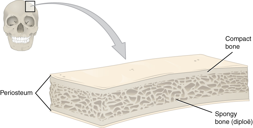
The skull is a bone protective cavity for the brain. The skull is composed of four types of bone i.e., cranial bones, facial bones, ear ossicles and hyoid bone. However two parts are more prominent: the cranium and the mandible. In humans, these two parts are the neurocranium and the viscerocranium ( facial skeleton) that includes the mandible as its largest bone. The skull forms the anterior-most portion of the skeleton and is a product of cephalisation—housing the brain, and several sensory structures such as the eyes, ears, nose, and mouth. In humans these sensory structures are part of the facial skeleton. Functions of the skull include protection of the brain, fixing the distance between the eyes to allow stereoscopic vision, and fixing the position of the ears to enable sound localisation of the direction and distance of sounds. In some animals, such as horned ungulates (mammals with hooves), the skull also has a defensive function by providing the mount (on the front ... [...More Info...] [...Related Items...] OR: [Wikipedia] [Google] [Baidu] |
Obliquus Capitis Superior Muscle
The obliquus capitis superior muscle () is a small muscle in the upper back part of the neck and is one of the suboccipital muscles and part of the suboccipital triangle. It arises from the lateral mass of the atlas bone. It passes superiorly and posteriorly to insert into the lateral half of the inferior nuchal line on the external surface of the occipital bone. The muscle is innervated by the suboccipital nerve, the dorsal ramus of the first spinal nerve. It acts at the atlanto-occipital joint to extend the head and flex the head to the ipsilateral side. Additional images File:Obliquus capitis superior muscle - animation04.gif, Position of obliquus capitis superior (shown in red). Animation. File:Obliquus capitis superior muscle05.png, Still image. Posterior view. File:Obliquus capitis superior.png, Deep muscles of the back (obliquus capitis superior labeled at upper left) File:Gray129.png, Occipital bone. Outer surface. Muscle attachments are shown as red circles. File ... [...More Info...] [...Related Items...] OR: [Wikipedia] [Google] [Baidu] |
Rectus Capitis Posterior Major Muscle
The rectus capitis posterior major (or rectus capitis posticus major, both being Latin for ''larger posterior straight muscle of the head'') arises by a pointed tendon from the spinous process of the axis, and, becoming broader as it ascends, is inserted into the lateral part of the inferior nuchal line of the occipital bone and the surface of the bone immediately below the line. A soft tissue connection bridging from the rectus capitis posterior major to the cervical dura mater was described in 2011. Various clinical manifestations may be linked to this anatomical relationship. It has also been postulated that this connection serves as a monitor of dural tension along with the rectus capitis posterior minor and the obliquus capitis inferior. As the muscles of the two sides pass upward and lateralward, they leave between them a triangular space, in which the rectus capitis posterior minor is seen. Its main actions are to extend and rotate the atlanto-occipital joint. See also * ... [...More Info...] [...Related Items...] OR: [Wikipedia] [Google] [Baidu] |
Rectus Capitis Lateralis
The rectus capitis lateralis, a short, flat muscle, arises from the upper surface of the transverse process of the atlas, and is inserted into the under surface of the jugular process of the occipital bone. Additional images File:Rectus capitis lateralis muscle - animation01.gif, Position of rectus capitis lateralis muscle (shown in red). Animation. File:Rectus capitis lateralis muscle - animation05.gif, Close up. Skull has been removed (except occipital bone). File:Rectus capitis lateralis muscle03.png, Lateral view. Still image. File:Gray129.png, Occipital bone. Outer surface. File:Gray187.png, Base of skull. Inferior surface. See also * Atlanto-occipital joint * Rectus capitis posterior major muscle * Rectus capitis posterior minor muscle * Rectus capitis anterior muscle The rectus capitis anterior (rectus capitis anticus minor) is a short, flat muscle, situated immediately behind the upper part of the Longus capitis. It arises from the anterior surface of the lateral ... [...More Info...] [...Related Items...] OR: [Wikipedia] [Google] [Baidu] |
Atlanto-occipital Joint
The atlanto-occipital joint (''Capsula articularis atlantooccipitalis'') is an articulation between the atlas bone and the occipital bone. It consists of a pair of condyloid joints. It is a synovial joint. Structure The atlanto-occipital joint is an articulation between the atlas bone and the occipital bone. It consists of a pair of condyloid joints. It is a synovial joint. Ligaments The ligaments connecting the bones are: * Two articular capsules * Posterior atlanto-occipital membrane * Anterior atlanto-occipital membrane Capsule The capsules of the atlantooccipital articulation surround the condyles of the occipital bone, and connect them with the articular processes of the atlas: they are thin and loose. Function The movements permitted in this joint are: * (a) flexion and extension around the mediolateral axis, which give rise to the ordinary forward and backward nodding of the head. * (b) slight lateral motion, lateroflexion, to one or other side around the anteroposter ... [...More Info...] [...Related Items...] OR: [Wikipedia] [Google] [Baidu] |
Human Skull
The skull is a bone protective cavity for the brain. The skull is composed of four types of bone i.e., cranial bones, facial bones, ear ossicles and hyoid bone. However two parts are more prominent: the cranium and the mandible. In humans, these two parts are the neurocranium and the viscerocranium ( facial skeleton) that includes the mandible as its largest bone. The skull forms the anterior-most portion of the skeleton and is a product of cephalisation—housing the brain, and several sensory structures such as the eyes, ears, nose, and mouth. In humans these sensory structures are part of the facial skeleton. Functions of the skull include protection of the brain, fixing the distance between the eyes to allow stereoscopic vision, and fixing the position of the ears to enable sound localisation of the direction and distance of sounds. In some animals, such as horned ungulates (mammals with hooves), the skull also has a defensive function by providing the mount (on the front ... [...More Info...] [...Related Items...] OR: [Wikipedia] [Google] [Baidu] |
Cervicogenic Headache
Cervicogenic headache is a type of headache characterized by chronic hemicranial pain referred to the head from either the cervical spine or soft tissues within the neck. The main symptoms of cervicogenic headaches include pain originating in the neck that can travel to the head or face, headaches that get worse with neck movement, and limited ability to move the neck. Diagnostic imaging can display lesions of the cervical spine or soft tissue of the neck that can be indicative of a cervicogenic headache. When being evaluated for cervicogenic headaches, it is important to rule out a history of migraines and traumatic brain injuries. Studies show that combining interventions such as moist heat applied to the area of pain, spinal and cervical manipulations, and neck massages all help reduce or relieve symptoms. Neck exercises are also beneficial. Specifically, craniocervical flexion, or forward bending of the neck, against light resistance helps increase muscular stability of the he ... [...More Info...] [...Related Items...] OR: [Wikipedia] [Google] [Baidu] |
Cervical Nerve
A spinal nerve is a mixed nerve, which carries motor, sensory, and autonomic signals between the spinal cord and the body. In the human body there are 31 pairs of spinal nerves, one on each side of the vertebral column. These are grouped into the corresponding cervical, thoracic, lumbar, sacral and coccygeal regions of the spine. There are eight pairs of cervical nerves, twelve pairs of thoracic nerves, five pairs of lumbar nerves, five pairs of sacral nerves, and one pair of coccygeal nerves. The spinal nerves are part of the peripheral nervous system. Structure Each spinal nerve is a mixed nerve, formed from the combination of nerve fibers from its dorsal and ventral roots. The dorsal root is the afferent sensory root and carries sensory information to the brain. The ventral root is the efferent motor root and carries motor information from the brain. The spinal nerve emerges from the spinal column through an opening (intervertebral foramen) between adjacent vertebrae. Th ... [...More Info...] [...Related Items...] OR: [Wikipedia] [Google] [Baidu] |
Atlanto-occipital Joint
The atlanto-occipital joint (''Capsula articularis atlantooccipitalis'') is an articulation between the atlas bone and the occipital bone. It consists of a pair of condyloid joints. It is a synovial joint. Structure The atlanto-occipital joint is an articulation between the atlas bone and the occipital bone. It consists of a pair of condyloid joints. It is a synovial joint. Ligaments The ligaments connecting the bones are: * Two articular capsules * Posterior atlanto-occipital membrane * Anterior atlanto-occipital membrane Capsule The capsules of the atlantooccipital articulation surround the condyles of the occipital bone, and connect them with the articular processes of the atlas: they are thin and loose. Function The movements permitted in this joint are: * (a) flexion and extension around the mediolateral axis, which give rise to the ordinary forward and backward nodding of the head. * (b) slight lateral motion, lateroflexion, to one or other side around the anteroposter ... [...More Info...] [...Related Items...] OR: [Wikipedia] [Google] [Baidu] |
Rectus Capitis Posterior Major
The rectus capitis posterior major (or rectus capitis posticus major, both being Latin for ''larger posterior straight muscle of the head'') arises by a pointed tendon from the spinous process of the axis, and, becoming broader as it ascends, is inserted into the lateral part of the inferior nuchal line of the occipital bone and the surface of the bone immediately below the line. A soft tissue connection bridging from the rectus capitis posterior major to the cervical dura mater was described in 2011. Various clinical manifestations may be linked to this anatomical relationship. It has also been postulated that this connection serves as a monitor of dural tension along with the rectus capitis posterior minor and the obliquus capitis inferior. As the muscles of the two sides pass upward and lateralward, they leave between them a triangular space, in which the rectus capitis posterior minor is seen. Its main actions are to extend and rotate the atlanto-occipital joint. See also * ... [...More Info...] [...Related Items...] OR: [Wikipedia] [Google] [Baidu] |
Back
The human back, also called the dorsum, is the large posterior area of the human body, rising from the top of the buttocks to the back of the neck. It is the surface of the body opposite from the chest and the abdomen. The vertebral column runs the length of the back and creates a central area of recession. The breadth of the back is created by the shoulders at the top and the pelvis at the bottom. Back pain is a common medical condition, generally benign in origin. Structure The central feature of the human back is the vertebral column, specifically the length from the top of the thoracic vertebrae to the bottom of the lumbar vertebrae, which houses the spinal cord in its spinal canal, and which generally has some curvature that gives shape to the back. The ribcage extends from the spine at the top of the back (with the top of the ribcage corresponding to the T1 vertebra), more than halfway down the length of the back, leaving an area with less protection between the bottom ... [...More Info...] [...Related Items...] OR: [Wikipedia] [Google] [Baidu] |
Anatomical Terms Of Muscle
Anatomical terminology is used to uniquely describe aspects of skeletal muscle, cardiac muscle, and smooth muscle such as their actions, structure, size, and location. Types There are three types of muscle tissue in the body: skeletal, smooth, and cardiac. Skeletal muscle Skeletal muscle, or "voluntary muscle", is a striated muscle tissue that primarily joins to bone with tendons. Skeletal muscle enables movement of bones, and maintains posture. The widest part of a muscle that pulls on the tendons is known as the belly. Muscle slip A muscle slip is a slip of muscle that can either be an anatomical variant, or a branching of a muscle as in rib connections of the serratus anterior muscle. Smooth muscle Smooth muscle is involuntary and found in parts of the body where it conveys action without conscious intent. The majority of this type of muscle tissue is found in the digestive and urinary systems where it acts by propelling forward food, chyme, and feces in the forme ... [...More Info...] [...Related Items...] OR: [Wikipedia] [Google] [Baidu] |


