|
Punctate Inner Choroiditis
Punctate inner choroiditis (PIC) is an inflammatory choroiditis which occurs mainly in young women. Symptoms include blurred vision and scotomata. Yellow lesions are mainly present in the posterior pole and are between 100 and 300 micrometres in size. PIC is one of the so-called White Dot Syndromes. PIC has only been recognised as a distinct condition as recently as 1984 when Watzke identified 10 patients who appeared to make up a distinct group within the White Dot Syndromes. Signs and symptoms • Typically affects short sighted (myopic) women. (90% of cases are female). • The average age of patients with PIC is 27 years with a range of 16–40 years. • Patients are otherwise healthy and there is usually no illness, which triggers the condition or precedes it. • The inflammation is confined to the back of the eye (posterior). There is no inflammation in the front of the eye (anterior chamber) or vitreous (the clear jelly inside the eye). This is an important distingui ... [...More Info...] [...Related Items...] OR: [Wikipedia] [Google] [Baidu] |
Photopsia
Photopsia is the presence of perceived flashes of light in the Visual field, field of vision. It is most commonly associated with: * posterior vitreous detachment * migraine aura (ocular migraine / retinal migraine) * acephalgic migraine, migraine aura without headache * scintillating scotoma * retinal detachment, retinal break or detachment * occipital lobe infarction (similar to stroke, occipital stroke) * sensory deprivation (ophthalmopathic hallucinations) * Macular degeneration, age-related macular degeneration * vertebrobasilar insufficiency * optic neuritis * visual snow syndrome Vitreous shrinkage or liquefaction, which are the most common causes of photopsia, cause a pull in vitreoretinal attachments, irritating the retina and causing it to discharge electrical impulses. These impulses are interpreted by the brain as flashes. This condition has also been identified as a common initial symptom of punctate inner choroiditis (punctate inner choroiditis, PIC), a rare retin ... [...More Info...] [...Related Items...] OR: [Wikipedia] [Google] [Baidu] |
Choroiditis
Chorioretinitis is an inflammation of the choroid (thin pigmented vascular coat of the eye) and retina of the eye. It is a form of posterior uveitis. If only the choroid is inflamed, not the retina, the condition is termed choroiditis. The ophthalmologist's goal in treating these potentially blinding conditions is to eliminate the inflammation and minimize the potential risk of therapy to the patient. Symptoms Symptoms may include the presence of floating black spots, blurred vision, pain or redness in the eye, sensitivity to light, or excessive tearing. Causes Chorioretinitis is often caused by toxoplasmosis and cytomegalovirus infections (mostly seen in immunodeficient subjects such as people with HIV/AIDS or on immunosuppressant drugs). Congenital toxoplasmosis via transplacental transmission can also lead to sequelae such as chorioretinitis along with hydrocephalus and cerebral calcifications. Other possible causes of chorioretinitis are syphilis, sarcoidosis, tuberculosis, Be ... [...More Info...] [...Related Items...] OR: [Wikipedia] [Google] [Baidu] |
Scotoma
A scotoma is an area of partial alteration in the field of vision consisting of a partially diminished or entirely degenerated visual acuity that is surrounded by a field of normal – or relatively well-preserved – vision. Every normal mammalian eye has a scotoma in its field of vision, usually termed its blind spot. This is a location with no photoreceptor cells, where the retinal ganglion cell axons that compose the optic nerve exit the retina. This location is called the optic disc. There is no direct conscious awareness of visual scotomas. They are simply regions of reduced information within the visual field. Rather than recognizing an incomplete image, patients with scotomas report that things "disappear" on them. The presence of the blind spot scotoma can be demonstrated subjectively by covering one eye, carefully holding fixation with the open eye, and placing an object (such as one's thumb) in the lateral and horizontal visual field, about 15 degrees from fix ... [...More Info...] [...Related Items...] OR: [Wikipedia] [Google] [Baidu] |
White Dot Syndromes
White dot syndromes are inflammatory diseases characterized by the presence of white dots on the fundus (eye), fundus, the interior surface of the eye. Tewari A, Elliot D. White Dot Syndromes. 2007. Emedicine from WebMD. The majority of individuals affected with white dot syndromes are younger than fifty years of age. Some symptoms include blurred vision and visual field loss.Quillen DA, Davis JB, Gottlieb JL, Blodi BA, Callanan DG, Chang TS, et al. The white dot syndromes. American Journal of Ophthalmology. 2004;137(3):538-50. There are many theories for the etiology of white dot syndromes including infectious, viral, genetics and autoimmune. Classically recognized white dot syndromes include:Forrester JV, IOIS, Okada AA, BenEzra D. Posterior segment intraocular inflammation: guidelines. 1998:184. [...More Info...] [...Related Items...] OR: [Wikipedia] [Google] [Baidu] |
Ophthalmologist
Ophthalmology ( ) is a surgery, surgical subspecialty within medicine that deals with the diagnosis and treatment of eye disorders. An ophthalmologist is a physician who undergoes subspecialty training in medical and surgical eye care. Following a medical degree, a doctor specialising in ophthalmology must pursue additional postgraduate residency (medicine), residency training specific to that field. This may include a one-year integrated internship that involves more general medical training in other fields such as internal medicine or general surgery. Following residency, additional specialty training (or fellowship) may be sought in a particular aspect of eye pathology. Ophthalmologists prescribe medications to treat eye diseases, implement laser therapy, and perform surgery when needed. Ophthalmologists provide both primary and specialty eye care - medical and surgical. Most ophthalmologists participate in academic research on eye diseases at some point in their training an ... [...More Info...] [...Related Items...] OR: [Wikipedia] [Google] [Baidu] |
Visual Field Test
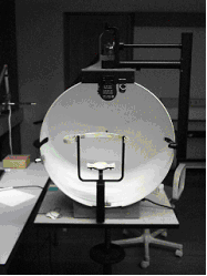
A visual field test is an eye examination that can detect dysfunction in central and peripheral vision which may be caused by various medical conditions such as glaucoma, stroke, pituitary disease, brain tumours or other neurological deficits. Visual field testing can be performed clinically by keeping the subject's gaze fixed while presenting objects at various places within their visual field. Simple manual equipment can be used such as in the tangent screen test or the Amsler grid. When dedicated machinery is used it is called a perimeter. The exam may be performed by a technician in one of several ways. The test may be performed by a technician directly, with the assistance of a machine, or completely by an automated machine. Machine-based tests aid diagnostics by allowing a detailed printout of the patient's visual field. Other names for this test may include perimetry, Tangent screen exam, Automated perimetry exam, Goldmann visual field exam, or brand names such as Hen ... [...More Info...] [...Related Items...] OR: [Wikipedia] [Google] [Baidu] |
Choroidal Neovascularization
Choroidal neovascularization (CNV) is the creation of new blood vessels in the choroid layer of the eye. Choroidal neovascularization is a common cause of neovascular degenerative maculopathy (i.e. 'wet' macular degeneration) commonly exacerbated by extreme myopia, malignant myopic degeneration, or age-related developments. Causes CNV can occur rapidly in individuals with defects in Bruch's membrane, the innermost layer of the choroid. It is also associated with excessive amounts of vascular endothelial growth factor (VEGF). As well as in wet macular degeneration, CNV can also occur frequently with the rare genetic disease pseudoxanthoma elasticum and rarely with the more common optic disc drusen. CNV has also been associated with extreme myopia or malignant myopic degeneration, where in choroidal neovascularization occurs primarily in the presence of cracks within the retinal (specifically) macular tissue known as lacquer cracks. Symptoms CNV can create a sudden deterioration o ... [...More Info...] [...Related Items...] OR: [Wikipedia] [Google] [Baidu] |
VEGF
Vascular endothelial growth factor (VEGF, ), originally known as vascular permeability factor (VPF), is a signal protein produced by many cells that stimulates the formation of blood vessels. To be specific, VEGF is a sub-family of growth factors, the platelet-derived growth factor family of cystine-knot growth factors. They are important signaling proteins involved in both vasculogenesis (the '' de novo'' formation of the embryonic circulatory system) and angiogenesis (the growth of blood vessels from pre-existing vasculature). It is part of the system that restores the oxygen supply to tissues when blood circulation is inadequate such as in hypoxic conditions. Serum concentration of VEGF is high in bronchial asthma and diabetes mellitus. VEGF's normal function is to create new blood vessels during embryonic development, new blood vessels after injury, muscle following exercise, and new vessels (collateral circulation) to bypass blocked vessels. It can contribute to disease. Sol ... [...More Info...] [...Related Items...] OR: [Wikipedia] [Google] [Baidu] |
Bevacizumab
Bevacizumab, sold under the brand name Avastin among others, is a medication used to treat a number of types of cancers and a specific eye disease. For cancer, it is given by slow injection into a vein (intravenous) and used for colon cancer, lung cancer, glioblastoma, and renal-cell carcinoma. In many of these diseases it is used as a first-line therapy. For age-related macular degeneration it is given by injection into the eye (intravitreal). Common side effects when used for cancer include nose bleeds, headache, high blood pressure, and rash. Other severe side effects include gastrointestinal perforation, bleeding, allergic reactions, blood clots, and an increased risk of infection. When used for eye disease side effects can include vision loss and retinal detachment. Bevacizumab is a monoclonal antibody that functions as an angiogenesis inhibitor. It works by slowing the growth of new blood vessels by inhibiting vascular endothelial growth factor A (VEGF-A), in other word ... [...More Info...] [...Related Items...] OR: [Wikipedia] [Google] [Baidu] |
Ranibizumab
Ranibizumab, sold under the brand name Lucentis among others, is a monoclonal antibody fragment ( Fab) created from the same parent mouse antibody as bevacizumab. It is an anti-angiogenic that is approved to treat the "wet" type of age-related macular degeneration (AMD, also ARMD), diabetic retinopathy, and macular edema due to branch retinal vein occlusion or central retinal vein occlusion. Ranibizumab was developed by Genentech and marketed by them in the United States, and elsewhere by Novartis, under the brand name Lucentis. Ranibizumab (Lucentis) was approved for medical use in the United States in June 2006. Ranibizumab (Susvimo) was approved for medical use in the United States in October 2021. Medical uses In the United States, ranibizumab is indicated for the treatment of neovascular (wet) age-related macular degeneration, macular edema following retinal vein occlusion, diabetic macular edema, diabetic retinopathy, and myopic choroidal neovascularization. In the Europ ... [...More Info...] [...Related Items...] OR: [Wikipedia] [Google] [Baidu] |


