|
Pulmonary Injury
A chest injury, also known as chest trauma, is any form of physical injury to the chest including the ribs, heart and lungs. Chest injuries account for 25% of all deaths from traumatic injury. Typically chest injuries are caused by blunt mechanisms such as direct, indirect, compression, contusion, deceleration, or blasts- caused by motor vehicle collisions or penetrating mechanisms such as stabbings. Classification Chest injuries can be classified as blunt or penetrating. Blunt and penetrating injuries have different pathophysiologies and clinical courses. Specific types of injuries include: * Injuries to the chest wall ** Chest wall contusions or hematomas. ** Rib fractures ** Flail chest ** Sternal fractures ** Fractures of the shoulder girdle * Pulmonary injury (injury to the lung) and injuries involving the pleural space ** Pulmonary contusion ** Pulmonary laceration ** Pneumothorax ** Hemothorax ** Hemopneumothorax * Injury to the airways ** Tracheobronchial tear * Car ... [...More Info...] [...Related Items...] OR: [Wikipedia] [Google] [Baidu] |
Chest X-ray
A chest radiograph, called a chest X-ray (CXR), or chest film, is a projection radiograph of the chest used to diagnose conditions affecting the chest, its contents, and nearby structures. Chest radiographs are the most common film taken in medicine. Like all methods of radiography, chest radiography employs ionizing radiation in the form of X-rays to generate images of the chest. The mean radiation dose to an adult from a chest radiograph is around 0.02 mSv (2 mrem) for a front view (PA, or posteroanterior) and 0.08 mSv (8 mrem) for a side view (LL, or latero-lateral). Together, this corresponds to a background radiation equivalent time of about 10 days. Medical uses Conditions commonly identified by chest radiography * Pneumonia * Pneumothorax * Interstitial lung disease * Heart failure * Bone fracture * Hiatal hernia Chest radiographs are used to diagnose many conditions involving the chest wall, including its bones, and also structures contained within the thoracic c ... [...More Info...] [...Related Items...] OR: [Wikipedia] [Google] [Baidu] |
Sternal Fracture
A sternal fracture is a fracture of the sternum (the breastbone), located in the center of the chest. The injury, which occurs in 5–8% of people who experience significant blunt chest trauma, may occur in vehicle accidents, when the still-moving chest strikes a steering wheel or dashboard or is injured by a seatbelt. Cardiopulmonary resuscitation (CPR), has also been known to cause thoracic injury, including sternum and rib fractures. Sternal fractures may also occur as a pathological fracture, in people who have weakened bone in their sternum, due to another disease process. Sternal fracture can interfere with breathing by making it more painful; however, its primary significance is that it can indicate the presence of serious associated internal injuries, especially to the heart and lungs. Signs and symptoms Signs and symptoms include crepitus (a crunching sound made when broken bone ends rub together), pain, tenderness, bruising, and swelling over the fracture site. The f ... [...More Info...] [...Related Items...] OR: [Wikipedia] [Google] [Baidu] |
Hemopericardium
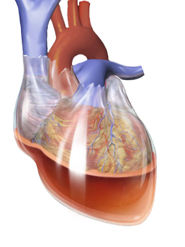
Hemopericardium refers to blood in the pericardial sac of the heart. It is clinically similar to a pericardial effusion, and, depending on the volume and rapidity with which it develops, may cause cardiac tamponade. The condition can be caused by full-thickness necrosis (death) of the myocardium (heart muscle) after myocardial infarction, chest trauma, and by over-prescription of anticoagulants. Other causes include ruptured aneurysm of sinus of Valsalva and other aneurysms of the aortic arch.Gray's Anatomy, 1902 ed. Hemopericardium can be diagnosed with a chest X-ray or a chest ultrasound, and is most commonly treated with pericardiocentesis. While hemopericardium itself is not deadly, it can lead to cardiac tamponade, a condition that is fatal if left untreated. Symptoms and signs Symptoms of hemopericardium often include difficulty breathing, abnormally rapid breathing, and fatigue, each of which can be a sign of a serious medical condition not limited to hemopericardium ... [...More Info...] [...Related Items...] OR: [Wikipedia] [Google] [Baidu] |
Traumatic Arrest
Traumatic cardiac arrest (TCA) is a condition in which the heart has ceased to beat due to blunt or penetrating trauma, such as a stab wound to the thoracic area. It is a medical emergency which will always result in death without prompt advanced medical care. Even with prompt medical intervention, survival without neurological complications is rare. In recent years, protocols have been proposed to improve survival rate in patients with traumatic cardiac arrest, though the variable causes of this condition as well as many coexisting injuries can make these protocols difficult to standardize. Traumatic cardiac arrest is a complex form of cardiac arrest often derailing from advanced cardiac life support in the sense that the emergency team must first establish the cause of the traumatic arrest and reverse these effects, for example hypovolemia and haemorrhagic shock due to a penetrating injury. Mechanism Traumatic cardiac arrest can occur in patients following any severe blunt o ... [...More Info...] [...Related Items...] OR: [Wikipedia] [Google] [Baidu] |
Myocardial Contusion
A blunt cardiac injury is an injury to the heart as the result of blunt trauma, typically to the anterior chest wall. It can result in a variety of specific injuries to the heart, the most common of which is a myocardial contusion, which is a term for a bruise (contusion) to the heart after an injury. Other injuries which can result include septal defects and valvular failures. The right ventricle is thought to be most commonly affected due to its anatomic location as the most anterior surface of the heart. Myocardial contusion is not a specific diagnosis and the extent of the injury can vary greatly. Usually, there are other chest injuries seen with a myocardial contusion such as rib fractures, pneumothorax, and heart valve injury. When a myocardial contusion is suspected, consideration must be given to any other chest injuries, which will likely be determined by clinical signs, tests, and imaging. The signs and symptoms of a myocardial contusion can manifest in different ways in ... [...More Info...] [...Related Items...] OR: [Wikipedia] [Google] [Baidu] |
Pericardial Tamponade
Cardiac tamponade, also known as pericardial tamponade (), is the buildup of fluid in the pericardium (the sac around the heart), resulting in compression of the heart. Onset may be rapid or gradual. Symptoms typically include those of obstructive shock including shortness of breath, weakness, lightheadedness, and cough. Other symptoms may relate to the underlying cause. Common causes of cardiac tamponade include cancer, kidney failure, chest trauma, myocardial infarction, and pericarditis. Other causes include connective tissues diseases, hypothyroidism, aortic rupture, autoimmune disease, and complications of cardiac surgery. In Africa, tuberculosis is a relatively common cause. Diagnosis may be suspected based on low blood pressure, jugular venous distension, or quiet heart sounds (together known as Beck's triad). A pericardial rub may be present in cases due to inflammation. The diagnosis may be further supported by specific electrocardiogram (ECG) changes, chest ... [...More Info...] [...Related Items...] OR: [Wikipedia] [Google] [Baidu] |
Tracheobronchial Tear
Tracheobronchial injury is damage to the tracheobronchial tree (the airway structure involving the trachea and bronchi). It can result from blunt or penetrating trauma to the neck or chest, inhalation of harmful fumes or smoke, or aspiration of liquids or objects. Though rare, TBI is a serious condition; it may cause obstruction of the airway with resulting life-threatening respiratory insufficiency. Other injuries accompany TBI in about half of cases. Of those people with TBI who die, most do so before receiving emergency care, either from airway obstruction, exsanguination, or from injuries to other vital organs. Of those who do reach a hospital, the mortality rate may be as high as 30%. TBI is frequently difficult to diagnose and treat. Early diagnosis is important to prevent complications, which include stenosis (narrowing) of the airway, respiratory tract infection, and damage to the lung tissue. Diagnosis involves procedures such as bronchoscopy, radiography, and x-ra ... [...More Info...] [...Related Items...] OR: [Wikipedia] [Google] [Baidu] |
Hemopneumothorax
Hemopneumothorax, or haemopneumothorax, is the condition of having both air (pneumothorax) and blood (hemothorax) in the chest cavity. A hemothorax, pneumothorax, or the combination of both can occur due to an injury to the lung or chest. Cause The pleural space is located anatomically between the visceral membrane, which is firmly attached to the lungs, and the parietal membrane which is firmly attached to the chest wall (a.k.a. ribcage and intercostal muscles, muscles between the ribs). The pleural space contains pleural fluid. This fluid holds the two membranes together by surface tension, as much as a drop of water between two sheets of glass prevents them from separating. Because of this, when the intercostal muscles move the ribcage outward, the lungs are pulled out as well, dropping the pressure in the lungs and pulling air into the bronchi, when we 'breathe in'. The pleural space is maintained in a constant state of negative pressure (in comparison to atmospheric pressu ... [...More Info...] [...Related Items...] OR: [Wikipedia] [Google] [Baidu] |
Hemothorax
A hemothorax (derived from hemo- lood+ thorax hest plural ''hemothoraces'') is an accumulation of blood within the pleural cavity. The symptoms of a hemothorax may include chest pain and difficulty breathing, while the clinical signs may include reduced breath sounds on the affected side and a rapid heart rate. Hemothoraces are usually caused by an injury, but they may occur spontaneously due to cancer invading the pleural cavity, as a result of a blood clotting disorder, as an unusual manifestation of endometriosis, in response to a collapsed lung, or rarely in association with other conditions. Hemothoraces are usually diagnosed using a chest X-ray, but they can be identified using other forms of imaging including ultrasound, a CT scan, or an MRI. They can be differentiated from other forms of fluid within the pleural cavity by analysing a sample of the fluid, and are defined as having a hematocrit of greater than 50% that of the person's blood. Hemothoraces may be tre ... [...More Info...] [...Related Items...] OR: [Wikipedia] [Google] [Baidu] |
Pneumothorax
A pneumothorax is an abnormal collection of air in the pleural space between the lung and the chest wall. Symptoms typically include sudden onset of sharp, one-sided chest pain and shortness of breath. In a minority of cases, a one-way valve is formed by an area of damaged tissue, and the amount of air in the space between chest wall and lungs increases; this is called a tension pneumothorax. This can cause a steadily worsening oxygen shortage and low blood pressure. This leads to a type of shock called obstructive shock, which can be fatal unless reversed. Very rarely, both lungs may be affected by a pneumothorax. It is often called a "collapsed lung", although that term may also refer to atelectasis. A primary spontaneous pneumothorax is one that occurs without an apparent cause and in the absence of significant lung disease. A secondary spontaneous pneumothorax occurs in the presence of existing lung disease. Smoking increases the risk of primary spontaneous pneumothora ... [...More Info...] [...Related Items...] OR: [Wikipedia] [Google] [Baidu] |
Pulmonary Laceration
A pulmonary laceration is a chest injury in which lung tissue is torn or cut. An injury that is potentially more serious than pulmonary contusion, pulmonary laceration involves disruption of the architecture of the lung, while pulmonary contusion does not. Pulmonary laceration is commonly caused by penetrating trauma but may also result from forces involved in blunt trauma such as shear stress. A cavity filled with blood, air, or both can form. The injury is diagnosed when collections of air or fluid are found on a CT scan of the chest. Surgery may be required to stitch the laceration, to drain blood, or even to remove injured parts of the lung. The injury commonly heals quickly with few problems if it is given proper treatment; however it may be associated with scarring of the lung or other complications. Presentation Complications Complications are not common but include infection, lung abscess, and bronchopleural fistula (a fistula between the pleural space and the bronchi ... [...More Info...] [...Related Items...] OR: [Wikipedia] [Google] [Baidu] |
Pulmonary Contusion
A pulmonary contusion, also known as lung contusion, is a bruise of the lung, caused by chest trauma. As a result of damage to capillaries, blood and other fluids accumulate in the lung tissue. The excess fluid interferes with gas exchange, potentially leading to inadequate oxygen levels ( hypoxia). Unlike pulmonary laceration, another type of lung injury, pulmonary contusion does not involve a cut or tear of the lung tissue. A pulmonary contusion is usually caused directly by blunt trauma but can also result from explosion injuries or a shock wave associated with penetrating trauma. With the use of explosives during World Wars I and II, pulmonary contusion resulting from blasts gained recognition. In the 1960s its occurrence in civilians began to receive wider recognition, in which cases it is usually caused by traffic accidents. The use of seat belts and airbags reduces the risk to vehicle occupants. Diagnosis is made by studying the cause of the injury, physical examinati ... [...More Info...] [...Related Items...] OR: [Wikipedia] [Google] [Baidu] |


.jpg)
