|
Pterygoid Canal
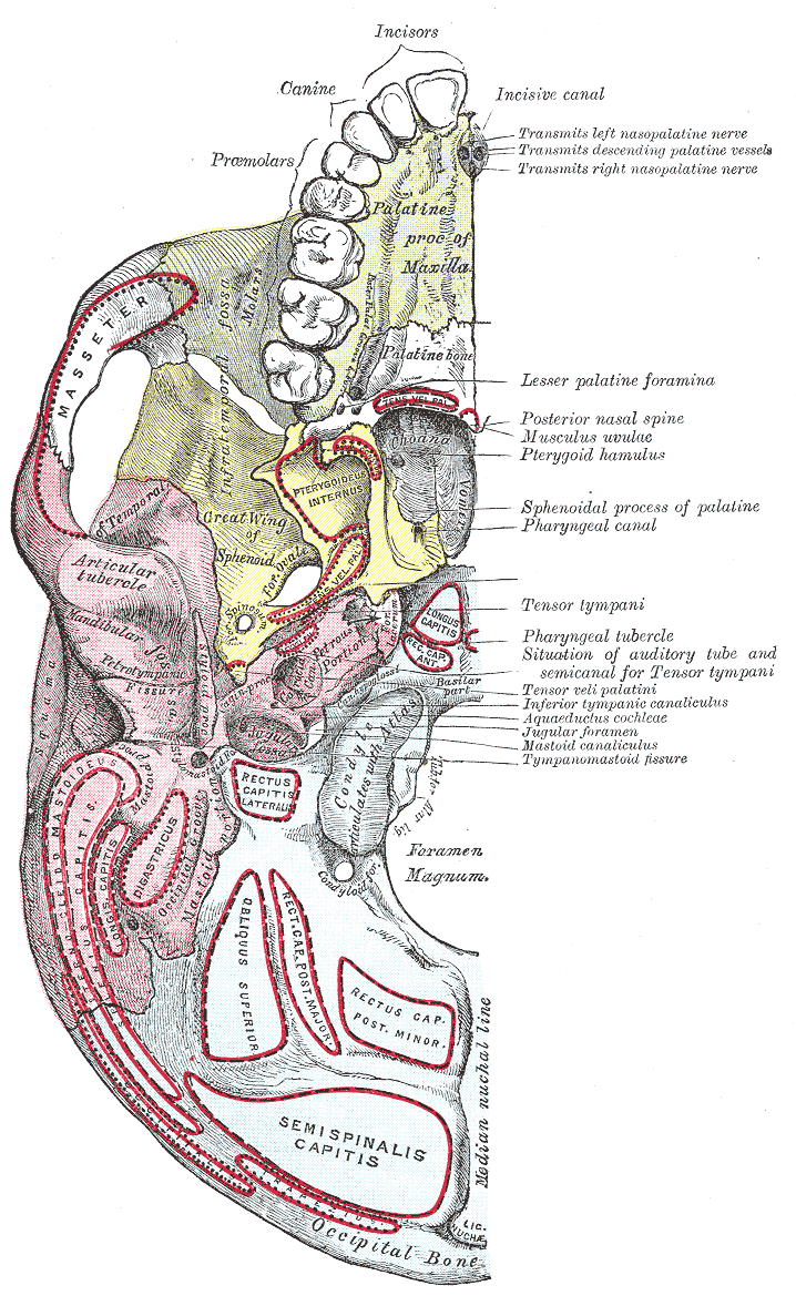
The pterygoid canal (also vidian canal) is a passage in the sphenoid bone of the skull leading from just anterior to the foramen lacerum in the middle cranial fossa to the pterygopalatine fossa. Structure The pterygoid canal runs through the medial pterygoid plate of the sphenoid bone to the back wall of the pterygopalatine fossa. Contents It transmits the nerve of pterygoid canal, (Vidian nerve), the artery of the pterygoid canal The artery of the pterygoid canal (or Vidian artery) is an artery in the pterygoid canal, in the head. It usually arises from the external carotid artery, but can arise from either the internal or external carotid artery or serve as an anastomosi ... (Vidian artery), and the vein of the pterygoid canal (Vidian vein). Additional images File:Gray192.png, Medial wall of left orbit. External links * * () References Foramina of the skull {{musculoskeletal-stub ... [...More Info...] [...Related Items...] OR: [Wikipedia] [Google] [Baidu] |
Base Of Skull
The base of skull, also known as the cranial base or the cranial floor, is the most inferior area of the skull. It is composed of the endocranium and the lower parts of the calvaria. Structure Structures found at the base of the skull are for example: Bones There are five bones that make up the base of the skull: *Ethmoid bone *Sphenoid bone *Occipital bone *Frontal bone *Temporal bone Sinuses *Occipital sinus *Superior sagittal sinus *Superior petrosal sinus Foramina of the skull * Foramen cecum *Optic foramen *Foramen lacerum *Foramen rotundum *Foramen magnum * Foramen ovale *Jugular foramen *Internal auditory meatus *Mastoid foramen *Sphenoidal emissary foramen *Foramen spinosum Sutures *Frontoethmoidal suture *Sphenofrontal suture *Sphenopetrosal suture *Sphenoethmoidal suture * Petrosquamous suture *Sphenosquamosal suture Other *Sphenoidal lingula *Subarcuate fossa *Dorsum sellae *Jugular process *Petro-occipital fissure *Condylar canal *Jugular tubercle *Tuberc ... [...More Info...] [...Related Items...] OR: [Wikipedia] [Google] [Baidu] |
Artery Of The Pterygoid Canal
The artery of the pterygoid canal (or Vidian artery) is an artery in the pterygoid canal, in the head. It usually arises from the external carotid artery, but can arise from either the internal or external carotid artery or serve as an anastomosis between the two. The eponym, Vidian artery, is derived from the Italian surgeon and anatomist Vidus Vidius. From external carotid artery In this case; the artery passes backward along the pterygoid canal with the corresponding nerve. It is distributed to the upper part of the pharynx and to the auditory tube, sending into the tympanic cavity a small branch which anastomoses with the other tympanic arteries. It can end in the oropharynx. From internal carotid artery In this case; the artery passes inferiorly through foramen lacerum towards the oropharynx, with its main trunk continuing anteriorly through the pterygoid canal to anastomose with the pterygopalatine part of the maxillary artery. The artery is small and inconstant, passing ... [...More Info...] [...Related Items...] OR: [Wikipedia] [Google] [Baidu] |
Nerve Of Pterygoid Canal
The nerve of the pterygoid canal (Vidian nerve) is formed by the junction of the greater petrosal nerve and deep petrosal nerve, which passes from the foramen lacerum to the pterygopalatine fossa through the pterygoid canal. Structure The nerve of the pterygoid canal forms from the junction of the greater petrosal nerve and the deep petrosal nerve within the foreamen lacerum. This combined nerve exits the foramen lacerum and travels to the pterygopalatine fossa through the pterygoid canal in the sphenoid. The nerve of the pterygoid canal contains axons of both sympathetic and parasympathetic axons, specifically; *preganglonic parasympathetic axons from the greater petrosal nerve, a branch of the facial nerve (cell bodies are located in the superior salivatory nucleus) *postganglionic sympathetic axons from the deep petrosal nerve, a branch of the internal carotid plexus (cell bodies are located in the superior cervical ganglion) Function The preganglionic parasympathetic axon ... [...More Info...] [...Related Items...] OR: [Wikipedia] [Google] [Baidu] |
Human Skull
The skull is a bone protective cavity for the brain. The skull is composed of four types of bone i.e., cranial bones, facial bones, ear ossicles and hyoid bone. However two parts are more prominent: the cranium and the mandible. In humans, these two parts are the neurocranium and the viscerocranium ( facial skeleton) that includes the mandible as its largest bone. The skull forms the anterior-most portion of the skeleton and is a product of cephalisation—housing the brain, and several sensory structures such as the eyes, ears, nose, and mouth. In humans these sensory structures are part of the facial skeleton. Functions of the skull include protection of the brain, fixing the distance between the eyes to allow stereoscopic vision, and fixing the position of the ears to enable sound localisation of the direction and distance of sounds. In some animals, such as horned ungulates (mammals with hooves), the skull also has a defensive function by providing the mount (on the front ... [...More Info...] [...Related Items...] OR: [Wikipedia] [Google] [Baidu] |
Anterior
Standard anatomical terms of location are used to unambiguously describe the anatomy of animals, including humans. The terms, typically derived from Latin or Greek roots, describe something in its standard anatomical position. This position provides a definition of what is at the front ("anterior"), behind ("posterior") and so on. As part of defining and describing terms, the body is described through the use of anatomical planes and anatomical axes. The meaning of terms that are used can change depending on whether an organism is bipedal or quadrupedal. Additionally, for some animals such as invertebrates, some terms may not have any meaning at all; for example, an animal that is radially symmetrical will have no anterior surface, but can still have a description that a part is close to the middle ("proximal") or further from the middle ("distal"). International organisations have determined vocabularies that are often used as standard vocabularies for subdisciplines of anatomy ... [...More Info...] [...Related Items...] OR: [Wikipedia] [Google] [Baidu] |
Foramen Lacerum
The foramen lacerum ( la, lacerated piercing) is a triangular hole in the base of skull. It is located between the sphenoid bone, the apex of the petrous part of the temporal bone, and the basilar part of the occipital bone. Structure The foramen lacerum ( la, lacerated piercing) is a triangular hole in the base of skull. It is located between 3 bones: * the sphenoid bone, forming the anterior border. * the apex of petrous part of the temporal bone, forming the posterolateral border. * the basilar part of occipital bone, forming the posteromedial border. It is the junction point of 3 sutures of the skull: * the petroclival (petrooccipital) suture. * the sphenopetrosal suture. * the sphenooccipital suture. It is situated anteromedial to the carotid canal. Development The foramen lacerum fills with cartilage after birth. Function The foramen lacerum transmits many structures, including: * the artery of the pterygoid canal. * the recurrent artery of the foramen laceru ... [...More Info...] [...Related Items...] OR: [Wikipedia] [Google] [Baidu] |
Middle Cranial Fossa
The middle cranial fossa, deeper than the anterior cranial fossa, is narrow medially and widens laterally to the sides of the skull. It is separated from the posterior fossa by the clivus and the petrous crest. It is bounded in front by the posterior margins of the lesser wings of the sphenoid bone, the anterior clinoid processes, and the ridge forming the anterior margin of the chiasmatic groove; behind, by the superior angles of the petrous portions of the temporal bones and the dorsum sellæ; laterally by the temporal squamæ, sphenoidal angles of the parietals, and greater wings of the sphenoid. It is traversed by the squamosal, sphenoparietal, sphenosquamosal, and sphenopetrosal sutures. It houses the temporal lobes of the brain and the pituitary gland. A middle fossa craniotomy is one means to surgically remove acoustic neuromas (vestibular schwannoma) growing within the internal auditory canal of the temporal bone. Middle part The middle part of the fossa presents, i ... [...More Info...] [...Related Items...] OR: [Wikipedia] [Google] [Baidu] |
Pterygopalatine Fossa
In human anatomy, the pterygopalatine fossa (sphenopalatine fossa) is a fossa in the skull. A human skull contains two pterygopalatine fossae—one on the left side, and another on the right side. Each fossa is a cone-shaped paired depression deep to the infratemporal fossa and posterior to the maxilla on each side of the skull, located between the pterygoid process and the maxillary tuberosity close to the apex of the orbit. It is the indented area medial to the pterygomaxillary fissure leading into the sphenopalatine foramen. It communicates with the nasal and oral cavities, infratemporal fossa, orbit, pharynx, and middle cranial fossa through eight foramina. Structure Boundaries It has the following boundaries: * ''anterior'': superomedial part of the infratemporal surface of maxilla * ''posterior'': root of the pterygoid process and adjoining anterior surface of the greater wing of sphenoid bone * ''medial'': perpendicular plate of the palatine bone and its orbital and sph ... [...More Info...] [...Related Items...] OR: [Wikipedia] [Google] [Baidu] |
Medial Pterygoid Plate
The pterygoid processes of the sphenoid (from Greek ''pteryx'', ''pterygos'', "wing"), one on either side, descend perpendicularly from the regions where the body and the greater wings of the sphenoid bone unite. Each process consists of a medial pterygoid plate and a lateral pterygoid plate, the latter of which serve as the origins of the medial and lateral pterygoid muscles. The medial pterygoid, along with the masseter allows the jaw to move in a vertical direction as it contracts and relaxes. The lateral pterygoid allows the jaw to move in a horizontal direction during mastication (chewing). Fracture of either plate are used in clinical medicine to distinguish the Le Fort fracture classification for high impact injuries to the sphenoid and maxillary bones. The superior portion of the pterygoid processes are fused anteriorly; a vertical groove, the pterygopalatine fossa, descends on the front of the line of fusion. The plates are separated below by an angular cleft, the pt ... [...More Info...] [...Related Items...] OR: [Wikipedia] [Google] [Baidu] |
Sphenoid Bone
The sphenoid bone is an unpaired bone of the neurocranium. It is situated in the middle of the skull towards the front, in front of the basilar part of occipital bone, basilar part of the occipital bone. The sphenoid bone is one of the seven bones that articulate to form the orbit (anatomy), orbit. Its shape somewhat resembles that of a butterfly or bat with its wings extended. Structure It is divided into the following parts: * a median portion, known as the body of sphenoid bone, containing the sella turcica, which houses the pituitary gland as well as the paired paranasal sinuses, the sphenoidal sinuses * two Greater wing of sphenoid bone, greater wings on the lateral side of the body and two Lesser wing of sphenoid bone, lesser wings from the anterior side. * Pterygoid processes of the sphenoides, directed downwards from the junction of the body and the greater wings. Two sphenoidal conchae are situated at the anterior and inferior part of the body. Intrinsic ligaments of ... [...More Info...] [...Related Items...] OR: [Wikipedia] [Google] [Baidu] |


