|
Posterior Superior Alveolar Nerve
The posterior superior alveolar branches (posterior superior dental branches) arise from the trunk of the maxillary nerve just before it enters the infraorbital groove; they are generally two in number, but sometimes arise by a single trunk. They descend on the tuberosity of the maxilla and give off several twigs to the gums and neighboring parts of the mucous membrane of the cheek. They then enter the alveolar canals on the infratemporal surface of the maxilla, and, passing from behind forward in the substance of the bone, communicate with the middle superior alveolar nerve, and give off branches to the lining membrane of the maxillary sinus and gingival and dental branches to each molar tooth from a superior dental plexus; these branches enter the apical foramina at the roots of the teeth. The posterior superior alveolar nerve innervates the second and third maxillary molars, and two of the three roots of the maxillary first molar (all but the mesiobuccal root). When giving a ... [...More Info...] [...Related Items...] OR: [Wikipedia] [Google] [Baidu] |
Maxillary Nerve
In neuroanatomy, the maxillary nerve (V) is one of the three branches or divisions of the trigeminal nerve, the fifth (CN V) cranial nerve. It comprises the principal functions of sensation from the maxilla, nasal cavity, sinuses, the palate and subsequently that of the mid-face, and is intermediate, both in position and size, between the ophthalmic nerve and the mandibular nerve.Illustrated Anatomy of the Head and Neck, Fehrenbach and Herring, Elsevier, 2012, page 180 Structure It begins at the middle of the trigeminal ganglion as a flattened plexiform band then it passes through the lateral wall of the cavernous sinus. It leaves the skull through the foramen rotundum, where it becomes more cylindrical in form, and firmer in texture. After leaving foramen rotundum it gives two branches to the pterygopalatine ganglion. It then crosses the pterygopalatine fossa, inclines lateralward on the back of the maxilla, and enters the orbit through the inferior orbital fissure. It then r ... [...More Info...] [...Related Items...] OR: [Wikipedia] [Google] [Baidu] |
Mucous Membrane
A mucous membrane or mucosa is a membrane that lines various cavities in the body of an organism and covers the surface of internal organs. It consists of one or more layers of epithelial cells overlying a layer of loose connective tissue. It is mostly of endodermal origin and is continuous with the skin at body openings such as the eyes, eyelids, ears, inside the nose, inside the mouth, lips, the genital areas, the urethral opening and the anus. Some mucous membranes secrete mucus, a thick protective fluid. The function of the membrane is to stop pathogens and dirt from entering the body and to prevent bodily tissues from becoming dehydrated. Structure The mucosa is composed of one or more layers of epithelial cells that secrete mucus, and an underlying lamina propria of loose connective tissue. The type of cells and type of mucus secreted vary from organ to organ and each can differ along a given tract. Mucous membranes line the digestive, respiratory and reproductive trac ... [...More Info...] [...Related Items...] OR: [Wikipedia] [Google] [Baidu] |
Middle Superior Alveolar Nerve
The middle superior alveolar nerve is a nerve that drops from the infraorbital portion of the maxillary nerve to supply the sinus mucosa, the roots of the maxillary premolars, and the mesiobuccal root of the first maxillary molar. It is not always present; in 72% of cases it is non existent with the anterior superior alveolar nerve innervating the premolars and the posterior superior alveolar nerve The posterior superior alveolar branches (posterior superior dental branches) arise from the trunk of the maxillary nerve just before it enters the infraorbital groove; they are generally two in number, but sometimes arise by a single trunk. They ... innervating the molars, including the mesiobuccal root of the first molar. External links * Maxillary nerve {{Neuroanatomy-stub ... [...More Info...] [...Related Items...] OR: [Wikipedia] [Google] [Baidu] |
Apical Foramina
In dental anatomy, the apical foramen, literally translated "small opening of the apex," is the tooth's natural opening, found at the root's very tip—that is, the root's apex—whereby an artery, vein, and nerve enter the tooth and commingle with the tooth's internal soft tissue, called pulp. Additionally, the apical foramen is the point where the pulp meets the periodontal tissues, the connective tissues that surround and support the tooth. Characteristics The average size of the orifice is 0.3 to 0.4 mm in diameter. There can be two or more foramina separated by a portion of dentin and cementum or by cementum only. If more than one foramen is present on each root, the largest one is designated as the apical foramen and the rest are considered accessory foramina. Apical delta Apical delta refers to the branching pattern of small accessory canals and minor foramina seen at the tip or apex of some tooth roots. The pattern is said to be reminiscent of a river delta when se ... [...More Info...] [...Related Items...] OR: [Wikipedia] [Google] [Baidu] |
Superior Dental Plexus
The superior dental plexus is a nerve plexus which supplies the upper jaw. Formed by posterior superior alveolar nerve, middle superior alveolar nerve, and anterior superior alveolar nerve. See also * Inferior dental plexus The inferior dental plexus is a nerve plexus which supplies the lower jaw. It is branches off of the inferior alveolar nerve and functions as innervation to the mandibular molars, first bicuspid, and part of the second bicuspid. The inferior denta ... Maxillary nerve {{Neuroanatomy-stub ... [...More Info...] [...Related Items...] OR: [Wikipedia] [Google] [Baidu] |
Molar Tooth
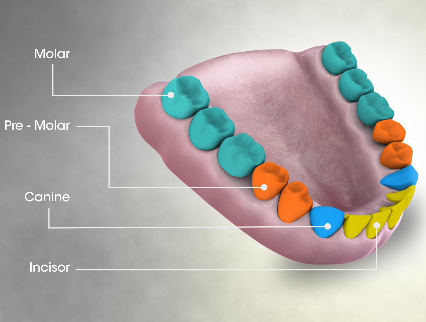
The molars or molar teeth are large, flat teeth at the back of the mouth. They are more developed in mammals. They are used primarily to grind food during chewing. The name ''molar'' derives from Latin, ''molaris dens'', meaning "millstone tooth", from ''mola'', millstone and ''dens'', tooth. Molars show a great deal of diversity in size and shape across mammal groups. The third molar of humans is sometimes vestigial. Human anatomy In humans, the molar teeth have either four or five cusps. Adult humans have 12 molars, in four groups of three at the back of the mouth. The third, rearmost molar in each group is called a wisdom tooth. It is the last tooth to appear, breaking through the front of the gum at about the age of 20, although this varies from individual to individual. Race can also affect the age at which this occurs, with statistical variations between groups. In some cases, it may not even erupt at all. The human mouth contains upper (maxillary) and lower (mandibul ... [...More Info...] [...Related Items...] OR: [Wikipedia] [Google] [Baidu] |
Middle Superior Alveolar Nerve
The middle superior alveolar nerve is a nerve that drops from the infraorbital portion of the maxillary nerve to supply the sinus mucosa, the roots of the maxillary premolars, and the mesiobuccal root of the first maxillary molar. It is not always present; in 72% of cases it is non existent with the anterior superior alveolar nerve innervating the premolars and the posterior superior alveolar nerve The posterior superior alveolar branches (posterior superior dental branches) arise from the trunk of the maxillary nerve just before it enters the infraorbital groove; they are generally two in number, but sometimes arise by a single trunk. They ... innervating the molars, including the mesiobuccal root of the first molar. External links * Maxillary nerve {{Neuroanatomy-stub ... [...More Info...] [...Related Items...] OR: [Wikipedia] [Google] [Baidu] |
Infratemporal Surface Of The Body Of The Maxilla
The maxilla (plural: ''maxillae'' ) in vertebrates is the upper fixed (not fixed in Neopterygii) bone of the jaw formed from the fusion of two maxillary bones. In humans, the upper jaw includes the hard palate in the front of the mouth. The two maxillary bones are fused at the intermaxillary suture, forming the anterior nasal spine. This is similar to the mandible (lower jaw), which is also a fusion of two mandibular bones at the mandibular symphysis. The mandible is the movable part of the jaw. Structure In humans, the maxilla consists of: * The body of the maxilla * Four processes ** the zygomatic process ** the frontal process of maxilla ** the alveolar process ** the palatine process * three surfaces – anterior, posterior, medial * the Infraorbital foramen * the maxillary sinus * the incisive foramen Articulations Each maxilla articulates with nine bones: * two of the cranium: the frontal and ethmoid * seven of the face: the nasal, zygomatic, lacrimal, inferior ... [...More Info...] [...Related Items...] OR: [Wikipedia] [Google] [Baidu] |
Alveolar Canals
The alveolar canals are apertures in the center of the infratemporal surface of the maxilla. The alveolar canals transmit the posterior superior alveolar vessels and nerves A nerve is an enclosed, cable-like bundle of nerve fibers (called axons) in the peripheral nervous system. A nerve transmits electrical impulses. It is the basic unit of the peripheral nervous system. A nerve provides a common pathway for the e .... References Foramina of the skull {{musculoskeletal-stub ... [...More Info...] [...Related Items...] OR: [Wikipedia] [Google] [Baidu] |
Cheek
The cheeks ( la, buccae) constitute the area of the face below the eyes and between the nose and the left or right ear. "Buccal" means relating to the cheek. In humans, the region is innervated by the buccal nerve. The area between the inside of the cheek and the teeth and gums is called the vestibule or buccal pouch or buccal cavity and forms part of the mouth. In other animals the cheeks may also be referred to as jowls. Structure Humans Cheeks are fleshy in humans, the skin being suspended by the chin and the jaws, and forming the lateral wall of the human mouth, visibly touching the cheekbone below the eye. The inside of the cheek is lined with a mucous membrane (buccal mucosa, part of the oral mucosa). During mastication (chewing), the cheeks and tongue between them serve to keep the food between the teeth. Other animals The cheeks are covered externally by hairy skin, and internally by stratified squamous epithelium. This is mostly smooth, but may have caudally di ... [...More Info...] [...Related Items...] OR: [Wikipedia] [Google] [Baidu] |
Gums
The gums or gingiva (plural: ''gingivae'') consist of the mucosal tissue that lies over the mandible and maxilla inside the mouth. Gum health and disease can have an effect on general health. Structure The gums are part of the soft tissue lining of the mouth. They surround the teeth and provide a seal around them. Unlike the soft tissue linings of the lips and cheeks, most of the gums are tightly bound to the underlying bone which helps resist the friction of food passing over them. Thus when healthy, it presents an effective barrier to the barrage of periodontal insults to deeper tissue. Healthy gums are usually coral pink in light skinned people, and may be naturally darker with melanin pigmentation. Changes in color, particularly increased redness, together with swelling and an increased tendency to bleed, suggest an inflammation that is possibly due to the accumulation of bacterial plaque. Overall, the clinical appearance of the tissue reflects the underlying histology, bo ... [...More Info...] [...Related Items...] OR: [Wikipedia] [Google] [Baidu] |
Mandibular Nerve
In neuroanatomy, the mandibular nerve (V) is the largest of the three divisions of the trigeminal nerve, the fifth cranial nerve (CN V). Unlike the other divisions of the trigeminal nerve (ophthalmic nerve, maxillary nerve) which contain only afferent fibers, the mandibular nerve contains both afferent and efferent fibers. These nerve fibers innervate structures of the lower jaw and face, such as the tongue, lower lip, and chin. The mandibular nerve also innervates the muscles of mastication. Structure The large sensory root emerges from the lateral part of the trigeminal ganglion and exits the cranial cavity through the foramen ovale. Portio minor, the small motor root of the trigeminal nerve, passes under the trigeminal ganglion and through the foramen ovale to unite with the sensory root just outside the skull. The mandibular nerve immediately passes between tensor veli palatini, which is medial, and lateral pterygoid, which is lateral, and gives off a meningeal branch (n ... [...More Info...] [...Related Items...] OR: [Wikipedia] [Google] [Baidu] |


