|
Posterior Spinal Artery
The posterior spinal artery (dorsal spinal arteries) arises from the vertebral artery in 25% of humans or the posterior inferior cerebellar artery in 75% of humans, adjacent to the medulla oblongata. It supplies the grey and white posterior columns of the spinal cord. Structure It passes posteriorly to descend the medulla passing in front of the posterior roots of the spinal nerves. Along its course it is reinforced by a succession of segmental or radiculopial branches, which enter the vertebral canal through the intervertebral foramina, forming a plexus called the vasocorona with the anterior vertebral arteries. Below the medulla spinalis and upper cervical spine, the posterior spinal arteries are rather discontinuous; unlike the anterior spinal artery, which can be traced as a distinct channel throughout its course, the posterior spinal arteries are seen as somewhat larger longitudinal channels of an extensive pial arterial meshwork. At the level of the conus medullaris, the post ... [...More Info...] [...Related Items...] OR: [Wikipedia] [Google] [Baidu] |
Posterior Spinal Veins
Posterior spinal veins are small veins which receive blood from the dorsal spinal cord The spinal cord is a long, thin, tubular structure made up of nervous tissue, which extends from the medulla oblongata in the brainstem to the lumbar region of the vertebral column (backbone). The backbone encloses the central canal of the sp .... References External links * http://sci.rutgers.edu/index.php?page=viewarticle&afile=10_January_2002@SCIschemia.html Veins of the torso {{circulatory-stub ... [...More Info...] [...Related Items...] OR: [Wikipedia] [Google] [Baidu] |
Medulla Spinalis
The spinal cord is a long, thin, tubular structure made up of nervous tissue, which extends from the medulla oblongata in the brainstem to the lumbar region of the vertebral column (backbone). The backbone encloses the central canal of the spinal cord, which contains cerebrospinal fluid. The brain and spinal cord together make up the central nervous system (CNS). In humans, the spinal cord begins at the occipital bone, passing through the foramen magnum and then enters the spinal canal at the beginning of the cervical vertebrae. The spinal cord extends down to between the first and second lumbar vertebrae, where it ends. The enclosing bony vertebral column protects the relatively shorter spinal cord. It is around long in adult men and around long in adult women. The diameter of the spinal cord ranges from in the cervical and lumbar regions to in the thoracic area. The spinal cord functions primarily in the transmission of nerve signals from the motor cortex to the body, an ... [...More Info...] [...Related Items...] OR: [Wikipedia] [Google] [Baidu] |
Proprioception
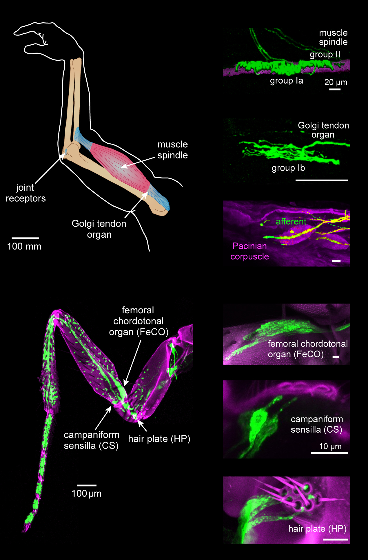
Proprioception ( ), also referred to as kinaesthesia (or kinesthesia), is the sense of self-movement, force, and body position. It is sometimes described as the "sixth sense". Proprioception is mediated by proprioceptors, mechanosensory neurons located within muscles, tendons, and joints. Most animals possess multiple subtypes of proprioceptors, which detect distinct kinematic parameters, such as joint position, movement, and load. Although all mobile animals possess proprioceptors, the structure of the sensory organs can vary across species. Proprioceptive signals are transmitted to the central nervous system, where they are integrated with information from other sensory systems, such as the visual system and the vestibular system, to create an overall representation of body position, movement, and acceleration. In many animals, sensory feedback from proprioceptors is essential for stabilizing body posture and coordinating body movement. System overview In vertebrates, limb ve ... [...More Info...] [...Related Items...] OR: [Wikipedia] [Google] [Baidu] |
Sensory Decussation
In neuroanatomy, the sensory decussation or decussation of the lemnisci is a decussation (i.e. crossover) of axons from the gracile nucleus and cuneate nucleus, which are responsible for fine touch, vibration, proprioception and two-point discrimination of the body. The fibres of this decussation are called the internal arcuate fibres and are found at the superior aspect of the closed medulla superior to the motor decussation. It is part of the second neuron in the posterior column–medial lemniscus pathway. Structure At the level of the closed medulla in the posterior white column, two large nuclei namely the gracile nucleus and the cuneate nucleus can be found. The two nuclei receive the impulse from the two ascending tracts: fasciculus gracilis and fasciculus cuneatus. After the two tracts terminate upon these nuclei, the heavily myelinated fibres arise and ascend anteromedially around the periaqueductal gray as internal arcuate fibres. These fibres decussate (cross) ... [...More Info...] [...Related Items...] OR: [Wikipedia] [Google] [Baidu] |
Cuneate Nucleus
In neuroanatomy, the dorsal column nuclei are a pair of nuclei in the dorsal columns in the brainstem. The name refers collectively to the cuneate nucleus and gracile nucleus, which are present at the bottom of the medulla oblongata. Both nuclei contain second-order neurons of the dorsal column–medial lemniscus pathway, which carries fine touch and proprioceptive information from the body to the brain. Fibres reach the thalamus. Structure Nerve pathways The dorsal column nuclei each have an associated nerve tract in the spinal cord, the gracile fasciculus and the cuneate fasciculus. Both dorsal column nuclei contain synapses from afferent nerve fibers that have travelled in the spinal cord. They then send on second-order neurons of the dorsal column–medial lemniscal pathway. Neurons of the dorsal column nuclei eventually reach the midbrain and the thalamus. They send axons that form the internal arcuate fibers. These cross over at the sensory decussation to form ... [...More Info...] [...Related Items...] OR: [Wikipedia] [Google] [Baidu] |
Cuneate Fasciculus , a tract from the spinal cord into the brainstem
{{disambiguation ...
Cuneate means "wedge-shaped", and can apply to: * Cuneate leaf, a leaf shape * Cuneate nucleus, a part of the brainstem * Cuneate fasciculus Cuneate means "wedge-shaped", and can apply to: * Cuneate leaf The following is a list of terms which are used to describe leaf morphology in the description and taxonomy of plants. Leaves may be simple (a single leaf blade or lamina) or compou ... [...More Info...] [...Related Items...] OR: [Wikipedia] [Google] [Baidu] |
Gracile Nucleus
In neuroanatomy, the dorsal column nuclei are a pair of nuclei in the dorsal columns in the brainstem. The name refers collectively to the cuneate nucleus and gracile nucleus, which are present at the bottom of the medulla oblongata. Both nuclei contain second-order neurons of the dorsal column–medial lemniscus pathway, which carries fine touch and proprioceptive information from the body to the brain. Fibres reach the thalamus. Structure Nerve pathways The dorsal column nuclei each have an associated nerve tract in the spinal cord, the gracile fasciculus and the cuneate fasciculus. Both dorsal column nuclei contain synapses from afferent nerve fibers that have travelled in the spinal cord. They then send on second-order neurons of the dorsal column–medial lemniscal pathway. Neurons of the dorsal column nuclei eventually reach the midbrain and the thalamus. They send axons that form the internal arcuate fibers. These cross over at the sensory decussation to form th ... [...More Info...] [...Related Items...] OR: [Wikipedia] [Google] [Baidu] |
Gracile Fasciculus
Gracility is slenderness, the condition of being gracile, which means slender. It derives from the Latin adjective ''gracilis'' (masculine or feminine), or ''gracile'' ( neuter), which in either form means slender, and when transferred for example to discourse takes the sense of "without ornament", "simple" or various similar connotations. In ''Glossary of Botanic Terms'', B. D. Jackson speaks dismissively of an entry in earlier dictionary of A. A. Crozier as follows: ''Gracilis (Lat.), slender. Crozier has the needless word "gracile"''. However, his objection would be hard to sustain in current usage; apart from the fact that ''gracile'' is a natural and convenient term, it is hardly a neologism. The ''Shorter Oxford English Dictionary'' gives the source date for that usage as 1623 and indicates the word is misused (through association with ''grace'') for Gracefully slender." This misuse is unfortunate at least, because the terms ''gracile'' and ''grace'' are unrelated: the etym ... [...More Info...] [...Related Items...] OR: [Wikipedia] [Google] [Baidu] |
Fourth Ventricle
The fourth ventricle is one of the four connected fluid-filled cavities within the human brain. These cavities, known collectively as the ventricular system, consist of the left and right lateral ventricles, the third ventricle, and the fourth ventricle. The fourth ventricle extends from the cerebral aqueduct (''aqueduct of Sylvius'') to the obex, and is filled with cerebrospinal fluid (CSF). The fourth ventricle has a characteristic diamond shape in cross-sections of the human brain. It is located within the pons or in the upper part of the medulla oblongata. CSF entering the fourth ventricle through the cerebral aqueduct can exit to the subarachnoid space of the spinal cord through two lateral apertures and a single, midline median aperture. Boundaries The fourth ventricle has a roof at its ''upper'' (posterior) surface and a floor at its ''lower'' (anterior) surface, and side walls formed by the cerebellar peduncles (nerve bundles joining the structure on the posterior sid ... [...More Info...] [...Related Items...] OR: [Wikipedia] [Google] [Baidu] |
Posterior Roots Of The Spinal Nerves
The dorsal root of spinal nerve (or posterior root of spinal nerve or sensory root) is one of two "roots" which emerge from the spinal cord. It emerges directly from the spinal cord, and travels to the dorsal root ganglion. Nerve fibres with the ventral root then combine to form a spinal nerve. The dorsal root transmits sensory information, forming the afferent sensory root of a spinal nerve. Structure The root emerges from the posterior part of the spinal cord and travels to the dorsal root ganglion. The dorsal root ganglia contain the pseudo-unipolar cell bodies of the nerve fibres which travel from the ganglia through the root into the spinal cord. The lateral division of the dorsal root contains lightly myelinated and unmyelinated fibres of small diameter. These carry pain and temperature sensation. These fibers cross through the anterior white commissure to form the anterolateral system in the lateral funiculus. The medial division of the dorsal root contains mye ... [...More Info...] [...Related Items...] OR: [Wikipedia] [Google] [Baidu] |
Anastomosis
An anastomosis (, plural anastomoses) is a connection or opening between two things (especially cavities or passages) that are normally diverging or branching, such as between blood vessels, leaf#Veins, leaf veins, or streams. Such a connection may be normal (such as the foramen ovale (heart), foramen ovale in a fetus's heart) or abnormal (such as the atrial septal defect#Patent foramen ovale, patent foramen ovale in an adult's heart); it may be acquired (such as an arteriovenous fistula) or innate (such as the arteriovenous shunt of a metarteriole); and it may be natural (such as the aforementioned examples) or artificial (such as a surgical anastomosis). The reestablishment of an anastomosis that had become blocked is called a reanastomosis. Anastomoses that are abnormal, whether congenital disorder, congenital or acquired, are often called fistulas. The term is used in medicine, biology, mycology, geology, and geography. Etymology Anastomosis: medical or Modern Latin, from Gre ... [...More Info...] [...Related Items...] OR: [Wikipedia] [Google] [Baidu] |
Cauda Equina
The cauda equina () is a bundle of spinal nerves and spinal nerve rootlets, consisting of the second through fifth lumbar nerve pairs, the first through fifth sacral nerve pairs, and the coccygeal nerve, all of which arise from the lumbar enlargement and the conus medullaris of the spinal cord. The cauda equina occupies the lumbar cistern, a subarachnoid space inferior to the conus medullaris. The nerves that compose the cauda equina innervate the pelvic organs and lower limbs to include motor innervation of the hips, knees, ankles, feet, internal anal sphincter and external anal sphincter. In addition, the cauda equina extends to sensory innervation of the perineum and, partially, parasympathetic innervation of the bladder. Structure In adulthood, the cauda equina is made of lumbosacral spinal nerve roots. Development In humans, the spinal cord stops growing in infancy and the end of the spinal cord is about the level of the third lumbar vertebra, or L3, at birth. Because the ... [...More Info...] [...Related Items...] OR: [Wikipedia] [Google] [Baidu] |


