|
Posterior Chamber Of Eyeball
The posterior chamber is a narrow space behind the peripheral part of the iris, and in front of the suspensory ligament of the lens and the ciliary processes. The posterior chamber consists of small space directly posterior to the iris but anterior to the lens. The posterior chamber is part of the anterior segment and should not be confused with the vitreous chamber (in the posterior segment). Posterior chamber is an important structure involved in production and circulation of aqueous humor. Aqueous humor produced by the epithelium of the ciliary body is secreted into the posterior chamber, from which it flows through the pupil to enter the anterior chamber. The hypermature cataractous lens or, the intraocular lens implanted after cataract surgery may obstruct the aqueous flow through the pupil. The block in flow of aqueous from the posterior to the anterior chamber will lead to a condition known as Iris bombe. In this condition, pressure in the posterior chamber rises, result ... [...More Info...] [...Related Items...] OR: [Wikipedia] [Google] [Baidu] |
Human Eye
The human eye is a sensory organ, part of the sensory nervous system, that reacts to visible light and allows humans to use visual information for various purposes including seeing things, keeping balance, and maintaining circadian rhythm. The eye can be considered as a living optical device. It is approximately spherical in shape, with its outer layers, such as the outermost, white part of the eye (the sclera) and one of its inner layers (the pigmented choroid) keeping the eye essentially light tight except on the eye's optic axis. In order, along the optic axis, the optical components consist of a first lens (the cornea—the clear part of the eye) that accomplishes most of the focussing of light from the outside world; then an aperture (the pupil) in a diaphragm (the iris—the coloured part of the eye) that controls the amount of light entering the interior of the eye; then another lens (the crystalline lens) that accomplishes the remaining focussing of light in ... [...More Info...] [...Related Items...] OR: [Wikipedia] [Google] [Baidu] |
Trabecular Meshwork
The trabecular meshwork is an area of tissue in the eye located around the base of the cornea, near the ciliary body, and is responsible for draining the aqueous humor from the eye via the anterior chamber (the chamber on the front of the eye covered by the cornea). The tissue is spongy and lined by trabeculocytes; it allows fluid to drain into a set of tubes called Schlemm's canal which is lined by endothelium with blood and lymphatic properties that allow aqueous humor to flow into the blood system. Structure The meshwork is divided up into three parts, with characteristically different ultrastructures: #''Inner uveal meshwork'' - Closest to the anterior chamber angle, contains thin cord-like trabeculae, orientated predominantly in a radial fashion, enclosing trabeculae spaces larger than the corneoscleral meshwork. #''Corneoscleral meshwork'' - Contains a large amount of elastin, arranged as a series of thin, flat, perforated sheets arranged in a laminar pattern; c ... [...More Info...] [...Related Items...] OR: [Wikipedia] [Google] [Baidu] |
Cataract Surgery
Cataract surgery, also called lens replacement surgery, is the removal of the natural lens of the eye (also called "crystalline lens") that has developed an opacification, which is referred to as a cataract, and its replacement with an intraocular lens. Metabolic changes of the crystalline lens fibers over time lead to the development of the cataract, causing impairment or loss of vision. Some infants are born with congenital cataracts, and certain environmental factors may also lead to cataract formation. Early symptoms may include strong glare from lights and small light sources at night, and reduced acuity at low light levels. During cataract surgery, a patient's cloudy natural cataract lens is removed, either by emulsification in place or by cutting it out. An artificial intraocular lens (IOL) is implanted in its place. Cataract surgery is generally performed by an ophthalmologist in an ambulatory setting at a surgical center or hospital rather than an inpatient setting. ... [...More Info...] [...Related Items...] OR: [Wikipedia] [Google] [Baidu] |
Aqueous Humour
The aqueous humour is a transparent water-like fluid similar to plasma, but containing low protein concentrations. It is secreted from the ciliary body, a structure supporting the lens of the eyeball. It fills both the anterior and the posterior chambers of the eye, and is not to be confused with the vitreous humour, which is located in the space between the lens and the retina, also known as the posterior cavity or vitreous chamber. Blood cannot normally enter the eyeball. Structure Composition * Amino acids: transported by ciliary muscles *98% water * Electrolytes ( pH = 7.4 -one source gives 7.1) **Sodium = 142.09 **Potassium = 2.2 - 4.0 **Calcium = 1.8 **Magnesium = 1.1 **Chloride = 131.6 **HCO3- = 20.15 **Phosphate = 0.62 ** Osm = 304 *Ascorbic acid *Glutathione * Immunoglobulins Function *Maintains the intraocular pressure and inflates the globe of the eye. It is this hydrostatic pressure that keeps the eyeball in a roughly spherical shape and keeps the walls of the eye ... [...More Info...] [...Related Items...] OR: [Wikipedia] [Google] [Baidu] |
Anterior Chamber
The anterior chamber ( AC) is the aqueous humor-filled space inside the eye between the iris and the cornea's innermost surface, the endothelium. Hyphema, anterior uveitis and glaucoma are three main pathologies in this area. In hyphema, blood fills the anterior chamber as a result of a hemorrhage, most commonly after a blunt eye injury. Anterior uveitis is an inflammatory process affecting the iris and ciliary body, with resulting inflammatory signs in the anterior chamber. In glaucoma, blockage of the trabecular meshwork prevents the normal outflow of aqueous humour, resulting in increased intraocular pressure, progressive damage to the optic nerve head, and eventually blindness. The depth of the anterior chamber of the eye varies between 1.5 and 4.0 mm, averaging 3.0 mm. It tends to become shallower at older age and in eyes with hypermetropia (far sightedness). As depth decreases below 2.5 mm, the risk for angle closure glaucoma increases. Clinical significan ... [...More Info...] [...Related Items...] OR: [Wikipedia] [Google] [Baidu] |
Iridectomy
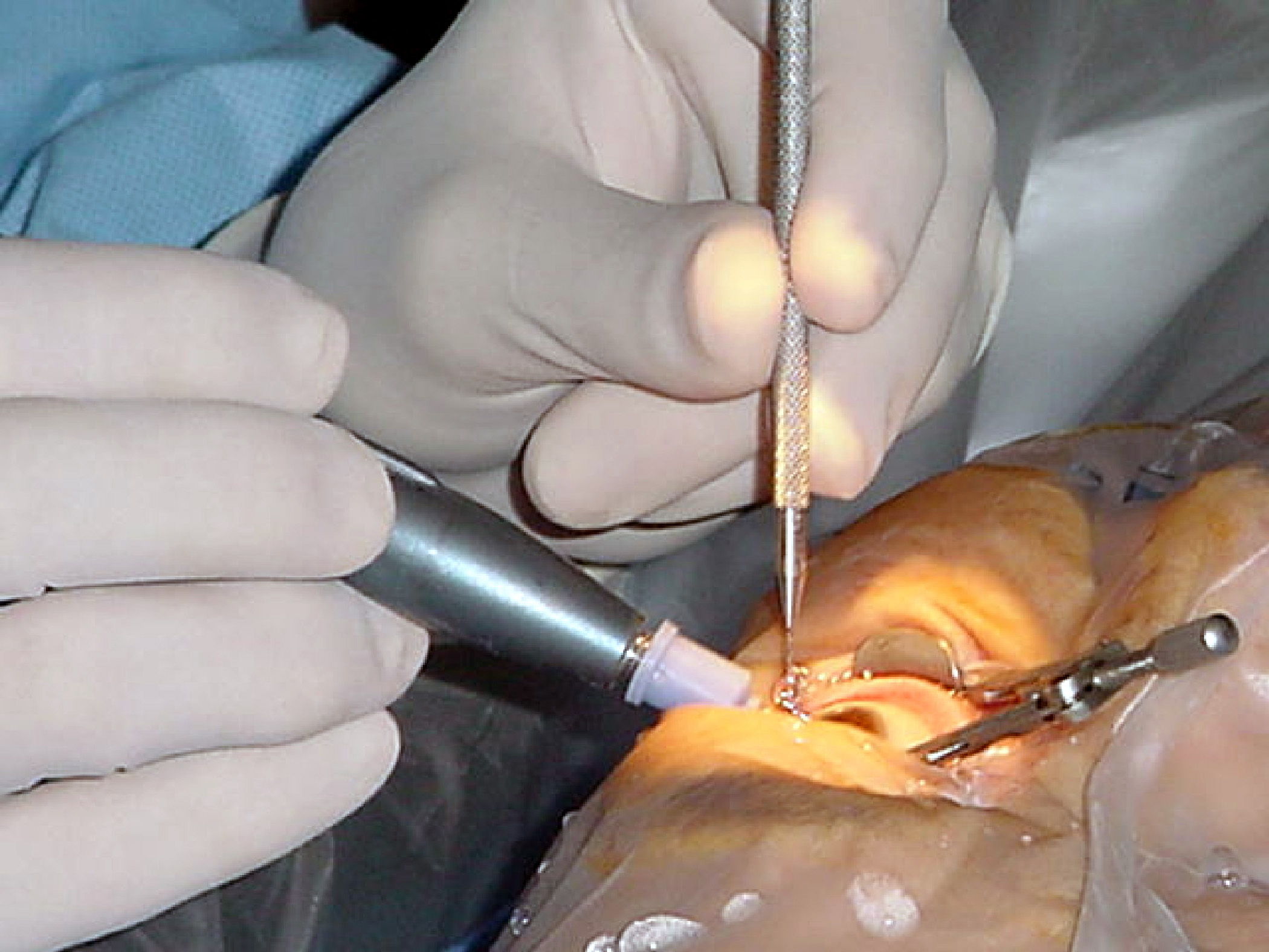
An iridectomy, also known as a surgical iridectomy or corectomy, is the surgical removal of part of the iris.Cline D; Hofstetter HW; Griffin JR. ''Dictionary of Visual Science''. 4th ed. Butterworth-Heinemann, Boston 1997. Surgery Encyclopedia - "Iridectomy." Encyclopedia of Surgery: A Guide for Patients and Caregivers. Retrieved May 20, 2006. These procedures are most frequently performed in the treatment of closed-angle glaucoma and iris melanoma. Comparison with Nd:YAG laser iridotomy In |
Argon Laser
An ion laser is a gas laser that uses an ionized gas as its lasing medium. Like other gas lasers, ion lasers feature a sealed cavity containing the laser medium and mirrors forming a Fabry–Pérot resonator. Unlike helium–neon lasers, the energy level transitions that contribute to laser action come from ions. Because of the large amount of energy required to excite the ionic transitions used in ion lasers, the required current is much greater, and as a result all but the smallest ion lasers are water-cooled. A small air-cooled ion laser might produce, for example, 130 milliwatts of output light with a tube current of about 10 amperes and a voltage of 105 volts. Since one ampere times one volt is one watt, this is an electrical power input of about one kilowatt. Subtracting the (desirable) light output of 130 mW from power input, this leaves the large amount of waste heat of nearly one kW. This has to be dissipated by the cooling system. In other words, the power efficienc ... [...More Info...] [...Related Items...] OR: [Wikipedia] [Google] [Baidu] |
Glaucoma Surgery
Glaucoma is a group of diseases affecting the optic nerve that results in vision loss and is frequently characterized by raised intraocular pressure (IOP). There are many glaucoma surgeries, and variations or combinations of those surgeries, that facilitate the escape of excess aqueous humor from the eye to lower intraocular pressure, and a few that lower IOP by decreasing the production of aqueous humor. Procedures that facilitate outflow of aqueous humor Laser trabeculoplasty A trabeculoplasty is a modification of the trabecular meshwork. Laser trabeculoplasty (LTP) is the application of a laser beam to burn areas of the trabecular meshwork, located near the base of the iris, to increase fluid outflow. LTP is used in the treatment of various open-angle glaucomas. The two types of laser trabeculoplasty are argon laser trabeculoplasty (ALT) and selective laser trabeculoplasty (SLT). As its name suggests, argon laser trabeculoplasty uses an argon laser to create tiny burns on the t ... [...More Info...] [...Related Items...] OR: [Wikipedia] [Google] [Baidu] |
Angle Closure Glaucoma
Glaucoma is a group of eye diseases that result in damage to the optic nerve (or retina) and cause vision loss. The most common type is open-angle (wide angle, chronic simple) glaucoma, in which the drainage angle for fluid within the eye remains open, with less common types including closed-angle (narrow angle, acute congestive) glaucoma and normal-tension glaucoma. Open-angle glaucoma develops slowly over time and there is no pain. Peripheral vision may begin to decrease, followed by central vision, resulting in blindness if not treated. Closed-angle glaucoma can present gradually or suddenly. The sudden presentation may involve severe eye pain, blurred vision, mid-dilated pupil, redness of the eye, and nausea. Vision loss from glaucoma, once it has occurred, is permanent. Eyes affected by glaucoma are referred to as being glaucomatous. Risk factors for glaucoma include increasing age, high pressure in the eye, a family history of glaucoma, and use of steroid medication. F ... [...More Info...] [...Related Items...] OR: [Wikipedia] [Google] [Baidu] |
Cataract Surgery
Cataract surgery, also called lens replacement surgery, is the removal of the natural lens of the eye (also called "crystalline lens") that has developed an opacification, which is referred to as a cataract, and its replacement with an intraocular lens. Metabolic changes of the crystalline lens fibers over time lead to the development of the cataract, causing impairment or loss of vision. Some infants are born with congenital cataracts, and certain environmental factors may also lead to cataract formation. Early symptoms may include strong glare from lights and small light sources at night, and reduced acuity at low light levels. During cataract surgery, a patient's cloudy natural cataract lens is removed, either by emulsification in place or by cutting it out. An artificial intraocular lens (IOL) is implanted in its place. Cataract surgery is generally performed by an ophthalmologist in an ambulatory setting at a surgical center or hospital rather than an inpatient setting. ... [...More Info...] [...Related Items...] OR: [Wikipedia] [Google] [Baidu] |
Iris (anatomy)
In humans and most mammals and birds, the iris (plural: ''irides'' or ''irises'') is a thin, annular structure in the eye, responsible for controlling the diameter and size of the pupil, and thus the amount of light reaching the retina. Eye color is defined by the iris. In optical terms, the pupil is the eye's aperture, while the iris is the diaphragm. Structure The iris consists of two layers: the front pigmented fibrovascular layer known as a stroma and, beneath the stroma, pigmented epithelial cells. The stroma is connected to a sphincter muscle ( sphincter pupillae), which contracts the pupil in a circular motion, and a set of dilator muscles (dilator pupillae), which pull the iris radially to enlarge the pupil, pulling it in folds. The sphincter pupillae is the opposing muscle of the dilator pupillae. The pupil's diameter, and thus the inner border of the iris, changes size when constricting or dilating. The outer border of the iris does not change size. The constr ... [...More Info...] [...Related Items...] OR: [Wikipedia] [Google] [Baidu] |


