|
Posterior Atlantoaxial Ligament
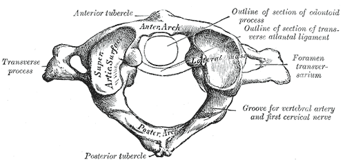
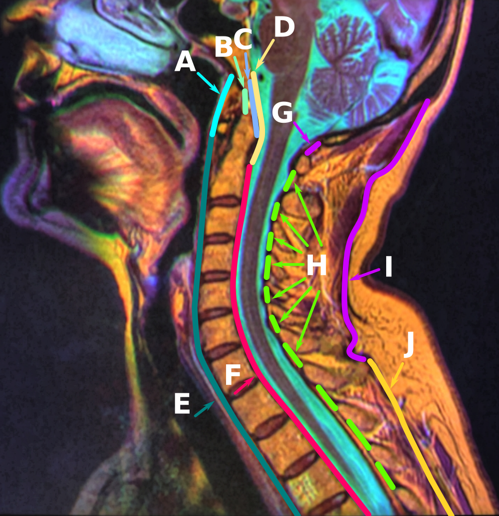
The posterior atlantoaxial ligament is a broad, thin membrane attached, above, to the lower border of the posterior arch of the Atlas (anatomy), atlas; below, to the upper edges of the laminæ of the Axis (anatomy), axis. It supplies the place of the ligamenta flava, and is in relation, behind, with the Obliqui capitis inferiores. See also * Atlanto-axial joint References External links Description at spineuniverse.com Ligaments of the head and neck Bones of the vertebral column {{Portal bar, Anatomy ... [...More Info...] [...Related Items...] OR: [Wikipedia] [Google] [Baidu] |
Atlas (anatomy)
In anatomy, the atlas (C1) is the most superior (first) cervical vertebra of the spine and is located in the neck. It is named for Atlas of Greek mythology because, just as Atlas supported the globe, it supports the entire head. The atlas is the topmost vertebra and, with the axis (the vertebra below it), forms the joint connecting the skull and spine. The atlas and axis are specialized to allow a greater range of motion than normal vertebrae. They are responsible for the nodding and rotation movements of the head. The atlanto-occipital joint allows the head to nod up and down on the vertebral column. The dens acts as a pivot that allows the atlas and attached head to rotate on the axis, side to side. The atlas's chief peculiarity is that it has no body. It is ring-like and consists of an anterior and a posterior arch and two lateral masses. The atlas and axis are important neurologically because the brainstem extends down to the axis. Structure Anterior arch The anterio ... [...More Info...] [...Related Items...] OR: [Wikipedia] [Google] [Baidu] |
Axis (anatomy)
In anatomy, the axis (from Latin ''axis'', "axle") or epistropheus is the second cervical vertebra (C2) of the spine, immediately inferior to the atlas, upon which the head rests. The axis' defining feature is its strong odontoid process (bony protrusion) known as the dens, which rises dorsally from the rest of the bone. Structure The body is deeper in front or in the back and is prolonged downward anteriorly to overlap the upper and front part of the third vertebra. It presents a median longitudinal ridge in front, separating two lateral depressions for the attachment of the longus colli muscles. Odontoid Process of Axis (Dens) The dens, also called the odontoid process or the peg, is the most pronounced projecting feature of the axis. The dens exhibits a slight constriction where it joins the main body of the vertebra. The condition where the dens is separated from the body of the axis is called ''os odontoideum'' and may cause nerve and circulation compression syndrome. ... [...More Info...] [...Related Items...] OR: [Wikipedia] [Google] [Baidu] |
Ligamenta Flava
The ligamenta flava (singular, ''ligamentum flavum'', Latin for ''yellow ligament'') are a series of ligaments that connect the ventral parts of the lamina of the vertebral arch, laminae of adjacent vertebrae. They help to preserve Bipedalism, upright posture, preventing Anatomical terms of motion, hyperflexion, and ensuring that the vertebral column straightens after flexion. Hypertrophy can cause spinal stenosis. Structure Each ligamentum flavum connects the Lamina of the vertebral arch, laminae two adjacent vertebrae. They begin with the junction of the Axis (anatomy), axis and third cervical vertebra, continuing down to the junction of the fifth lumbar vertebra and the sacrum. They are best seen from the interior of the vertebral canal. when looked at from the outer surface they appear short, being overlapped by the lamina of the vertebral arch. Each ligament consists of two Anatomical terms of location#Left and right (lateral), and medial, lateral portions which commence on ... [...More Info...] [...Related Items...] OR: [Wikipedia] [Google] [Baidu] |
Obliqui Capitis Inferiores
The obliquus capitis inferior muscle () is the larger of the two oblique muscles of the neck. It arises from the apex of the spinous process of the axis and passes laterally and slightly upward, to be inserted into the lower and back part of the transverse process of the atlas. It lies deep to the semispinalis capitis and trapezius muscles. The muscle is responsible for rotation of the head and first cervical vertebra (atlanto-axial joint). It forms the lower boundary of the suboccipital triangle of the neck. The naming of this muscle may be confusing, as it is the only capitis (L. "head") muscle that does NOT attach to the cranium. Function The obliquus capitis inferior muscle, like the other suboccipital muscles, has an important role in proprioception. This muscle has a very high density of Golgi organs and muscle spindles which accounts for this. It is believed that proprioception may be the primary role of the inferior oblique (and indeed the other suboccipital muscles) a ... [...More Info...] [...Related Items...] OR: [Wikipedia] [Google] [Baidu] |
Atlanto-axial Joint
The atlanto-axial joint is a joint in the upper part of the neck between the atlas bone and the axis bone, which are the first and second cervical vertebrae. It is a pivot joint. Structure The atlanto-axial joint is a joint between the atlas bone and the axis bone, which are the first and second cervical vertebrae. It is a pivot joint. There is a pivot articulation between the odontoid process of the axis and the ring formed by the anterior arch and the transverse ligament of the atlas. Lateral and median joints There are three atlanto-axial joints: one median and two lateral: * The median atlanto-axial joint is sometimes considered a triple joint: ** one between the posterior surface of the anterior arch of atlas and the front of the odontoid process ** one between the anterior surface of the ligament and the back of the odontoid process * The lateral atlantoaxial joint involves the lateral mass of atlas and axis. Between the articular processes of the two bones there is o ... [...More Info...] [...Related Items...] OR: [Wikipedia] [Google] [Baidu] |
Ligaments Of The Head And Neck
A ligament is the fibrous connective tissue that connects bones to other bones. It is also known as ''articular ligament'', ''articular larua'', ''fibrous ligament'', or ''true ligament''. Other ligaments in the body include the: * Peritoneal ligament: a fold of peritoneum or other membranes. * Fetal remnant ligament: the remnants of a fetal tubular structure. * Periodontal ligament: a group of fibers that attach the cementum of teeth to the surrounding alveolar bone. Ligaments are similar to tendons and fasciae as they are all made of connective tissue. The differences among them are in the connections that they make: ligaments connect one bone to another bone, tendons connect muscle to bone, and fasciae connect muscles to other muscles. These are all found in the skeletal system of the human body. Ligaments cannot usually be regenerated naturally; however, there are periodontal ligament stem cells located near the periodontal ligament which are involved in the adult regenerat ... [...More Info...] [...Related Items...] OR: [Wikipedia] [Google] [Baidu] |


