|
Postdentary Trough
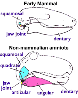
The postdentary trough is a skeletal feature seen in Mesozoic mammals. It is found on the inside of the lower jaw (dentary), at the back behind the molar teeth. It is the hollow in which the postdentary bones and Meckel's cartilage sit. These bones form the middle ear in later mammal groups (see Evolution of mammalian auditory ossicles); they include the incus ( quadrate), malleus (articular The articular bone is part of the lower jaw of most vertebrates, including most jawed fish, amphibians, birds and various kinds of reptiles, as well as ancestral mammals. Anatomy In most vertebrates, the articular bone is connected to two oth ...), ectotympanic ( angular) and prearticular.Zhe-Xi Luo 201Developmental patterns in Mesozoic evolution of mammal ears Annual Review of Ecology, Evolution, and Systematics, 42, 355-380 In Mesozoic mammals these bones gradually change position and size until they are incorporated in the middle ear. References Mammal anatomy Evolution of mamma ... [...More Info...] [...Related Items...] OR: [Wikipedia] [Google] [Baidu] |
Evolution Of Mammals
The evolution of mammals has passed through many stages since the first appearance of their synapsid ancestors in the Pennsylvanian sub-period of the late Carboniferous period. By the mid-Triassic, there were many synapsid species that looked like mammals. The lineage leading to today's mammals split up in the Jurassic; synapsids from this period include '' Dryolestes'', more closely related to extant placentals and marsupials than to monotremes, as well as '' Ambondro'', more closely related to monotremes. Later on, the eutherian and metatherian lineages separated; the metatherians are the animals more closely related to the marsupials, while the eutherians are those more closely related to the placentals. Since '' Juramaia'', the earliest known eutherian, lived 160 million years ago in the Jurassic, this divergence must have occurred in the same period. After the Cretaceous–Paleogene extinction event wiped out the non-avian dinosaurs (birds being the only surviving dinosaurs ... [...More Info...] [...Related Items...] OR: [Wikipedia] [Google] [Baidu] |
Dentary
In anatomy, the mandible, lower jaw or jawbone is the largest, strongest and lowest bone in the human facial skeleton. It forms the lower jaw and holds the lower tooth, teeth in place. The mandible sits beneath the maxilla. It is the only movable bone of the skull (discounting the ossicles of the middle ear). It is connected to the temporal bones by the temporomandibular joints. The bone is formed prenatal development, in the fetus from a fusion of the left and right mandibular prominences, and the point where these sides join, the mandibular symphysis, is still visible as a faint ridge in the midline. Like other symphyses in the body, this is a midline articulation where the bones are joined by fibrocartilage, but this articulation fuses together in early childhood.Illustrated Anatomy of the Head and Neck, Fehrenbach and Herring, Elsevier, 2012, p. 59 The word "mandible" derives from the Latin word ''mandibula'', "jawbone" (literally "one used for chewing"), from ''wikt:mandere ... [...More Info...] [...Related Items...] OR: [Wikipedia] [Google] [Baidu] |
Meckel's Cartilage
In humans, the cartilaginous bar of the mandibular arch is formed by what are known as Meckel's cartilages (right and left) also known as Meckelian cartilages; above this the incus and malleus are developed. Meckel's cartilage arises from the first pharyngeal arch. The dorsal end of each cartilage is connected with the ear-capsule and is ossified to form the malleus; the ventral ends meet each other in the region of the symphysis menti, and are usually regarded as undergoing ossification to form that portion of the mandible which contains the incisor teeth. The intervening part of the cartilage disappears; the portion immediately adjacent to the malleus is replaced by fibrous membrane, which constitutes the sphenomandibular ligament, while from the connective tissue covering the remainder of the cartilage the greater part of the mandible is ossified. Johann Friedrich Meckel, the Younger discovered this cartilage in 1820. Evolution Meckel's cartilage is a piece of cartilage from ... [...More Info...] [...Related Items...] OR: [Wikipedia] [Google] [Baidu] |
Evolution Of Mammalian Auditory Ossicles
The evolution of mammalian auditory ossicles was an evolutionary event that resulted in the formation of the bones of the mammalian middle ear. These bones, or ossicles, are a defining characteristic of all mammals. The event is well-documented and important as a demonstration of transitional forms and exaptation, the re-purposing of existing structures during evolution. The ossicles evolved from skull bones present in most tetrapods, including the reptilian lineage. The reptilian quadrate bone, articular bone, and columella evolved into the mammalian incus, malleus, and stapes (anvil, hammer, and stirrup), respectively. In reptiles, the eardrum is connected to the inner ear via a single bone, the columella, while the upper and lower jaws contain several bones not found in mammals. Over the course of the evolution of mammals, one bone from the lower and one from the upper jaw (the articular and quadrate bones) lost their purpose in the jaw joint and migrated to the middle ea ... [...More Info...] [...Related Items...] OR: [Wikipedia] [Google] [Baidu] |
Incus
The ''incus'' (plural incudes) or anvil is a bone in the middle ear. The anvil-shaped small bone is one of three ossicles in the middle ear. The ''incus'' receives vibrations from the ''malleus'', to which it is connected laterally, and transmits these to the ''stapes'' medially. The ''incus'' is so-called because of its resemblance to an anvil ( la, Incus). Structure The incus is the second of the ossicles, three bones in the middle ear which act to transmit sound. It is shaped like an anvil, and has a long and short crus extending from the body, which articulates with the malleus. The short crus attaches to the posterior ligament of the incus. The long crus articulates with the stirrup at the lenticular process. The superior ligament of the incus attaches at the body of the incus to the roof of the tympanic cavity. Function Vibrations in the middle ear are received via the tympanic membrane. The malleus, resting on the membrane, conveys vibrations to the incus. This in tu ... [...More Info...] [...Related Items...] OR: [Wikipedia] [Google] [Baidu] |
Quadrate Bone
The quadrate bone is a skull bone in most tetrapods, including amphibians, sauropsids (reptiles, birds), and early synapsids. In most tetrapods, the quadrate bone connects to the quadratojugal and squamosal bones in the skull, and forms upper part of the jaw joint. The lower jaw articulates at the articular bone, located at the rear end of the lower jaw. The quadrate bone forms the lower jaw articulation in all classes except mammals. Evolutionarily, it is derived from the hindmost part of the primitive cartilaginous upper jaw. Function in reptiles In certain extinct reptiles, the variation and stability of the morphology of the quadrate bone has helped paleontologists in the species-level taxonomy and identification of mosasaur squamates and spinosaurine dinosaurs. In some lizards and dinosaurs, the quadrate is articulated at both ends and movable. In snakes, the quadrate bone has become elongated and very mobile, and contributes greatly to their ability to swallow very ... [...More Info...] [...Related Items...] OR: [Wikipedia] [Google] [Baidu] |
Malleus
The malleus, or hammer, is a hammer-shaped small bone or ossicle of the middle ear. It connects with the incus, and is attached to the inner surface of the eardrum. The word is Latin for 'hammer' or 'mallet'. It transmits the sound vibrations from the eardrum to the ''incus'' (anvil). Structure The malleus is a bone situated in the middle ear. It is the first of the three ossicles, and attached to the tympanic membrane. The head of the malleus is the large protruding section, which attaches to the incus. The head connects to the neck of malleus. The bone continues as the handle (or manubrium) of malleus, which connects to the tympanic membrane. Between the neck and handle of the malleus, lateral and anterior processes emerge from the bone. The bone is oriented so that the head is superior and the handle is inferior. Development Embryologically, the malleus is derived from the first pharyngeal arch along with the ''incus''. It grows from Meckel's cartilage. Function The malleu ... [...More Info...] [...Related Items...] OR: [Wikipedia] [Google] [Baidu] |
Articular
The articular bone is part of the lower jaw of most vertebrates, including most jawed fish, amphibians, birds and various kinds of reptiles, as well as ancestral mammals. Anatomy In most vertebrates, the articular bone is connected to two other lower jaw bones, the suprangular and the angular. Developmentally, it originates from the embryonic mandibular cartilage. The most caudal portion of the mandibular cartilage ossifies to form the articular bone, while the remainder of the mandibular cartilage either remains cartilaginous or disappears. In snakes In snakes, the articular, surangular, and prearticular bones have fused to form the compound bone. The mandible is suspended from the quadrate bone and articulates at this compound bone. Function In amphibians and reptiles In most tetrapods, the articular bone forms the lower portion of the jaw joint. The upper jaw articulates at the quadrate bone. In mammals In mammals, the articular bone evolves to form the malle ... [...More Info...] [...Related Items...] OR: [Wikipedia] [Google] [Baidu] |
Angular Bone
The angular is a large bone in the lower jaw (mandible) of amphibians and reptiles (birds included), which is connected to all other lower jaw bones: the dentary (which is the entire lower jaw in mammals), the splenial, the suprangular, and the articular. It is homologous to the tympanic bone in mammals, due to the incorporation of several jaw bones into the mammalian middle ear early in mammal evolution. In therapsids (mammal ancestors and their kin), the lower jaw is made up of the dentary (the mandible in mammals) and a group of smaller "postdentary" bones near the jaw joint. As the dentary increased in size over million of years, two of these postdentary bones, the articular and angular, became increasingly reduced and the dentary eventually made direct contact with the upper jaw. These postdentary bones, even before their articular function was lost, probably transmitted sound vibrations to the stapes and, in some therapsids, a bent plate that might have supported a membrane ... [...More Info...] [...Related Items...] OR: [Wikipedia] [Google] [Baidu] |
Mammal Anatomy
Mammals () are a group of vertebrate animals constituting the class Mammalia (), characterized by the presence of mammary glands which in females produce milk for feeding (nursing) their young, a neocortex (a region of the brain), fur or hair, and three middle ear bones. These characteristics distinguish them from reptiles (including birds) from which they diverged in the Carboniferous, over 300 million years ago. Around 6,400 extant species of mammals have been described divided into 29 orders. The largest orders, in terms of number of species, are the rodents, bats, and Eulipotyphla (hedgehogs, moles, shrews, and others). The next three are the Primates (including humans, apes, monkeys, and others), the Artiodactyla ( cetaceans and even-toed ungulates), and the Carnivora (cats, dogs, seals, and others). In terms of cladistics, which reflects evolutionary history, mammals are the only living members of the Synapsida (synapsids); this clade, together wit ... [...More Info...] [...Related Items...] OR: [Wikipedia] [Google] [Baidu] |


