|
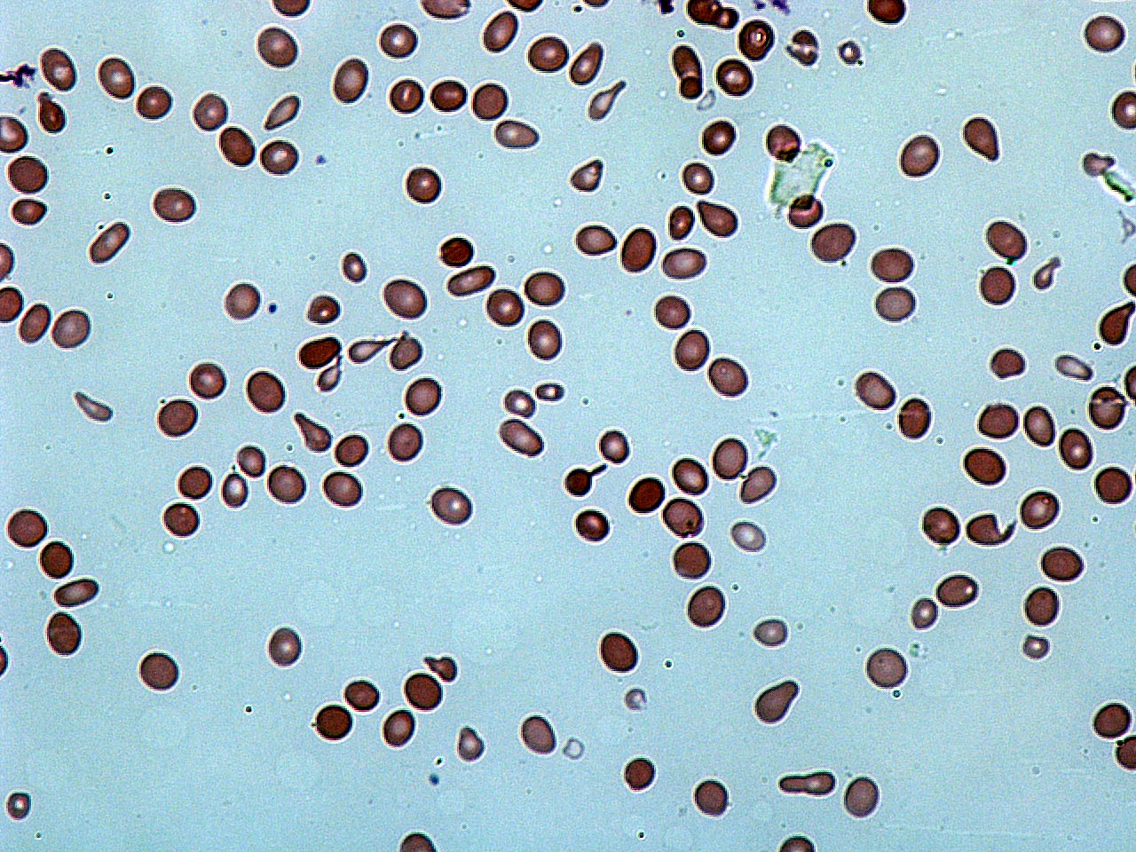
Poikilocyte
Poikilocytosis is variation in the shapes of red blood cells. Poikilocytes may be oval, teardrop-shaped, sickle-shaped or irregularly contracted. Normal red blood cells are round, flattened disks that are thinner in the middle than at the edges. A ''poikilocyte'' is an abnormally-shaped red blood cell. Generally, poikilocytosis can refer to an increase in abnormal red blood cells of any shape, where they make up 10% or more of the total population of red blood cells. Types Membrane abnormalities # Acanthocytes or Spur/Spike cells # Codocytes or Target cells # Echinocytes and Burr cells # Elliptocytes and Ovalocytes # Spherocytosis, Spherocytes # Stomatocytes or Mouth cells # Drepanocytosis, Drepanocytes or Sickle Cells # Degmacytes or "bite cells" Trauma # Dacrocytes or Teardrop Cells # Keratocytes # Microspherocytes and Pyropoikilocytes # Schistocytes # Semilunar body, Semilunar bodies Diagnosis Poikilocytosis may be diagnosed with a test called a Blood film, blood smear. Durin ... [...More Info...] [...Related Items...] OR: [Wikipedia] [Google] [Baidu] |
Dacrocyte
A dacrocyte (or dacryocyte) is a type of poikilocyte that is shaped like a Tears, teardrop (a "teardrop cell"). A marked increase of dacrocytes is known as dacrocytosis. These tear drop cells are found primarily in diseases with bone marrow fibrosis, such as: primary myelofibrosis, myelodysplastic syndromes during the late course of the disease, rare form of acute leukemias and Myelophthisic anemia, myelophthisis caused by metastatic cancers. Rare causes are myelofibrosis associated with post-irradiation, toxins, autoimmune diseases, metabolic conditions, inborn hemolytic anemias, iron-deficiency anemia or β-thalassemia. Etiology One theory regarding dacrocyte formation is that red blood cells containing various inclusions undergo "pitting" by the spleen to remove these inclusions, and in the process, they can be stretched too far to return to their original shape. It is also thought that this can similarly occur when red blood cells with large inclusions are obstructed from pass ... [...More Info...] [...Related Items...] OR: [Wikipedia] [Google] [Baidu] |
Keratocyte
Corneal keratocytes (corneal fibroblasts) are specialized fibroblasts residing in the stroma. This corneal layer, representing about 85-90% of corneal thickness, is built up from highly regular collagenous lamellae and extracellular matrix components. Keratocytes play the major role in keeping it transparent, healing its wounds, and synthesizing its components. In the unperturbed cornea keratocytes stay dormant, coming into action after any kind of injury or inflammation. Some keratocytes underlying the site of injury, even a light one, undergo apoptosis immediately after the injury. Any glitch in the precisely orchestrated process of healing may cloud the cornea, while excessive keratocyte apoptosis may be a part of the pathological process in the degenerative corneal disorders such as keratoconus, and these considerations prompt the ongoing research into the function of these cells. Origin and functions Keratocytes are developmentally derived from the cranial population of neural ... [...More Info...] [...Related Items...] OR: [Wikipedia] [Google] [Baidu] |
Anisocytosis
Anisocytosis is a medical term meaning that a patient's red blood cells are of unequal size. This is commonly found in anemia and other blood conditions. False diagnostic flagging may be triggered on a complete blood count by an elevated WBC count, agglutinated RBCs, RBC fragments, giant platelets or platelet clumps. In addition, it is a characteristic feature of bovine blood. The red cell distribution width (RDW) is a measurement of anisocytosis and is calculated as a coefficient of variation of the distribution of RBC volumes divided by the mean corpuscular volume ( MCV). Types Anisocytosis is identified by RDW and is classified according to the size of RBC measured by MCV. According to this, it can be divided into *Anisocytosis with microcytosis – Iron deficiency, sickle cell anemia *Anisocytosis with macrocytosis – Folate or vitamin B12 deficiency, autoimmune hemolytic anemia, cytotoxic chemotherapy, chronic liver disease, myelodysplastic syndrome Increased RDW is s ... [...More Info...] [...Related Items...] OR: [Wikipedia] [Google] [Baidu] |
Celiac Disease
Coeliac disease (British English) or celiac disease (American English) is a long-term autoimmune disorder, primarily affecting the small intestine, where individuals develop intolerance to gluten, present in foods such as wheat, rye and barley. Classic symptoms include gastrointestinal problems such as chronic diarrhoea, abdominal distention, malabsorption, loss of appetite, and among children failure to grow normally. This often begins between six months and two years of age. Non-classic symptoms are more common, especially in people older than two years. There may be mild or absent gastrointestinal symptoms, a wide number of symptoms involving any part of the body, or no obvious symptoms. Coeliac disease was first described in childhood; however, it may develop at any age. It is associated with other autoimmune diseases, such as Type 1 diabetes mellitus and Hashimoto's thyroiditis, among others. Coeliac disease is caused by a reaction to gluten, a group of various protei ... [...More Info...] [...Related Items...] OR: [Wikipedia] [Google] [Baidu] |
Folic Acid
Folate, also known as vitamin B9 and folacin, is one of the B vitamins. Manufactured folic acid, which is converted into folate by the body, is used as a dietary supplement and in food fortification as it is more stable during processing and storage. Folate is required for the body to make DNA and RNA and metabolise amino acids necessary for cell division. As humans cannot make folate, it is required in the diet, making it an essential nutrient. It occurs naturally in many foods. The recommended adult daily intake of folate in the U.S. is 400 micrograms from foods or dietary supplements. Folate in the form of folic acid is used to treat anemia caused by folate deficiency. Folic acid is also used as a supplement by women during pregnancy to reduce the risk of neural tube defects (NTDs) in the baby. Low levels in early pregnancy are believed to be the cause of more than half of babies born with NTDs. More than 80 countries use either mandatory or voluntary fortification of c ... [...More Info...] [...Related Items...] OR: [Wikipedia] [Google] [Baidu] |
Vitamin B12
Vitamin B12, also known as cobalamin, is a water-soluble vitamin involved in metabolism. It is one of eight B vitamins. It is required by animals, which use it as a cofactor in DNA synthesis, in both fatty acid and amino acid metabolism. It is important in the normal functioning of the nervous system via its role in the synthesis of myelin, and in the circulatory system in the maturation of red blood cells in the bone marrow. Plants do not need cobalamin and carry out the reactions with enzymes that are not dependent on it. Vitamin B12 is the most chemically complex of all vitamins, and for humans, the only vitamin that must be sourced from animal-derived foods or from supplements. Only some archaea and bacteria can synthesize vitamin B12. Most people in developed countries get enough B12 from the consumption of meat or foods with animal sources. Foods containing vitamin B12 include meat, clams, liver, fish, poultry, eggs, and dairy products. Many breakfast cereals are ... [...More Info...] [...Related Items...] OR: [Wikipedia] [Google] [Baidu] |
Blood Film
A blood smear, peripheral blood smear or blood film is a thin layer of blood smeared on a glass microscope slide and then stained in such a way as to allow the various blood cells to be examined microscopically. Blood smears are examined in the investigation of hematological (blood) disorders and are routinely employed to look for blood parasites, such as those of malaria and filariasis. Preparation A blood smear is made by placing a drop of blood on one end of a slide, and using a ''spreader slide'' to disperse the blood over the slide's length. The aim is to get a region, called a monolayer, where the cells are spaced far enough apart to be counted and differentiated. The monolayer is found in the "feathered edge" created by the spreader slide as it draws the blood forward. The slide is left to air dry, after which the blood is fixed to the slide by immersing it briefly in methanol. The fixative is essential for good staining and presentation of cellular detail. After fixat ... [...More Info...] [...Related Items...] OR: [Wikipedia] [Google] [Baidu] |
Semilunar Body
Semilunar can refer to: * Semilunar valves * Semilunar ganglion, or the trigeminal ganglion * An older name for the Lunate bone The lunate bone (semilunar bone) is a carpal bone in the human hand. It is distinguished by its deep concavity and crescentic outline. It is situated in the center of the proximal row carpal bones, which lie between the ulna and radius and the h ... * In neurology, the semilunar fasciculus. {{Disambig ... [...More Info...] [...Related Items...] OR: [Wikipedia] [Google] [Baidu] |
Schistocyte
A schistocyte or schizocyte (from Greek for "divided" and for "hollow" or "cell") is a fragmented part of a red blood cell. Schistocytes are typically irregularly shaped, jagged, and have two pointed ends. Several microangiopathic diseases, including disseminated intravascular coagulation and thrombotic microangiopathies, generate fibrin strands that sever red blood cells as they try to move past a thrombus, creating schistocytes. Schistocytes are often seen in patients with hemolytic anemia. They are frequently a consequence of mechanical artificial heart valves, hemolytic uremic syndrome, and thrombotic thrombocytopenic purpura, among other causes. Excessive schistocytes present in blood can be a sign of microangiopathic hemolytic anemia (MAHA). Appearance Schistocytes are fragmented red blood cells that can take on different shapes. They can be found as triangular, helmet shaped, or comma shaped with pointed edges. Schistocytes are most often found to be microcytic with ... [...More Info...] [...Related Items...] OR: [Wikipedia] [Google] [Baidu] |
Red Blood Cells
Red blood cells (RBCs), also referred to as red cells, red blood corpuscles (in humans or other animals not having nucleus in red blood cells), haematids, erythroid cells or erythrocytes (from Greek language, Greek ''erythros'' for "red" and ''kytos'' for "hollow vessel", with ''-cyte'' translated as "cell" in modern usage), are the most common type of blood cell and the vertebrate's principal means of delivering oxygen (O2) to the body tissue (biology), tissues—via blood flow through the circulatory system. RBCs take up oxygen in the lungs, or in fish the gills, and release it into tissues while squeezing through the body's capillary, capillaries. The cytoplasm of a red blood cell is rich in hemoglobin, an iron-containing biomolecule that can bind oxygen and is responsible for the red color of the cells and the blood. Each human red blood cell contains approximately 270 million hemoglobin molecules. The cell membrane is composed of proteins and lipids, and this structure ... [...More Info...] [...Related Items...] OR: [Wikipedia] [Google] [Baidu] |


