|
Pleomorphism (cytology)
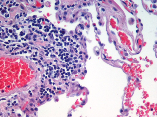
Pleomorphism is a term used in histology and cytopathology to describe variability in the size, shape and staining of cells and/or their nuclei. Several key determinants of cell and nuclear size, like ploidy and the regulation of cellular metabolism, are commonly disrupted in tumors. Therefore, cellular and nuclear pleomorphism is one of the earliest hallmarks of cancer progression and a feature characteristic of malignant neoplasms and dysplasia. Certain benign cell types may also exhibit pleomorphism, e.g. neuroendocrine cells, Arias-Stella reaction. A rare type of rhabdomyosarcoma that is found in adults is known as pleomorphic rhabdomyosarcoma. Despite the prevalence of pleomorphism in human pathology, its role in disease progression is unclear. In epithelial tissue, pleomorphism in cellular size can induce packing defects and disperse aberrant cells. But the consequence of atypical cell and nuclear morphology in other tissues is unknown. See also *Anaplasia *Cell growt ... [...More Info...] [...Related Items...] OR: [Wikipedia] [Google] [Baidu] |
Serous Carcinoma 2a - Cytology
In physiology, serous fluid or serosal fluid (originating from the Medieval Latin word ''serosus'', from Latin ''serum'') is any of various body fluids resembling Serum (blood), serum, that are typically pale yellow or transparent and of a benign nature. The fluid fills the inside of body cavity, body cavities. Serous fluid originates from serous glands, with secretions enriched with proteins and water. Serous fluid may also originate from mixed glands, which contain both mucous cell, mucous and serous cells. A common trait of serous fluids is their role in assisting digestion, excretion, and respiratory system, respiration. In medical fields, especially cytopathology, serous fluid is a synonym for effusion fluids from various body cavities. Examples of effusion fluid are pleural effusion and pericardial effusion. There are many causes of effusions which include involvement of the cavity by cancer. Cancer in a serous cavity is called a serous carcinoma. Cytopathology evaluation is ... [...More Info...] [...Related Items...] OR: [Wikipedia] [Google] [Baidu] |
Arias-Stella Reaction
Arias-Stella reaction, also Arias-Stella phenomenon, is a benign change in the endometrium associated with the presence of chorionic tissue. Arias-Stella reaction is due to progesterone primarily. Cytologically, it looks like a malignancy and, historically, it was diagnosed as endometrial cancer. Significance It is significant only because it can be misdiagnosed as a cancer. It may be seen in a completely normal pregnancy. Diagnosis It is characterized by nuclear enlargement and may also have any of the following: an irregular nuclear membrane, granular chromatin, centronuclear vacuolization, and pseudonuclear inclusions. Five subtypes are recognized: #Minimal atypia. #Early secretory pattern. #Secretory or hypersecretory pattern. #Regenerative, proliferative or nonsecretory pattern. #Monstrous cell pattern. History It was first described by Javier Arias Stella, a Peruvian pathologist, in 1954. See also * Choriocarcinoma * Chorioangioma * Herpes * Nuclear atypia Nucl ... [...More Info...] [...Related Items...] OR: [Wikipedia] [Google] [Baidu] |
Histology
Histology, also known as microscopic anatomy or microanatomy, is the branch of biology which studies the microscopic anatomy of biological tissues. Histology is the microscopic counterpart to gross anatomy, which looks at larger structures visible without a microscope. Although one may divide microscopic anatomy into ''organology'', the study of organs, ''histology'', the study of tissues, and ''cytology'', the study of cells, modern usage places all of these topics under the field of histology. In medicine, histopathology is the branch of histology that includes the microscopic identification and study of diseased tissue. In the field of paleontology, the term paleohistology refers to the histology of fossil organisms. Biological tissues Animal tissue classification There are four basic types of animal tissues: muscle tissue, nervous tissue, connective tissue, and epithelial tissue. All animal tissues are considered to be subtypes of these four principal tissue types ... [...More Info...] [...Related Items...] OR: [Wikipedia] [Google] [Baidu] |
Nuclear Atypia
Nuclear atypia refers to abnormal appearance of cell nuclei. It is a term used in cytopathology and histopathology. Atypical nuclei are often pleomorphic. Nuclear atypia can be seen in reactive changes, pre-neoplastic changes and malignancy. Severe nuclear atypia is, in most cases, considered an indicator of malignancy. See also * Arias-Stella reaction *NC ratio The nuclear-cytoplasmic ratio (also variously known as the nucleus:cytoplasm ratio, nucleus-cytoplasm ratio, N:C ratio, or N/C) is a measurement used in cell biology. It is a ratio of the size (i.e., volume) of the cell nucleus, nucleus of a cell ... * Nuclear pleomorphism {{pathology-stub Pathology ... [...More Info...] [...Related Items...] OR: [Wikipedia] [Google] [Baidu] |
Giant Cell Carcinoma Of The Lung
Giant-cell carcinoma of the lung (GCCL) is a rare histological form of large-cell lung carcinoma, a subtype of undifferentiated lung cancer, traditionally classified within the non-small-cell lung carcinomas (NSCLC). The characteristic feature of this highly lethal malignancy is the distinctive light microscopic appearance of its extremely large cells, which are bizarre and highly pleomorphic, and which often contain more than one huge, misshapen, pleomorphic nucleus ("syncytia"), which result from cell fusion. Although it is common in the lung cancer literature to refer to histologically mixed tumors containing significant numbers of malignant giant cells as "giant-cell carcinomas", technically a diagnosis of "giant-cell carcinoma" should be limited strictly to neoplasms containing ''only'' malignant giant cells (i.e. "pure" giant-cell carcinoma). Aside from the great heterogeneity seen in lung cancers (especially those occurring among tobacco smokers), the considerable varia ... [...More Info...] [...Related Items...] OR: [Wikipedia] [Google] [Baidu] |
Cytopathology
Cytopathology (from Greek , ''kytos'', "a hollow"; , ''pathos'', "fate, harm"; and , '' -logia'') is a branch of pathology that studies and diagnoses diseases on the cellular level. The discipline was founded by George Nicolas Papanicolaou in 1928. Cytopathology is generally used on samples of free cells or tissue fragments, in contrast to histopathology, which studies whole tissues. Cytopathology is frequently, less precisely, called "cytology", which means "the study of cells". Cytopathology is commonly used to investigate diseases involving a wide range of body sites, often to aid in the diagnosis of cancer but also in the diagnosis of some infectious diseases and other inflammatory conditions. For example, a common application of cytopathology is the Pap smear, a screening tool used to detect precancerous cervical lesions that may lead to cervical cancer. Cytopathologic tests are sometimes called smear tests because the samples may be smeared across a glass microscope slid ... [...More Info...] [...Related Items...] OR: [Wikipedia] [Google] [Baidu] |
Cell Growth
Cell growth refers to an increase in the total mass of a cell, including both cytoplasmic, nuclear and organelle volume. Cell growth occurs when the overall rate of cellular biosynthesis (production of biomolecules or anabolism) is greater than the overall rate of cellular degradation (the destruction of biomolecules via the proteasome, lysosome or autophagy, or catabolism). Cell growth is not to be confused with cell division or the cell cycle, which are distinct processes that can occur alongside cell growth during the process of cell proliferation, where a cell, known as the mother cell, grows and divides to produce two daughter cells. Importantly, cell growth and cell division can also occur independently of one another. During early embryonic development ( cleavage of the zygote to form a morula and blastoderm), cell divisions occur repeatedly without cell growth. Conversely, some cells can grow without cell division or without any progression of the cell cycle, such as g ... [...More Info...] [...Related Items...] OR: [Wikipedia] [Google] [Baidu] |
Anaplasia
Anaplasia (from grc, ἀνά ''ana'', "backward" + πλάσις ''plasis'', "formation") is a condition of cells with poor cellular differentiation, losing the morphological characteristics of mature cells and their orientation with respect to each other and to endothelial cells. The term also refers to a group of morphological changes in a cell (nuclear pleomorphism, altered nuclear-cytoplasmic ratio, presence of nucleoli, high proliferation index) that point to a possible malignant transformation. Such loss of structural differentiation is especially seen in most, but not all, malignant neoplasms. Sometimes, the term also includes an increased capacity for multiplication. Lack of differentiation is considered a hallmark of aggressive malignancies (for example, it differentiates leiomyosarcomas from leiomyomas). The term ''anaplasia'' literally means "to form backward". It implies dedifferentiation, or loss of structural and functional differentiation of normal cells. It is n ... [...More Info...] [...Related Items...] OR: [Wikipedia] [Google] [Baidu] |
Tissue (biology)
In biology, tissue is a biological organizational level between cells and a complete organ. A tissue is an ensemble of similar cells and their extracellular matrix from the same origin that together carry out a specific function. Organs are then formed by the functional grouping together of multiple tissues. The English word "tissue" derives from the French word "tissu", the past participle of the verb tisser, "to weave". The study of tissues is known as histology or, in connection with disease, as histopathology. Xavier Bichat is considered as the "Father of Histology". Plant histology is studied in both plant anatomy and physiology. The classical tools for studying tissues are the paraffin block in which tissue is embedded and then sectioned, the histological stain, and the optical microscope. Developments in electron microscopy, immunofluorescence, and the use of frozen tissue-sections have enhanced the detail that can be observed in tissues. With these tools, the c ... [...More Info...] [...Related Items...] OR: [Wikipedia] [Google] [Baidu] |
Epithelial
Epithelium or epithelial tissue is one of the four basic types of animal tissue, along with connective tissue, muscle tissue and nervous tissue. It is a thin, continuous, protective layer of compactly packed cells with a little intercellular matrix. Epithelial tissues line the outer surfaces of organs and blood vessels throughout the body, as well as the inner surfaces of cavities in many internal organs. An example is the epidermis, the outermost layer of the skin. There are three principal shapes of epithelial cell: squamous (scaly), columnar, and cuboidal. These can be arranged in a singular layer of cells as simple epithelium, either squamous, columnar, or cuboidal, or in layers of two or more cells deep as stratified (layered), or ''compound'', either squamous, columnar or cuboidal. In some tissues, a layer of columnar cells may appear to be stratified due to the placement of the nuclei. This sort of tissue is called pseudostratified. All glands are made up of epitheli ... [...More Info...] [...Related Items...] OR: [Wikipedia] [Google] [Baidu] |
Rhabdomyosarcoma
Rhabdomyosarcoma (RMS) is a highly aggressive form of cancer that develops from mesenchymal cells that have failed to fully differentiate into myocytes of skeletal muscle. Cells of the tumor are identified as rhabdomyoblasts. There are four subtypes – embryonal rhabdomyosarcoma, alveolar rhabdomyosarcoma, pleomorphic rhabdomyosarcoma, and spindle cell/sclerosing rhabdomyosarcoma. Embryonal, and alveolar are the main groups, and these types are the most common soft tissue sarcomas of childhood and adolescence. The pleomorphic type is usually found in adults. It is generally considered to be a disease of childhood, as the vast majority of cases occur in those below the age of 18. It is commonly described as one of the small-blue-round-cell tumors of childhood due to its appearance on an H&E stain. Despite being relatively rare, it accounts for approximately 40% of all recorded soft tissue sarcomas. RMS can occur in any soft tissue site in the body, but is primarily found in t ... [...More Info...] [...Related Items...] OR: [Wikipedia] [Google] [Baidu] |
Neuroendocrine Cell
Neuroendocrine cells are cells that receive neuronal input (through neurotransmitters released by nerve cells or neurosecretory cells) and, as a consequence of this input, release messenger molecules ( hormones) into the blood. In this way they bring about an integration between the nervous system and the endocrine system, a process known as neuroendocrine integration. An example of a neuroendocrine cell is a cell of the adrenal medulla (innermost part of the adrenal gland), which releases adrenaline to the blood. The adrenal medullary cells are controlled by the sympathetic division of the autonomic nervous system. These cells are modified postganglionic neurons. Autonomic nerve fibers lead directly to them from the central nervous system. The adrenal medullary hormones are kept in vesicles much in the same way neurotransmitters are kept in neuronal vesicles. Hormonal effects can last up to ten times longer than those of neurotransmitters. Sympathetic nerve fiber impulses sti ... [...More Info...] [...Related Items...] OR: [Wikipedia] [Google] [Baidu] |
.jpg)


.jpg)