|
Peripheral Blood Smear
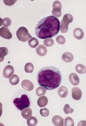
A blood smear, peripheral blood smear or blood film is a thin layer of blood smeared on a glass microscope slide and then stained in such a way as to allow the various blood cells to be examined microscopically. Blood smears are examined in the investigation of hematology, hematological (blood) disorders and are routinely employed to look for blood Apicomplexa, parasites, such as those of malaria and filariasis. Preparation A blood smear is made by placing a drop of blood on one end of a slide, and using a ''spreader slide'' to disperse the blood over the slide's length. The aim is to get a region, called a monolayer, where the cells are spaced far enough apart to be counted and differentiated. The monolayer is found in the "feathered edge" created by the spreader slide as it draws the blood forward. The slide is left to air dry, after which the blood is fixation (histology), fixed to the slide by immersing it briefly in methanol. The fixative is essential for good staining a ... [...More Info...] [...Related Items...] OR: [Wikipedia] [Google] [Baidu] |
Complete Blood Count
A complete blood count (CBC), also known as a full blood count (FBC), is a set of medical laboratory tests that provide cytometry, information about the cells in a person's blood. The CBC indicates the counts of white blood cells, red blood cells and platelets, the concentration of hemoglobin, and the hematocrit (the volume percentage of red blood cells). The red blood cell indices, which indicate the average size and hemoglobin content of red blood cells, are also reported, and a white blood cell differential, which counts the different types of white blood cells, may be included. The CBC is often carried out as part of a medical assessment and can be used to monitor health or diagnose diseases. The results are interpreted by comparing them to Reference ranges for blood tests, reference ranges, which vary with sex and age. Conditions like anemia and thrombocytopenia are defined by abnormal complete blood count results. The red blood cell indices can provide information about the ... [...More Info...] [...Related Items...] OR: [Wikipedia] [Google] [Baidu] |
Blood
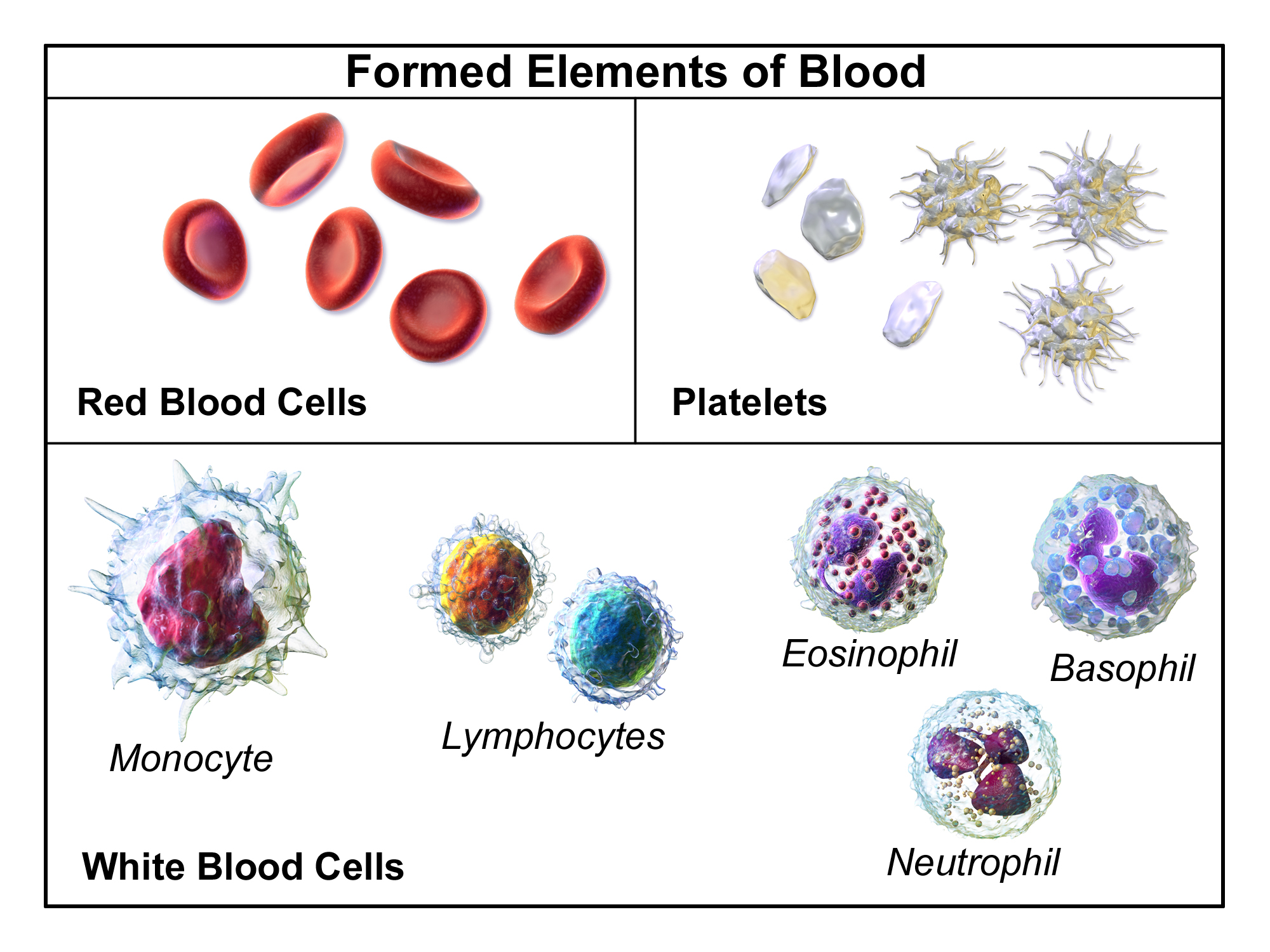
Blood is a body fluid in the circulatory system of humans and other vertebrates that delivers necessary substances such as nutrients and oxygen to the cells, and transports metabolic waste products away from those same cells. Blood in the circulatory system is also known as ''peripheral blood'', and the blood cells it carries, ''peripheral blood cells''. Blood is composed of blood cells suspended in blood plasma. Plasma, which constitutes 55% of blood fluid, is mostly water (92% by volume), and contains proteins, glucose, mineral ions, hormones, carbon dioxide (plasma being the main medium for excretory product transportation), and blood cells themselves. Albumin is the main protein in plasma, and it functions to regulate the colloidal osmotic pressure of blood. The blood cells are mainly red blood cells (also called RBCs or erythrocytes), white blood cells (also called WBCs or leukocytes) and platelets (also called thrombocytes). The most abundant cells in vertebrate blo ... [...More Info...] [...Related Items...] OR: [Wikipedia] [Google] [Baidu] |
Chronic Myeloid Leukemia
Chronic myelogenous leukemia (CML), also known as chronic myeloid leukemia, is a cancer of the white blood cells. It is a form of leukemia characterized by the increased and unregulated growth of myeloid cells in the bone marrow and the accumulation of these cells in the blood. CML is a clonal bone marrow stem cell disorder in which a proliferation of mature granulocytes (neutrophils, eosinophils and basophils) and their precursors is found. It is a type of myeloproliferative neoplasm associated with a characteristic chromosomal translocation called the Philadelphia chromosome. CML is largely treated with targeted drugs called tyrosine-kinase inhibitors (TKIs) which have led to dramatically improved long-term survival rates since 2001. These drugs have revolutionized treatment of this disease and allow most patients to have a good quality of life when compared to the former chemotherapy drugs. In Western countries, CML accounts for 15–25% of all adult leukemias and 14% of leuke ... [...More Info...] [...Related Items...] OR: [Wikipedia] [Google] [Baidu] |
Blast Cell
In cell biology, a precursor cell, also called a blast cell or simply blast, is a partially differentiated cell, usually referred to as a unipotent cell that has lost most of its stem cell properties. A precursor cell is also known as a progenitor cell but progenitor cells are multipotent. Precursor cells are known as the intermediate cell before they become differentiated after being a stem cell. Usually, a precursor cell is a stem cell with the capacity to differentiate into only one cell type. Sometimes, ''precursor cell'' is used as an alternative term for unipotent stem cells. In embryology, precursor cells are a group of cells that later differentiate into one organ. A blastoma is any cancer created by malignancies of precursor cells. Precursor cells, and progenitor cells, have many potential uses in medicine. , there is research being done to use these cells to build heart valves, blood vessels and other tissues, by using blood and muscle precursor, or progenitor cel ... [...More Info...] [...Related Items...] OR: [Wikipedia] [Google] [Baidu] |
Eosinophilia
Eosinophilia is a condition in which the eosinophil count in the peripheral blood exceeds . Hypereosinophilia is an elevation in an individual's circulating blood eosinophil count above 1.5 x 10⁹/ L (i.e. 1,500/μL). The hypereosinophilic syndrome is a sustained elevation in this count above 1.5 x 10⁹/L (i.e. 1,500/μL) that is also associated with evidence of eosinophil-based tissue injury. Eosinophils usually account for less than 7% of the circulating leukocytes. A marked increase in non-blood tissue eosinophil count noticed upon histopathologic examination is diagnostic for tissue eosinophilia. Several causes are known, with the most common being some form of allergic reaction or parasitic infection. Diagnosis of eosinophilia is via a complete blood count (CBC), but diagnostic procedures directed at the underlying cause vary depending on the suspected condition(s). An absolute eosinophil count is not generally needed if the CBC shows marked eosinophilia. The location of the c ... [...More Info...] [...Related Items...] OR: [Wikipedia] [Google] [Baidu] |
Neutrophilia
Neutrophilia (also called neutrophil leukocytosis or occasionally neutrocytosis) is leukocytosis of neutrophils, that is, a high number of neutrophils in the blood. Because neutrophils are the main type of granulocytes, mentions of granulocytosis often overlap in meaning with neutrophilia. The opposite of neutrophilia is neutropenia. Causes Neutrophils are the primary white blood cells that respond to a bacterial infection, so the most common cause of neutrophilia is a bacterial infection, especially pyogenic infections.Table 12-6 in: 8th edition. Neutrophils are also increased in any acute inflammation, so will be raised after a heart attack, other infarct or burns. Some drugs, such as prednisone, have the same effect as cortisol and adrenaline (epinephrine), causing marginated neutrophils to enter the blood stream. A neutrophilia might also be the result of a malignancy. Chronic myelogenous leukemia (CML or chronic myeloid leukaemia) is a disease where the blood cells pr ... [...More Info...] [...Related Items...] OR: [Wikipedia] [Google] [Baidu] |
White Blood Cell Differential
A white blood cell differential is a medical laboratory test that provides information about the types and amounts of white blood cells in a person's blood. The test, which is usually ordered as part of a complete blood count (CBC), measures the amounts of the five normal white blood cell typesneutrophils, lymphocytes, monocytes, eosinophils and basophilsas well as abnormal cell types if they are present. These results are reported as percentages and absolute values, and compared against reference ranges to determine whether the values are normal, low, or high. Changes in the amounts of white blood cells can aid in the diagnosis of many health conditions, including viral, bacterial, and parasitic infections and blood disorders such as leukemia. White blood cell differentials may be performed by an automated analyzera machine designed to run laboratory tests – or manually, by examining blood smears under a microscope. The test was performed manually until white blood cell dif ... [...More Info...] [...Related Items...] OR: [Wikipedia] [Google] [Baidu] |
White Blood Cells
White blood cells, also called leukocytes or leucocytes, are the cells of the immune system that are involved in protecting the body against both infectious disease and foreign invaders. All white blood cells are produced and derived from multipotent cells in the bone marrow known as hematopoietic stem cells. Leukocytes are found throughout the body, including the blood and lymphatic system. All white blood cells have nuclei, which distinguishes them from the other blood cells, the anucleated red blood cells (RBCs) and platelets. The different white blood cells are usually classified by cell lineage (myeloid cells or lymphoid cells). White blood cells are part of the body's immune system. They help the body fight infection and other diseases. Types of white blood cells are granulocytes (neutrophils, eosinophils, and basophils), and agranulocytes (monocytes, and lymphocytes (T cells and B cells)). Myeloid cells (myelocytes) include neutrophils, eosinophils, mast cells, bas ... [...More Info...] [...Related Items...] OR: [Wikipedia] [Google] [Baidu] |
Microangiopathic Hemolytic Anemia
Microangiopathic hemolytic anemia (MAHA) is a microangiopathic subgroup of hemolytic anemia (loss of red blood cells through destruction) caused by factors in the small blood vessels. It is identified by the finding of anemia and schistocytes on microscopy of the blood film. Signs and symptoms In diseases such as hemolytic uremic syndrome, disseminated intravascular coagulation, thrombotic thrombocytopenic purpura, and malignant hypertension, the endothelial layer of small vessels is damaged with resulting fibrin deposition and platelet aggregation. As red blood cells travel through these damaged vessels, they are fragmented resulting in intravascular hemolysis. The resulting schistocytes (red cell fragments) are also increasingly targeted for destruction by the reticuloendothelial system in the spleen, due to their narrow passage through obstructed vessel lumina. It is seen in systemic lupus erythematosus, where immune complexes aggregate with platelets, forming intravascular ... [...More Info...] [...Related Items...] OR: [Wikipedia] [Google] [Baidu] |
Megaloblastic Anemia
Megaloblastic anemia is a type of macrocytic anemia. An anemia is a red blood cell defect that can lead to an undersupply of oxygen. Megaloblastic anemia results from inhibition of DNA replication, DNA synthesis during red blood cell production. When DNA synthesis is impaired, the cell cycle cannot progress from the G2 growth stage to the mitosis (M) stage. This leads to continuing cell growth without division, which presents as macrocytosis. Megaloblastic anemia has a rather slow onset, especially when compared to that of other anemias. The defect in red cell DNA synthesis is most often due to hypovitaminosis, specifically vitamin B12 deficiency or folate deficiency. Loss of micronutrients may also be a cause. Megaloblastic anemia not due to hypovitaminosis may be caused by antimetabolites that poison DNA production directly, such as some chemotherapeutic or antimicrobial agents (for example azathioprine or trimethoprim). The pathological state of megaloblastosis is characteriz ... [...More Info...] [...Related Items...] OR: [Wikipedia] [Google] [Baidu] |
Sickle Cell Anemia
Sickle cell disease (SCD) is a group of blood disorders typically inherited from a person's parents. The most common type is known as sickle cell anaemia. It results in an abnormality in the oxygen-carrying protein haemoglobin found in red blood cells. This leads to a rigid, sickle-like shape under certain circumstances. Problems in sickle cell disease typically begin around 5 to 6 months of age. A number of health problems may develop, such as attacks of pain (known as a sickle cell crisis), anemia, swelling in the hands and feet, bacterial infections and stroke. Long-term pain may develop as people get older. The average life expectancy in the developed world is 40 to 60 years. Sickle cell disease occurs when a person inherits two abnormal copies of the β-globin gene (''HBB'') that makes haemoglobin, one from each parent. This gene occurs in chromosome 11. Several subtypes exist, depending on the exact mutation in each haemoglobin gene. An attack can be set off by tempera ... [...More Info...] [...Related Items...] OR: [Wikipedia] [Google] [Baidu] |
Iron Deficiency Anemia
Iron-deficiency anemia is anemia caused by a lack of iron. Anemia is defined as a decrease in the number of red blood cells or the amount of hemoglobin in the blood. When onset is slow, symptoms are often vague such as feeling tired, weak, short of breath, or having decreased ability to exercise. Anemia that comes on quickly often has more severe symptoms, including confusion, feeling like one is going to pass out or increased thirst. Anemia is typically significant before a person becomes noticeably pale. Children with iron deficiency anemia may have problems with growth and development. There may be additional symptoms depending on the underlying cause. Iron-deficiency anemia is caused by blood loss, insufficient dietary intake, or poor absorption of iron from food. Sources of blood loss can include heavy periods, childbirth, uterine fibroids, stomach ulcers, colon cancer, and urinary tract bleeding. Poor absorption of iron from food may occur as a result of an intestina ... [...More Info...] [...Related Items...] OR: [Wikipedia] [Google] [Baidu] |


