|
Percutaneous Nephrolithotomy
Percutaneous nephrolithotomy (PCNL) is a minimally-invasive procedure to remove stones from the kidney by a small puncture wound (up to about 1 cm) through the skin. It is most suitable to remove stones of more than 2 cm in size and which are present near the pelvic region. It is usually done under general anesthesia or spinal anesthesia. __TOC__ Procedure A retrograde pyelogram is done to locate the stone in the kidney. Contrast is diluted to the ratio of 1:3. If the ureteric catheter is placed in the upper pole of the kidney, it should be pulled down so that the tip is inside the renal pelvis. This helps for proper filling of the renal calyces. The contrast is injected slowly to prevent extravasation. Fluoroscopy monitoring should be continuous so that the sequence of calyces be filled can help to identify the position of posterior calyx. With a small 1 centimeter incision in the loin, the percutaneous nephrolithotomy (PCN) needle is passed into the pelvis of the ... [...More Info...] [...Related Items...] OR: [Wikipedia] [Google] [Baidu] |
Minimally-invasive Procedure
Minimally invasive procedures (also known as minimally invasive surgeries) encompass surgical techniques that limit the size of incisions needed, thereby reducing wound healing time, associated pain, and risk of infection. Surgery by definition is invasive and many operations requiring incisions of some size are referred to as ''open surgery''. Incisions made during open surgery can sometimes leave large wounds that may be painful and take a long time to heal. Advancements in medical technologies have enabled the development and regular use of minimally invasive procedures. For example, endovascular aneurysm repair, a minimally invasive surgery, has become the most common method of repairing abdominal aortic aneurysms in the US as of 2003. The procedure involves much smaller incisions than the corresponding open surgery procedure of open aortic surgery. Interventional radiologists were the forerunners of minimally invasive procedures. Using imaging techniques, radiologists were ... [...More Info...] [...Related Items...] OR: [Wikipedia] [Google] [Baidu] |
Kidney Stones
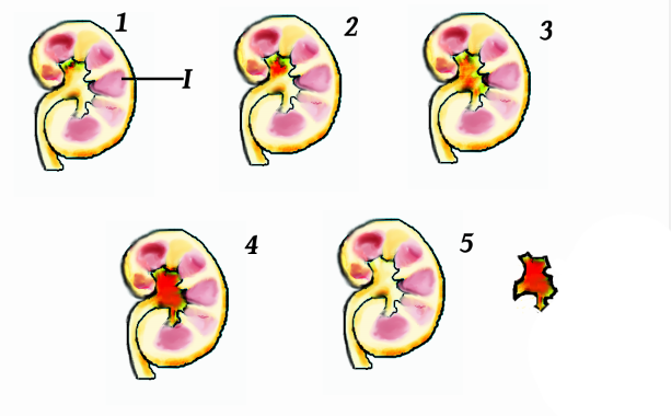
Kidney stone disease, also known as nephrolithiasis or urolithiasis, is a crystallopathy where a solid piece of material (kidney stone) develops in the urinary tract. Kidney stones typically form in the kidney and leave the body in the urine stream. A small stone may pass without causing symptoms. If a stone grows to more than , it can cause blockage of the ureter, resulting in sharp and severe pain in the lower back or abdomen. A stone may also result in blood in the urine, vomiting, or painful urination. About half of people who have had a kidney stone will have another within ten years. Most stones form by a combination of genetics and environmental factors. Risk factors include high urine calcium levels, obesity, certain foods, some medications, calcium supplements, hyperparathyroidism, gout and not drinking enough fluids. Stones form in the kidney when minerals in urine are at high concentration. The diagnosis is usually based on symptoms, urine testing, and medical ... [...More Info...] [...Related Items...] OR: [Wikipedia] [Google] [Baidu] |
Kidney
The kidneys are two reddish-brown bean-shaped organs found in vertebrates. They are located on the left and right in the retroperitoneal space, and in adult humans are about in length. They receive blood from the paired renal arteries; blood exits into the paired renal veins. Each kidney is attached to a ureter, a tube that carries excreted urine to the bladder. The kidney participates in the control of the volume of various body fluids, fluid osmolality, acid–base balance, various electrolyte concentrations, and removal of toxins. Filtration occurs in the glomerulus: one-fifth of the blood volume that enters the kidneys is filtered. Examples of substances reabsorbed are solute-free water, sodium, bicarbonate, glucose, and amino acids. Examples of substances secreted are hydrogen, ammonium, potassium and uric acid. The nephron is the structural and functional unit of the kidney. Each adult human kidney contains around 1 million nephrons, while a mouse kidney contains on ... [...More Info...] [...Related Items...] OR: [Wikipedia] [Google] [Baidu] |
Renal Pelvis
The renal pelvis or pelvis of the kidney is the funnel-like dilated part of the ureter in the kidney. It is formed by the covnvergence of the major calyces, acting as a funnel for urine flowing from the major calyces to the ureter. It has a mucous membrane and is covered with transitional epithelium and an underlying lamina propria of loose-to-dense connective tissue. The renal pelvis is situated within the renal sinus alongside the other structures of the renal sinus. The renal pelvis is the location of several kinds of kidney cancer and is affected by infection in pyelonephritis. Clinical significance The renal pelvis is the location of several kinds of kidney cancer and is affected by infection in pyelonephritis. A large "staghorn" kidney stone may block all or part of the renal pelvis. The size of the renal pelvis plays a major role in the grading of hydronephrosis. Normally, the anteroposterior diameter of the renal pelvis is less than 4 mm in fetuses up to 32 weeks ... [...More Info...] [...Related Items...] OR: [Wikipedia] [Google] [Baidu] |
General Anesthesia
General anaesthesia (UK) or general anesthesia (US) is a medically induced loss of consciousness that renders the patient unarousable even with painful stimuli. This effect is achieved by administering either intravenous or inhalational general anaesthetic medications, which often act in combination with an analgesic and neuromuscular blocking agent. Spontaneous ventilation is often inadequate during the procedure and intervention is often necessary to protect the airway. General anaesthesia is generally performed in an operating theater to allow surgical procedures that would otherwise be intolerably painful for a patient, or in an intensive care unit or emergency department to facilitate endotracheal intubation and mechanical ventilation in critically ill patients. A variety of drugs may be administered, with the overall goal of achieving unconsciousness, amnesia, analgesia, loss of reflexes of the autonomic nervous system, and in some cases paralysis of skeletal muscles. The ... [...More Info...] [...Related Items...] OR: [Wikipedia] [Google] [Baidu] |
Spinal Anesthesia
Spinal anaesthesia (or spinal anesthesia), also called spinal block, subarachnoid block, intradural block and intrathecal block, is a form of neuraxial regional anaesthesia involving the injection of a local anaesthetic or opioid into the subarachnoid space, generally through a fine needle, usually long. It is a safe and effective form of anesthesia usually performed by anesthesiologists that can be used as an alternative to general anesthesia commonly in surgeries involving the lower extremities and surgeries below the umbilicus. The local anesthetic with or without an opioid injected into the cerebrospinal fluid provides locoregional anaesthesia: true analgesia, motor, sensory and autonomic (sympathic) blockade. Administering analgesics (opioid, alpha2-adrenoreceptor agonist) in the cerebrospinal fluid without a local anaesthetic produces locoregional analgesia: markedly reduced pain sensation (incomplete analgesia), some autonomic blockade (parasympathetic plexi), but no sen ... [...More Info...] [...Related Items...] OR: [Wikipedia] [Google] [Baidu] |
Retrograde Pyelogram
Pyelogram (or pyelography or urography) is a form of imaging of the renal pelvis and ureter. Types include: * #Intravenous pyelogram, Intravenous pyelogram – In which a contrast solution is introduced through a vein into the circulatory system. * Retrograde pyelogram – Any pyelogram in which contrast medium is introduced from the lower urinary tract and flows toward the kidney (i.e. in a "retrograde" direction, against the normal flow of urine). * #Anterograde pyelogram, Anterograde pyelogram (also antegrade pyelogram) – A pyelogram where a contrast medium passes from the kidneys toward the bladder, mimicking the normal flow of urine. * Gas pyelogram – A pyelogram that uses a gaseous rather than liquid contrast medium. It may also form without the injection of a gas, when gas producing micro-organisms infect the most upper parts of urinary system. Intravenous pyelogram An intravenous pyelogram (IVP), also called an intravenous urogram (IVU), is a radiology, radiological pr ... [...More Info...] [...Related Items...] OR: [Wikipedia] [Google] [Baidu] |
Renal Pelvis
The renal pelvis or pelvis of the kidney is the funnel-like dilated part of the ureter in the kidney. It is formed by the covnvergence of the major calyces, acting as a funnel for urine flowing from the major calyces to the ureter. It has a mucous membrane and is covered with transitional epithelium and an underlying lamina propria of loose-to-dense connective tissue. The renal pelvis is situated within the renal sinus alongside the other structures of the renal sinus. The renal pelvis is the location of several kinds of kidney cancer and is affected by infection in pyelonephritis. Clinical significance The renal pelvis is the location of several kinds of kidney cancer and is affected by infection in pyelonephritis. A large "staghorn" kidney stone may block all or part of the renal pelvis. The size of the renal pelvis plays a major role in the grading of hydronephrosis. Normally, the anteroposterior diameter of the renal pelvis is less than 4 mm in fetuses up to 32 weeks ... [...More Info...] [...Related Items...] OR: [Wikipedia] [Google] [Baidu] |
Renal Calyx
The renal calyces are chambers of the kidney through which urine passes. The minor calyces surround the apex of the renal pyramids. Urine formed in the kidney passes through a renal papilla at the apex into the minor calyx; two or three minor calyces converge to form a major calyx, through which urine passes before continuing through the renal pelvis into the ureter. Function Peristalsis of the smooth muscle originating in pace-maker cells originating in the walls of the calyces propels urine through the renal pelvis and ureters to the bladder. The initiation is caused by the increase in volume that stretches the walls of the calyces. This causes them to fire impulses which stimulate rhythmical contraction and relaxation, called peristalsis. Parasympathetic innervation enhances the peristalsis while sympathetic innervation inhibits it. Clinical significance A "staghorn calculus" is a kidney stone that may extend into the renal calyces. A renal diverticulum is diverticulum of ... [...More Info...] [...Related Items...] OR: [Wikipedia] [Google] [Baidu] |
Loin
The loins, or lumbus, are the sides between the lower ribs and pelvis, and the lower part of the back. The term is used to describe the anatomy of humans and quadrupeds, such as horses, pigs, or cattle. The anatomical reference also applies to particular cuts of meat, including tenderloin or sirloin steak. Human anatomy In human anatomy the term "loin" or "loins" refers to the side of the human body below the rib cage to just above the pelvis. It is frequently used to reference the general area below the ribs. While the term "loin" is generally not used in medical science, some disorders do include the term. The lumbar region of the spinal column is located in the loin area of the body. Society and culture In contemporary usage the term appears primarily in two contexts: where loins are "girded" in preparation for a challenge, or else euphemistically referring to human genitals. In literature or poetry, to feel a "stirring" in one's loins may suggest sexual excitement. The ... [...More Info...] [...Related Items...] OR: [Wikipedia] [Google] [Baidu] |
Fluoroscopy
Fluoroscopy () is an imaging technique that uses X-rays to obtain real-time moving images of the interior of an object. In its primary application of medical imaging, a fluoroscope () allows a physician to see the internal structure and function of a patient, so that the pumping action of the heart or the motion of swallowing, for example, can be watched. This is useful for both diagnosis and therapy and occurs in general radiology, interventional radiology, and image-guided surgery. In its simplest form, a fluoroscope consists of an X-ray source and a fluorescent screen, between which a patient is placed. However, since the 1950s most fluoroscopes have included X-ray image intensifiers and cameras as well, to improve the image's visibility and make it available on a remote display screen. For many decades, fluoroscopy tended to produce live pictures that were not recorded, but since the 1960s, as technology improved, recording and playback became the norm. Fluoroscopy is s ... [...More Info...] [...Related Items...] OR: [Wikipedia] [Google] [Baidu] |
Ureteroscope
Ureteroscopy is an examination of the upper urinary tract, usually performed with a ureteroscope that is passed through the urethra and the bladder, and then directly into the ureter. The procedure is useful in the diagnosis and treatment of disorders such as kidney stones and urothelial carcinoma of the upper urinary tract. Smaller stones in the bladder or lower ureter can be removed in one piece, while bigger ones are usually broken before removal during ureteroscopy. The examination may be performed with either a flexible, semi-rigid or rigid device while the patient is under anesthesia. In specific cases, the patient is free to go home after the examination.Ureteropyeloscopy. Baylor College of Medicine. 2018 ccessed 2018 Mar 5br>/ref> In pyeloscopy, the endoscope is designed to reach all the way to the renal pelvis The renal pelvis or pelvis of the kidney is the funnel-like dilated part of the ureter in the kidney. It is formed by the covnvergence of the major calyces, acti ... [...More Info...] [...Related Items...] OR: [Wikipedia] [Google] [Baidu] |


