|
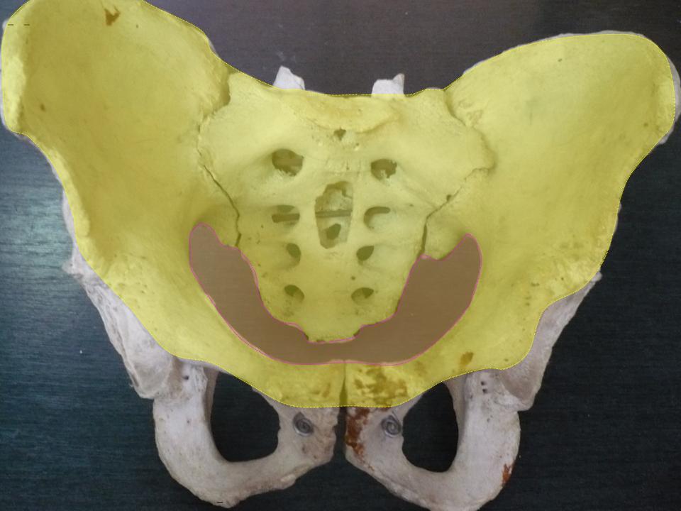
Pelvic Brim
The pelvic brim is the edge of the pelvic inlet. It is an approximately Mickey Mouse head-shaped line passing through the prominence of the sacrum, the arcuate and pectineal lines, and the upper margin of the pubic symphysis. Structure The pelvic brim is an approximately Mickey Mouse head-shaped line passing through the prominence of the sacrum, the arcuate and pectineal lines, and the upper margin of the pubic symphysis. The pelvic brim is obtusely pointed in front, diverging on either side, and encroached upon behind by the projection forward of the promontory of the sacrum. The oblique plane passing approximately through the pelvic brim divides the internal part of the pelvis (pelvic cavity) into the false or greater pelvis and the true or lesser pelvis. The false pelvis, which is above that plane, is sometimes considered to be a part of the abdominal cavity, rather than a part of the pelvic cavity. In this case, the pelvic cavity coincides with the true pelvis, which is ... [...More Info...] [...Related Items...] OR: [Wikipedia] [Google] [Baidu] |
Lesser Pelvis
The pelvic cavity is a body cavity that is bounded by the bones of the pelvis. Its oblique roof is the pelvic inlet (the superior opening of the pelvis). Its lower boundary is the pelvic floor. The pelvic cavity primarily contains the reproductive organs, urinary bladder, distal ureters, proximal urethra, terminal sigmoid colon, rectum, and anal canal. In females, the uterus, Fallopian tubes, ovaries and upper vagina occupy the area between the other viscera. The rectum is located at the back of the pelvis, in the curve of the sacrum and coccyx; the bladder is in front, behind the pubic symphysis. The pelvic cavity also contains major arteries, veins, muscles, and nerves. These structures coexist in a crowded space, and disorders of one pelvic component may impact upon another; for example, constipation may overload the rectum and compress the urinary bladder, or childbirth might damage the pudendal nerves and later lead to anal weakness. Structure The pelvis ha ... [...More Info...] [...Related Items...] OR: [Wikipedia] [Google] [Baidu] |
Abdominal Cavity
The abdominal cavity is a large body cavity in humans and many other animals that contains many organs. It is a part of the abdominopelvic cavity. It is located below the thoracic cavity, and above the pelvic cavity. Its dome-shaped roof is the thoracic diaphragm, a thin sheet of muscle under the lungs, and its floor is the pelvic inlet, opening into the pelvis. Structure Organs Organs of the abdominal cavity include the stomach, liver, gallbladder, spleen, pancreas, small intestine, kidneys, large intestine, and adrenal glands. Peritoneum The abdominal cavity is lined with a protective membrane termed the peritoneum. The inside wall is covered by the parietal peritoneum. The kidneys are located behind the peritoneum, in the retroperitoneum, outside the abdominal cavity. The viscera are also covered by visceral peritoneum. Between the visceral and parietal peritoneum is the peritoneal cavity, which is a potential space. It contains a serous fluid called peritone ... [...More Info...] [...Related Items...] OR: [Wikipedia] [Google] [Baidu] |
Pelvis Justo Major
''Pelvis justo major'' (also called "Giant Pelvis") is a rare condition of the adult female pelvis where the pelvis flairs above the Iliopectineal line. It is 1.5 or more times larger than an average pelvis in every direction and is at least 42 cm (16.5 inches) biiliac width. Even though this condition is classified as a congenital abnormality, it is not a medical disease or abnormality of the pelvis (as the pelvis is a true gynecoid shape, only larger). Women with this condition, at the time of delivery, may have a precipitous birth. There is virtually no resistance of the huge pelvic opening to the size of a newborn so only the soft parts resist the birth. With a huge ''Justo Major Pelvis'', there is no pelvic bone "molding" of the fetal head. With the average pelvic size (2/3 or less ''Justo Major'' size) the usual pelvic molding process slows the birth, resulting in a slow and gradual stretching of the vaginal opening for primiparous women. When a huge ''Justo Major ... [...More Info...] [...Related Items...] OR: [Wikipedia] [Google] [Baidu] |
Linea Terminalis
The linea terminalis or innominate line consists of the pubic crest, pectineal line (pecten pubis), the arcuate line, the sacral ala, and the sacral promontory. It is the pelvic brim, which is the edge of the pelvic inlet. The pelvic inlet is typically used to divide the abdominopelvic cavity into an abdominal (above the inlet) and a pelvic cavity (below the inlet). Sometimes, the pelvis cavity is considered to extend above the pelvic inlet, and in this case the pelvic inlet is used to divide the pelvic cavity into a false (above the inlet) and a true pelvis (below the inlet). Additional images File:Gray238.png, Diameters of superior aperture of lesser pelvis The pelvic cavity is a body cavity that is bounded by the bones of the pelvis. Its oblique roof is the pelvic inlet (the superior opening of the pelvis). Its lower boundary is the pelvic floor. The pelvic cavity primarily contains the reprod ... — female. File:Gray242.png, Female pelvis. References Exte ... [...More Info...] [...Related Items...] OR: [Wikipedia] [Google] [Baidu] |
Abdominal Surgery
The term abdominal surgery broadly covers surgical procedures that involve opening the abdomen (laparotomy). Surgery of each abdominal organ is dealt with separately in connection with the description of that organ (see stomach, kidney, liver, etc.) Diseases affecting the abdominal cavity are dealt with generally under their own names (e.g. appendicitis). Types The most common abdominal surgeries are described below. * Appendectomy: surgical opening of the abdominal cavity and removal of the appendix. Typically performed as definitive treatment for appendicitis, although sometimes the appendix is prophylactically removed incidental to another abdominal procedure. * Caesarean section (also known as C-section): a surgical procedure in which one or more incisions are made through a mother's abdomen (laparotomy) and uterus (hysterotomy) to deliver one or more babies, or, rarely, to remove a dead fetus. * Inguinal hernia surgery: the repair of an inguinal hernia. *Exploratory lapa ... [...More Info...] [...Related Items...] OR: [Wikipedia] [Google] [Baidu] |
Femoral Nerve
The femoral nerve is a nerve in the thigh that supplies skin on the upper thigh and inner leg, and the muscles that extend the knee. Structure The femoral nerve is the major nerve supplying the anterior compartment of the thigh. It is the largest branch of the lumbar plexus, and arises from the dorsal divisions of the ventral rami of the second, third, and fourth lumbar nerves (L2, L3, and L4). The nerve enters Scarpa's triangle by passing beneath the inguinal ligament, just lateral to the femoral artery. In the thigh, the nerve lies in a groove between iliacus muscle and psoas major muscle, outside the femoral sheath, and lateral to the femoral artery. After a short course of about 4 cm in the thigh, the nerve is divided into anterior and posterior divisions, separated by lateral femoral circumflex artery. The branches are shown below: Muscular branches * The nerve to the pectineus muscle arises immediately above the inguinal ligament from the medial side of the fem ... [...More Info...] [...Related Items...] OR: [Wikipedia] [Google] [Baidu] |
Sigmoid Colon
The sigmoid colon (or pelvic colon) is the part of the large intestine that is closest to the rectum and anus. It forms a loop that averages about in length. The loop is typically shaped like a Greek letter sigma (ς) or Latin letter S (thus '' sigma'' + '' -oid''). This part of the colon normally lies within the pelvis, but due to its freedom of movement it is liable to be displaced into the abdominal cavity. Structure The sigmoid colon begins at the superior aperture of the lesser pelvis, where it is continuous with the iliac colon, and passes transversely across the front of the sacrum to the right side of the pelvis. It then curves on itself and turns toward the left to reach the middle line at the level of the third piece of the sacrum, where it bends downward and ends in the rectum. Its function is to expel solid and gaseous waste from the gastrointestinal tract. The curving path it takes toward the anus allows it to store gas in the superior arched portion, enab ... [...More Info...] [...Related Items...] OR: [Wikipedia] [Google] [Baidu] |
Urinary Bladder
The urinary bladder, or simply bladder, is a hollow organ in humans and other vertebrates that stores urine from the kidneys before disposal by urination. In humans the bladder is a distensible organ that sits on the pelvic floor. Urine enters the bladder via the ureters and exits via the urethra. The typical adult human bladder will hold between 300 and (10.14 and ) before the urge to empty occurs, but can hold considerably more. The Latin phrase for "urinary bladder" is ''vesica urinaria'', and the term ''vesical'' or prefix ''vesico -'' appear in connection with associated structures such as vesical veins. The modern Latin word for "bladder" – ''cystis'' – appears in associated terms such as cystitis (inflammation of the bladder). Structure In humans, the bladder is a hollow muscular organ situated at the base of the pelvis. In gross anatomy, the bladder can be divided into a broad , a body, an apex, and a neck. The apex (also called the vertex) is directed fo ... [...More Info...] [...Related Items...] OR: [Wikipedia] [Google] [Baidu] |
Greater Pelvis
The pelvic cavity is a body cavity that is bounded by the bones of the pelvis. Its oblique roof is the pelvic inlet (the superior opening of the pelvis). Its lower boundary is the pelvic floor. The pelvic cavity primarily contains the reproductive organs, urinary bladder, distal ureters, proximal urethra, terminal sigmoid colon, rectum, and anal canal. In females, the uterus, Fallopian tubes, ovaries and upper vagina occupy the area between the other viscera. The rectum is located at the back of the pelvis, in the curve of the sacrum and coccyx; the bladder is in front, behind the pubic symphysis. The pelvic cavity also contains major arteries, veins, muscles, and nerves. These structures coexist in a crowded space, and disorders of one pelvic component may impact upon another; for example, constipation may overload the rectum and compress the urinary bladder, or childbirth might damage the pudendal nerves and later lead to anal weakness. Structure The pelvis has an antero ... [...More Info...] [...Related Items...] OR: [Wikipedia] [Google] [Baidu] |
Pelvic Inlet
The pelvic inlet or superior aperture of the pelvis is a planar surface which defines the boundary between the pelvic cavity and the abdominal cavity (or, according to some authors, between two parts of the pelvic cavity, called lesser pelvis and greater pelvis). It is a major target of measurements of pelvimetry. Its position and orientation relative to the skeleton of the pelvis is anatomically defined by its edge, the pelvic brim. The pelvic brim is an approximately apple-shaped line passing through the prominence of the sacrum, the arcuate and pectineal lines, and the upper margin of the pubic symphysis. Occasionally, the terms pelvic inlet and pelvic brim are used interchangeably. Boundaries The edge of the pelvic inlet (pelvic brim) is formed as follows: Diameters The diameters or conjugates of the pelvis are measured at the pelvic inlet and outlet and as oblique diameters. Two diameters may be measured from the outside of the body using a pelvimeter Additi ... [...More Info...] [...Related Items...] OR: [Wikipedia] [Google] [Baidu] |
Pelvic Cavity
The pelvic cavity is a body cavity that is bounded by the bones of the pelvis. Its oblique roof is the pelvic inlet (the superior opening of the pelvis). Its lower boundary is the pelvic floor. The pelvic cavity primarily contains the reproductive organs, urinary bladder, distal ureters, proximal urethra, terminal sigmoid colon, rectum, and anal canal. In females, the uterus, Fallopian tubes, ovaries and upper vagina occupy the area between the other viscera. The rectum is located at the back of the pelvis, in the curve of the sacrum and coccyx; the bladder is in front, behind the pubic symphysis. The pelvic cavity also contains major arteries, veins, muscles, and nerves. These structures coexist in a crowded space, and disorders of one pelvic component may impact upon another; for example, constipation may overload the rectum and compress the urinary bladder, or childbirth might damage the pudendal nerves and later lead to anal weakness. Structure The pelvis has a ... [...More Info...] [...Related Items...] OR: [Wikipedia] [Google] [Baidu] |
Pelvis
The pelvis (plural pelves or pelvises) is the lower part of the trunk, between the abdomen and the thighs (sometimes also called pelvic region), together with its embedded skeleton (sometimes also called bony pelvis, or pelvic skeleton). The pelvic region of the trunk includes the bony pelvis, the pelvic cavity (the space enclosed by the bony pelvis), the pelvic floor, below the pelvic cavity, and the perineum, below the pelvic floor. The pelvic skeleton is formed in the area of the back, by the sacrum and the coccyx and anteriorly and to the left and right sides, by a pair of hip bones. The two hip bones connect the spine with the lower limbs. They are attached to the sacrum posteriorly, connected to each other anteriorly, and joined with the two femurs at the hip joints. The gap enclosed by the bony pelvis, called the pelvic cavity, is the section of the body underneath the abdomen and mainly consists of the reproductive organs (sex organs) and the rectum, while the ... [...More Info...] [...Related Items...] OR: [Wikipedia] [Google] [Baidu] |


