|
Pectoralis Minor
Pectoralis minor muscle () is a thin, triangular muscle, situated at the upper part of the chest, beneath the pectoralis major in the human body. Structure Attachments Pectoralis minor muscle arises from the upper margins and outer surfaces of the third, fourth, and fifth ribs, near their costal cartilages and from the aponeuroses covering the intercostalis. The fibers pass superior and lateral and converge to form a flat tendon. This tendon inserts onto the medial border and upper surface of the coracoid process of the scapula. Relations Pectoralis minor muscle forms part of the anterior wall of the axilla. It is covered anteriorly (superficially) by the clavipectoral fascia. The medial pectoral nerve pierces the pectoralis minor and the clavipectoral fascia. In attaching to the coracoid process, the pectoralis minor forms a 'bridge' - structures passing into the upper limb from the thorax will pass directly underneath.http://www.teachmeanatomy.com/muscles-of-the-pector ... [...More Info...] [...Related Items...] OR: [Wikipedia] [Google] [Baidu] |
Shoulder Blade
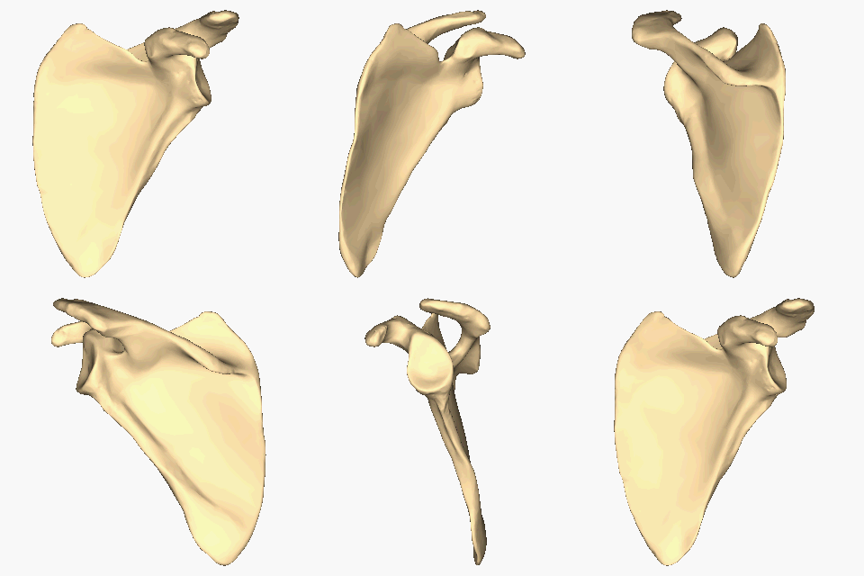
The scapula (plural scapulae or scapulas), also known as the shoulder blade, is the bone that connects the humerus (upper arm bone) with the clavicle (collar bone). Like their connected bones, the scapulae are paired, with each scapula on either side of the body being roughly a mirror image of the other. The name derives from the Classical Latin word for trowel or small shovel, which it was thought to resemble. In compound terms, the prefix omo- is used for the shoulder blade in medical terminology. This prefix is derived from ὦμος (ōmos), the Ancient Greek word for shoulder, and is cognate with the Latin , which in Latin signifies either the shoulder or the upper arm bone. The scapula forms the back of the shoulder girdle. In humans, it is a flat bone, roughly triangular in shape, placed on a posterolateral aspect of the thoracic cage. Structure The scapula is a thick, flat bone lying on the thoracic wall that provides an attachment for three groups of muscles: intrins ... [...More Info...] [...Related Items...] OR: [Wikipedia] [Google] [Baidu] |
Academic Press
Academic Press (AP) is an academic book publisher founded in 1941. It was acquired by Harcourt, Brace & World in 1969. Reed Elsevier bought Harcourt in 2000, and Academic Press is now an imprint of Elsevier. Academic Press publishes reference books, serials and online products in the subject areas of: * Communications engineering * Economics * Environmental science * Finance * Food science and nutrition * Geophysics * Life sciences * Mathematics and statistics * Neuroscience * Physical sciences * Psychology Well-known products include the ''Methods in Enzymology'' series and encyclopedias such as ''The International Encyclopedia of Public Health'' and the ''Encyclopedia of Neuroscience''. See also * Akademische Verlagsgesellschaft (AVG) — the German predecessor, founded in 1906 by Leo Jolowicz (1868–1940), the father of Walter Jolowicz Walter may refer to: People * Walter (name), both a surname and a given name * Little Walter, American blues harmonica player Marion Wa ... [...More Info...] [...Related Items...] OR: [Wikipedia] [Google] [Baidu] |
Pectoralis Minor Syndrome
Pectoralis minor syndrome (PMS) is a condition related to thoracic outlet syndrome (TOS) that results from the pectoralis minor muscle being too tight. PMS results from the brachial plexus being compressed under the pectoralis minor while TOS involves compression of the bundle above the clavicle. In most patients, the nerves are constricted resulting in neurogenic PMS, but venous compression (venous PMS) can also occur. PMS and TOS often, but not always, occur together. They share similar symptoms including tingling, pain, or weakness in the hand and arm, but in PMS there is also pain or tenderness in the chest wall The thoracic wall or chest wall is the boundary of the thoracic cavity. Structure The bone, bony human skeleton, skeletal part of the thoracic wall is the rib cage, and the rest is made up of muscle, skin, and fasciae. The chest wall has 10 lay ... where the pectoralis minor attaches to the scapula as well as in the armpit. One study of 100 patients diagnosed with ne ... [...More Info...] [...Related Items...] OR: [Wikipedia] [Google] [Baidu] |
Inferior Angle
The scapula (plural scapulae or scapulas), also known as the shoulder blade, is the bone that connects the humerus (upper arm bone) with the clavicle (collar bone). Like their connected bones, the scapulae are paired, with each scapula on either side of the body being roughly a mirror image of the other. The name derives from the Classical Latin word for trowel or small shovel, which it was thought to resemble. In compound terms, the prefix omo- is used for the shoulder blade in medical terminology. This prefix is derived from ὦμος (ōmos), the Ancient Greek word for shoulder, and is cognate with the Latin , which in Latin signifies either the shoulder or the upper arm bone. The scapula forms the back of the shoulder girdle. In humans, it is a flat bone, roughly triangular in shape, placed on a posterolateral aspect of the thoracic cage. Structure The scapula is a thick, flat bone lying on the thoracic wall that provides an attachment for three groups of muscles: intrins ... [...More Info...] [...Related Items...] OR: [Wikipedia] [Google] [Baidu] |
Thorax
The thorax or chest is a part of the anatomy of humans, mammals, and other tetrapod animals located between the neck and the abdomen. In insects, crustaceans, and the extinct trilobites, the thorax is one of the three main divisions of the creature's body, each of which is in turn composed of multiple segments. The human thorax includes the thoracic cavity and the thoracic wall. It contains organs including the heart, lungs, and thymus gland, as well as muscles and various other internal structures. Many diseases may affect the chest, and one of the most common symptoms is chest pain. Etymology The word thorax comes from the Greek θώραξ ''thorax'' "breastplate, cuirass, corslet" via la, thorax. Plural: ''thoraces'' or ''thoraxes''. Human thorax Structure In humans and other hominids, the thorax is the chest region of the body between the neck and the abdomen, along with its internal organs and other contents. It is mostly protected and supported by the rib cage, spi ... [...More Info...] [...Related Items...] OR: [Wikipedia] [Google] [Baidu] |
Shoulder
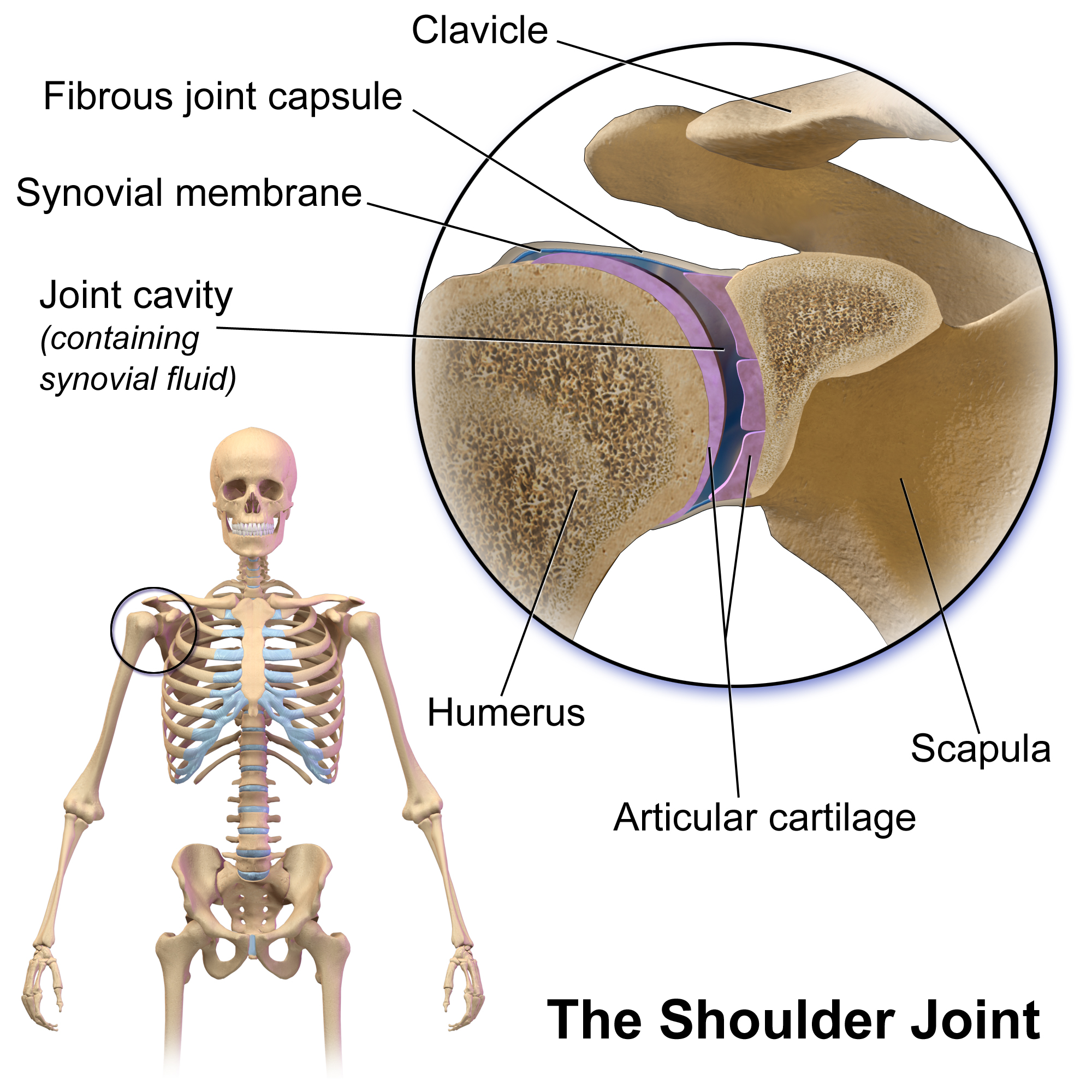
The human shoulder is made up of three bones: the clavicle (collarbone), the scapula (shoulder blade), and the humerus (upper arm bone) as well as associated muscles, ligaments and tendons. The articulations between the bones of the shoulder make up the shoulder joints. The shoulder joint, also known as the glenohumeral joint, is the major joint of the shoulder, but can more broadly include the acromioclavicular joint. In human anatomy, the shoulder joint comprises the part of the body where the humerus attaches to the scapula, and the head sits in the glenoid cavity. The shoulder is the group of structures in the region of the joint. The shoulder joint is the main joint of the shoulder. It is a ball and socket joint that allows the arm to rotate in a circular fashion or to hinge out and up away from the body. The joint capsule is a soft tissue envelope that encircles the glenohumeral joint and attaches to the scapula, humerus, and head of the biceps. It is lined by a thin, ... [...More Info...] [...Related Items...] OR: [Wikipedia] [Google] [Baidu] |
Depression (kinesiology)
Motion, the process of movement, is described using specific anatomical terms. Motion includes movement of organs, joints, limbs, and specific sections of the body. The terminology used describes this motion according to its direction relative to the anatomical position of the body parts involved. Anatomists and others use a unified set of terms to describe most of the movements, although other, more specialized terms are necessary for describing unique movements such as those of the hands, feet, and eyes. In general, motion is classified according to the anatomical plane it occurs in. ''Flexion'' and ''extension'' are examples of ''angular'' motions, in which two axes of a joint are brought closer together or moved further apart. ''Rotational'' motion may occur at other joints, for example the shoulder, and are described as ''internal'' or ''external''. Other terms, such as ''elevation'' and ''depression'', describe movement above or below the horizontal plane. Many anatomic ... [...More Info...] [...Related Items...] OR: [Wikipedia] [Google] [Baidu] |
Poland Syndrome
Poland syndrome is a birth defect characterized by an underdeveloped chest muscle and short webbed fingers on one side of the body. There may also be short ribs, less fat, and breast and nipple abnormalities on the same side of the body. Typically, the right side is involved. Those affected generally have normal movement and health. The cause of Poland syndrome is unknown. One theory is that it is due to disruption of blood flow during embryonic development. It is generally not inherited, and no genes that contribute to the disorder have been identified. Diagnosis of Poland syndrome is based on its symptoms. Often, those with the syndrome remain undiagnosed, and some may not realize they have it until puberty. Treatment of Poland syndrome depends on its severity and may include surgical correction. The syndrome affects about 1 in 20,000 newborns, and males are affected twice as often as females. It is named after English surgeon Sir Alfred Poland, who described the condition ... [...More Info...] [...Related Items...] OR: [Wikipedia] [Google] [Baidu] |
Medial Pectoral Nerve
The medial pectoral nerve (also known as the medial anterior thoracic nerve) arises from the medial cord (sometimes directly from the anterior division of the inferior trunk) of the brachial plexus, and through it from the eighth cervical and first thoracic roots (C8/T1). It passes behind the first part of the axillary artery, curves forward between the axillary artery and vein, and unites in front of the artery with a filament from the lateral nerve. It then enters the deep surface of the pectoralis minor muscle, where it divides into a number of branches, which supply the muscle. Two or three branches pierce the muscle and end in the sternocostal head of the pectoralis major muscle. The medial pectoral nerve pierces both the pectoralis minor and the sternocostal head of the pectoralis major. The lateral pectoral nerve pierces only the clavicular head of the pectoralis major. Clinical relevance The medial pectoral nerve can be used as a donor nerve when reconstructing a ... [...More Info...] [...Related Items...] OR: [Wikipedia] [Google] [Baidu] |
Clavipectoral Fascia
The clavipectoral fascia (costocoracoid membrane; coracoclavicular fascia) is a strong fascia situated under cover of the clavicular portion of the pectoralis major. It occupies the interval between the pectoralis minor and subclavius, and protects the axillary vein and artery, and axillary nerve. Traced upward, it splits to enclose the subclavius, and its two layers are attached to the clavicle, one in front of and the other behind the muscle; the deep layer fuses with the deep cervical fascia and with the sheath of the axillary vessels. Medially, it blends with the fascia covering the first two intercostal spaces, and is attached also to the first rib medial to the origin of the subclavius. Laterally, it is very thick and dense, and is attached to the coracoid process. The portion extending from the first rib to the coracoid process is often whiter and denser than the rest, and is sometimes called the costocoracoid membrane. Below this it is thin, and at the upper border of ... [...More Info...] [...Related Items...] OR: [Wikipedia] [Google] [Baidu] |
Axilla
The axilla (also, armpit, underarm or oxter) is the area on the human body directly under the shoulder joint. It includes the axillary space, an anatomical space within the shoulder girdle between the arm and the thoracic cage, bounded superiorly by the imaginary plane between the superior borders of the first rib, clavicle and scapula (above which are considered part of the neck), medially by the serratus anterior muscle and thoracolumbar fascia, anteriorly by the pectoral muscles and posteriorly by the subscapularis, teres major and latissimus dorsi muscle. The soft skin covering the lateral axilla contains many hair and sweat glands. In humans, the formation of body odor happens mostly in the axilla. These odorant substances have been suggested by some to serve as pheromones, which play a role related to mate selection, although this is a controversial topic within the scientific community. The underarms seem more important than the pubic area for emitting body odor, whi ... [...More Info...] [...Related Items...] OR: [Wikipedia] [Google] [Baidu] |


