|
Pathologic Fracture
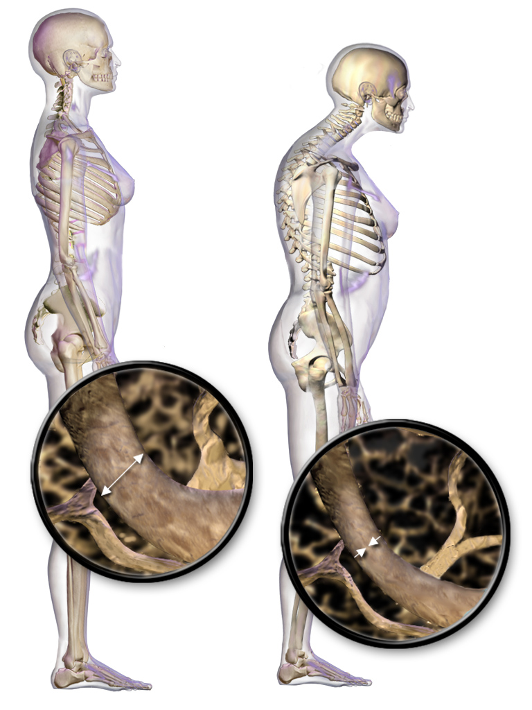
A pathologic fracture is a bone fracture caused by weakness of the bone structure that leads to decrease mechanical resistance to normal mechanical loads. This process is most commonly due to osteoporosis, but may also be due to other pathologies such as cancer, infection (such as osteomyelitis), inherited bone disorders, or a bone cyst. Only a small number of conditions are commonly responsible for pathological fractures, including osteoporosis, osteomalacia, Paget's disease, Osteitis, osteogenesis imperfecta, benign bone tumours and cysts, secondary malignant bone tumours and primary malignant bone tumours. Fragility fracture is a type of pathologic fracture that occurs as a result of an injury that would be insufficient to cause fracture in a normal bone. There are three fracture sites said to be typical of fragility fractures: vertebral fractures, fractures of the neck of the femur, and Colles fracture of the wrist. This definition arises because a normal human being ought to ... [...More Info...] [...Related Items...] OR: [Wikipedia] [Google] [Baidu] |
Metastasis
Metastasis is a pathogenic agent's spread from an initial or primary site to a different or secondary site within the host's body; the term is typically used when referring to metastasis by a cancerous tumor. The newly pathological sites, then, are metastases (mets). It is generally distinguished from cancer invasion, which is the direct extension and penetration by cancer cells into neighboring tissues. Cancer occurs after cells are genetically altered to proliferate rapidly and indefinitely. This uncontrolled proliferation by mitosis produces a primary heterogeneic tumour. The cells which constitute the tumor eventually undergo metaplasia, followed by dysplasia then anaplasia, resulting in a malignant phenotype. This malignancy allows for invasion into the circulation, followed by invasion to a second site for tumorigenesis. Some cancer cells known as circulating tumor cells acquire the ability to penetrate the walls of lymphatic or blood vessels, after which they are abl ... [...More Info...] [...Related Items...] OR: [Wikipedia] [Google] [Baidu] |
Compression Fracture
A compression fracture is a collapse of a vertebra. It may be due to trauma or due to a weakening of the vertebra (compare with burst fracture). This weakening is seen in patients with osteoporosis or osteogenesis imperfecta, lytic lesions from metastatic or primary tumors, or infection. In healthy patients, it is most often seen in individuals suffering extreme vertical shocks, such as ejecting from an ejection seat. Seen in lateral views in plain x-ray films, compression fractures of the spine characteristically appear as ''wedge deformities'', with greater loss of height anteriorly than posteriorly and intact pedicles in the anteroposterior view. Signs and symptoms Acute fractures will cause severe back pain. Compression fractures which develop gradually, such as in osteoporosis, may initially not cause any symptoms, but will later often lead to back pain and loss of height. Diagnosis Compression fractures are usually diagnosed on spinal radiographs, where a wedge-shaped vert ... [...More Info...] [...Related Items...] OR: [Wikipedia] [Google] [Baidu] |
Monostotic Fibrous Dysplasia
Monostotic fibrous dysplasia is a form of fibrous dysplasia where only one bone is involved. It comprises a majority of the cases of fibrous dysplasia (approximately 70–80%). It is a rare bone disease characterized by the replacement of normal elements of the bone by fibrous connective tissue, which can cause very painful swellings and bone deformities, and make bone abnormally fragile and prone to fracture. A congenital, noninherited, benign anomaly of bone development in a single bone, it consists of the replacement of normal marrow and cancellous bone by immature bone with fibrous stroma. Monostotic fibrous dysplasia occurs with equal frequency in both sexes and normally develops early in life, with lesions frequently identified late in the first and early second decades. Most patients are asymptomatic, with the diagnosis often established after an incidental finding or with pain, swelling, or fracture. Lesions usually enlarge in proportion to skeletal growth and the abnorm ... [...More Info...] [...Related Items...] OR: [Wikipedia] [Google] [Baidu] |
Juvenile Rheumatoid Arthritis
{{Disambiguation ...
Juvenile may refer to: *Juvenile status, or minor (law), prior to adulthood * Juvenile (organism) *Juvenile (rapper) (born 1975), American rapper * ''Juvenile'' (2000 film), Japanese film * ''Juvenile'' (2017 film) *Juvenile (greyhounds), a greyhound competition *Juvenile particles, a type of volcanic ejecta *A two-year-old horse in horse racing terminology See also *"The Juvenile", a song by Ace of Base *Juvenile novel **Any of "Heinlein juveniles" *Juvenile delinquency * Juvenilia, works by an author while a youth *Juvenal (other) Juvenal was a poet. Juvenal or Juvenals may also refer to: * Juvenal (name), and persons with the name * Juvenals, a student society * An immature bird {{disambiguation ... [...More Info...] [...Related Items...] OR: [Wikipedia] [Google] [Baidu] |
Juvenile Osteoporosis
Juvenile osteoporosis is osteoporosis in children and adolescents. Osteoporosis is rare in children and adolescents. When it occurs, it is usually secondary to some other condition, ''e.g.'' osteogenesis imperfecta, rickets Rickets is a condition that results in weak or soft bones in children, and is caused by either dietary deficiency or genetic causes. Symptoms include bowed legs, stunted growth, bone pain, large forehead, and trouble sleeping. Complications ma ..., eating disorders or arthritis. In some cases, there is no known cause and it is called idiopathic juvenile osteoporosis. Idiopathic juvenile osteoporosis usually goes away spontaneously. Also, child abuse should be suspected in recurring cases of bone fracture. Cause Diagnosis Treatment Treatment for secondary juvenile osteoporosis focuses on treating any underlying disorder. Treatment of Juvenile osteoporosis can also include maintaining a healthy lifestyle. This is accomplished by exercising, keeping a b ... [...More Info...] [...Related Items...] OR: [Wikipedia] [Google] [Baidu] |
Malignant Infantile Osteopetrosis
Malignant infantile osteopetrosis is a rare osteosclerosing type of skeletal dysplasia that typically presents in infancy and is characterized by a unique radiographic appearance of generalized hyperostosis (excessive growth of bone). The generalized increase in bone density has a special predilection to involve the medullary portion with relative sparing of the cortices.EL-Sobky TA, Elsobky E, Sadek I, Elsayed SM, Khattab MF (2016)"A case of infantile osteopetrosis: The radioclinical features with literature update' ''Bone Rep''. 4:11-16doi:10.1016/j.bonr.2015.11.002 Obliteration of bone marrow spaces and subsequent depression of the cellular function can result in serious hematologic complications. Optic atrophy and cranial nerve damage secondary to bony expansion can result in marked morbidity. The prognosis is extremely poor in ... [...More Info...] [...Related Items...] OR: [Wikipedia] [Google] [Baidu] |


